. 2012;7(12):e50367.
doi: 10.1371/journal.pone.0050367. Epub 2012 Dec 10.

Obstruction of photoinduced electron transfer from excited porphyrin to graphene oxide: a fluorescence turn-on sensing platform for iron (III) ions

Affiliations

Obstruction of photoinduced electron transfer from excited porphyrin to graphene oxide: a fluorescence turn-on sensing platform for iron (III) ions

Zhong De Liu et al. PLoS One. 2012.

Abstract

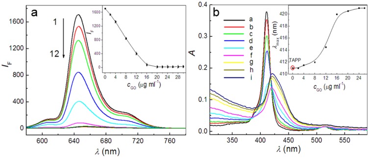
A comparative research of the assembly of different porphyrin molecules on graphene oxide (GO) and reduced graphene oxide (RGO) was carried out, respectively. Despite the cationic porphyrin molecules can be assembled onto the surfaces of graphene sheets, including GO and RGO, to form complexes through electrostatic and π-π stacking interactions, the more obvious fluorescence quenching and the larger red-shift of the Soret band of porphyrin molecule in RGO-bound states were observed than those in GO-bound states, due to the difference of molecular flattening in degree. Further, more interesting finding was that the complexes formed between cationic porphyrin and GO, rather than RGO sheets, can facilitate the incorporation of iron (III) ions into the porphyrin moieties, due to the presence of the oxygen-contained groups at the basal plane of GO sheets served as auxiliary coordination units, which can high-efficiently obstruct the electron transfer from excited porphyrin to GO sheets and result in the occurrence of fluorescence restoration. Thus, a fluorescence sensing platform has been developed for iron (III) ions detection in this contribution by using the porphyrin/GO nanohybrids as an optical probe, and our present one exhibited rapid and sensitive responses and high selectivity toward iron (III) ions.


Conflict of interest statement

Competing Interests: The authors have declared that no competing interests exist.

Figures

Figure 1
Figure 1. Schematic illustrations of PET from TAPP to GO sheets, and iron (III) ions selectively obstructing the process of PET.
Figure 2
Figure 2. Fluorescence and absorption spectra recorded during addition of different concentrations of GO suspension to TAPP solution.
The inset in Figure 2a shows that the variation of fluorescence intensity of TAPP at 645.0 nm varies with the increasing concentrations of GO. Concentrations: TAPP, 2.4 µM; GO from curve 2 to 12 (µg ml−1), 2.0, 4.0, 8.0, 12.0, 16.0, 20.0, 22.0, 24.0, 26.0, 28.0, 30.0. λ ex, 413.0 nm. The inset in Figure 2b shows that the variation of maximum absorption wavelength (λ max) of TAPP varies with the addition of GO. Concentration: TAPP, 2.4 µM; GO from curve b to i (µg ml−1), 2.0, 4.0, 8.0, 12.0, 16.0, 20.0, 24.0, 26.0. pH, 4.1.
Figure 3
Figure 3. Fluorescence and absorption spectra recorded during addition of different concentrations of RGO suspension to TAPP solution.
The inset in Figure 3a shows that the variation of fluorescence intensity of TAPP at 645.0 nm varies with the increasing concentrations of RGO. Concentrations: TAPP, 2.4 µM; RGO from curve 2 to 10 (µg ml−1), 2.2, 4.5, 6.8, 9.0, 11.2, 13.5, 15.8, 18.0, 22.5. λ ex, 413.0 nm. The concentration of RGO from curve b to f in Figure 3b (µg ml−1), 2.2, 4.5, 6.8, 9.0, 13.5. pH, 4.1.
Figure 4
Figure 4. AFM images of GO, TAPP/GO complex and that in presence of iron (III) ions on mica substrate together with section analysis along the scored line.
(a) GO; (b) TAPP/GO complex; (c) TAPP/GO complex in presence of iron (III) ions. Concentration: GO, 16.0 µg ml−1; TAPP, 2.4 µM; iron (III) ions, 8.0 µM.
Figure 5
Figure 5. Fluorescence and absorption spectra recorded during addition of the increasing concentrations of iron (III) ions to TAPP/GO complex solution.
The inset in Figure 5a shows that the enhanced fluorescence intensity at 645.0 nm varies with the increasing concentrations of the iron (III) ions. Concentration: TAPP, 2.4 µM; GO, 16.0 µg ml−1; iron (III) ions from curve 2 to 17 (µM), 0.3, 0.8, 1.0, 2.0, 3.0, 4.0, 5.0, 8.0, 10.0, 13.0, 15.0, 20.0, 30.0, 40.0, 50.0, 60.0. λ ex, 413.0 nm. The inset in Figure 5b is to intuitively display the blue-shift of Soret band of TAPP in GO-bound state with the addition of increasing concentration of iron (III) ions. Concentration: TAPP, 2.4 µM; GO except for curve a (µg ml−1), 16.0; iron (III) ions from curve c to j (µM), 0.3, 0.8, 1.0, 2.0, 4.0, 5.0, 8.0, 13.0. pH, 4.1.
Figure 6
Figure 6. Fluorescence spectra of the TAPP, TAPP/RGO complex and that in presence of iron (III) ions.
Concentrations: TAPP, 2.4 µM; RGO, 13.5 µg ml−1; iron (III) ions, 60.0 µM. λ ex, 413.0 nm, pH, 4.1.
Figure 7
Figure 7. XPS wide-scan survey of the fresh obtained TAPP/GO (black line) and TAPP/RGO (red line) complexes exposed to the contained iron (III) ions solution, respectively.
Inset shows detailed XPS survey of the Fe 2p region.
Figure 8
Figure 8. Time-resolved fluorescence Decays of the TAPP and TMPyP recorded at different GO concentrations and iron (III) ions.
Concentration: TAPP, 2.4 µM, TMPyP, 3.0 µM; GO from curve b to i (µg ml−1), 8.6, 13.0, 17.3, 21.6, 21.6, 21.6, 21.6, 21.6, 21.6; iron (III) ions from curve f to i (µM), 2.0, 4.0, 8.0, 16.0. pH, 4.1.
Figure 9
Figure 9. Fluorescence response of TAPP/GO complex to various metal ions.
Concentration: TAPP, 2.4 µM; GO, 16.0 µg ml−1; iron (III) ions, 15.0 µM; Other metal ions were all 30.0 µM. All data were collected at 645.0 nm.

References

    1. Li D, Kaner RB (2008) Graphene-based materials. Science 320: 1170–1171. - PubMed
    1. Dikin DA, Stankovich S, Zimney EJ, Piner RD, Dommett GHB, et al. (2007) Preparation and characterization of graphene oxide paper. Nature 448: 457–460. - PubMed
    1. Bunch SJ, van der Zande AM, Verbridge SS, Frank IW, Tanenbaum DM, et al. (2007) Electromechanical resonators from graphene sheets. Science 315: 490–493. - PubMed
    1. Geim AK, Novoselov KS (2007) The rise of graphene. Nat Mater 6: 183–191. - PubMed
    1. Xu YF, Liu ZB, Zhang XL, Wang Y, Tian JG, et al. (2009) A graphene hybrid material covalently functionalized with porphyrin: synthesis and optical limiting property. Adv Mater 21: 1275–1279.

Publication types