. 2012;7(12):e50804.
doi: 10.1371/journal.pone.0050804. Epub 2012 Dec 10.

Epithelial-stromal interactions in human breast cancer: effects on adhesion, plasma membrane fluidity and migration speed and directness

Affiliations

Epithelial-stromal interactions in human breast cancer: effects on adhesion, plasma membrane fluidity and migration speed and directness

Cristiana Angelucci et al. PLoS One. 2012.

Abstract

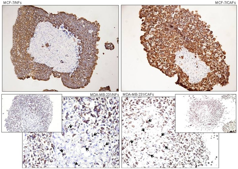
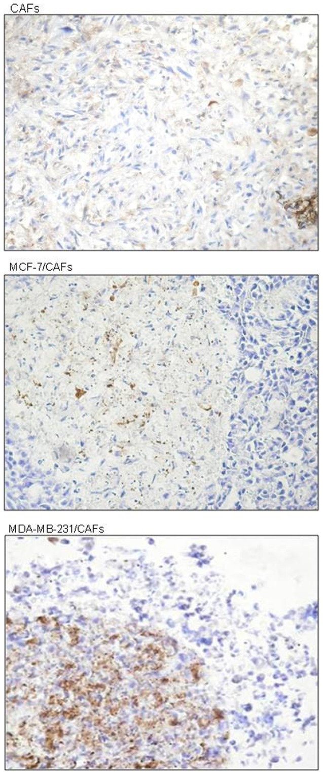
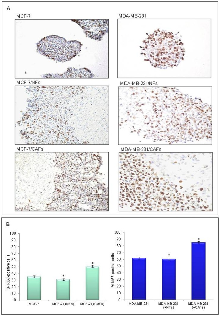
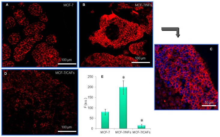
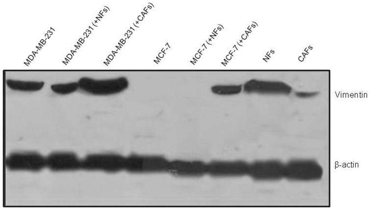
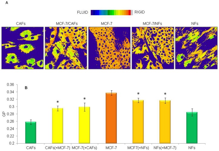
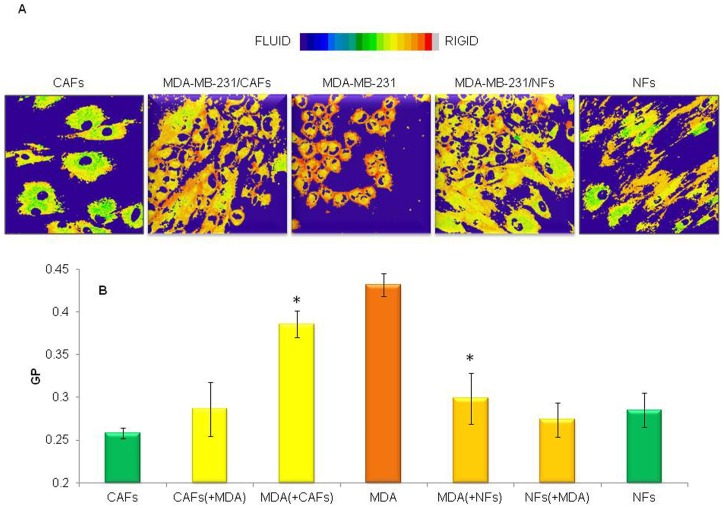
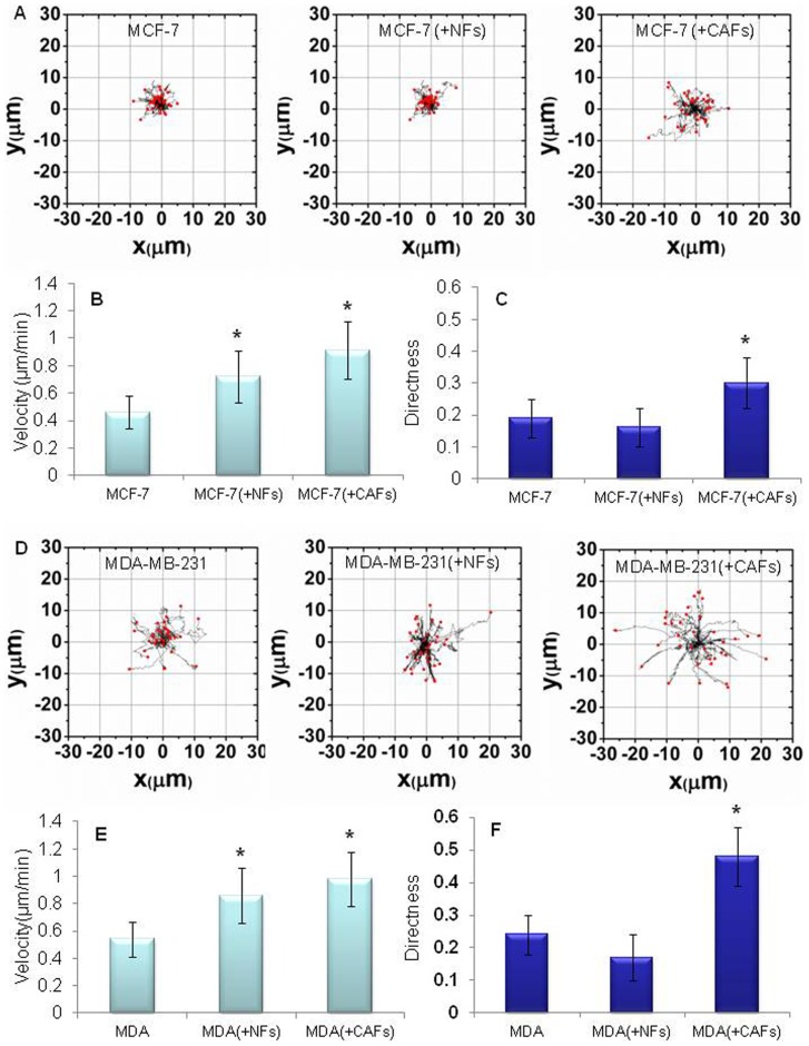
Interactions occurring between malignant cells and the stromal microenvironment heavily influence tumor progression. We investigated whether this cross-talk affects some molecular and functional aspects specifically correlated with the invasive phenotype of breast tumor cells (i.e. adhesion molecule expression, membrane fluidity, migration) by co-culturing mammary cancer cells exhibiting different degrees of metastatic potential (MDA-MB-231>MCF-7) with fibroblasts isolated from breast healthy skin (normal fibroblasts, NFs) or from breast tumor stroma (cancer-associated fibroblasts, CAFs) in 2D or 3D (nodules) cultures. Confocal immunofluorescence analysis of the epithelial adhesion molecule E-cadherin on frozen nodule sections demonstrated that NFs and CAFs, respectively, induced or inhibited its expression in MCF-7 cells. An increase in the mesenchymal adhesion protein N-cadherin was observed in CAFs, but not in NFs, as a result of the interaction with both kinds of cancer cells. CAFs, in turn, promoted N-cadherin up-regulation in MDA-MB-231 cells and its de novo expression in MCF-7 cells. Beyond promotion of "cadherin switching", another sign of the CAF-triggered epithelial-mesenchymal transition (EMT) was the induction of vimentin expression in MCF-7 cells. Plasma membrane labeling of monolayer cultures with the fluorescent probe Laurdan showed an enhancement of the membrane fluidity in cancer cells co-cultured with NFs or CAFs. An increase in lipid packing density of fibroblast membranes was promoted by MCF-7 cells. Time-lapsed cell tracking analysis of mammary cancer cells co-cultured with NFs or CAFs revealed an enhancement of tumor cell migration velocity, even with a marked increase in the directness induced by CAFs.Our results demonstrate a reciprocal influence of mammary cancer and fibroblasts on various adhesiveness/invasiveness features. Notably, CAFs' ability to promote EMT, reduction of cell adhesion, increase in membrane fluidity, and migration velocity and directness in mammary cancer cells can be viewed as an overall progression- and invasion-promoting effect.


Conflict of interest statement

Competing Interests: One of the authors Riccardo Masetti is the president of the Italian affiliate of the Susan G. Komen Breast Cancer Foundation. The other authors have declared that no competing interests exist.

Figures

Figure 1
Figure 1. Morphology of isolated normal and tumor-derived fibroblasts.
Light microscopic view showing the different morphology of fibroblasts isolated from human healthy mammary skin (A) or human breast cancer (B) samples after 6 days of culture. Original magnification, ×200.
Figure 2
Figure 2. Spatial distribution of breast tumor cells and fibroblasts in nodule co-cultures.
Sections (10 µm) of paraformaldehyde-fixed frozen nodule co-cultures of either MCF-7 or MDA-MB-231 breast tumor cells with fibroblasts (NFs or CAFs) at day 6 of culture are shown. Sections were stained immunohistochemically using an anti-cytokeratin (large spectrum, pankeratin) monoclonal antibody to identify the reciprocal location of the two cell types (tumor cells and fibroblasts) in the nodule co-cultures. The more invasive and less differentiated MDA-MB-231 cells showed a looser association as compared to MCF-7/fibroblast co-cultures, with various cells infiltrating the fibroblast nodule core (arrows). DAB for detection; hematoxylin counterstaining; original magnification for upper panels (MCF-7/NFs, MCF-7/CAFs) and lower insets, ×100; original magnification for lower panels (MDA-MB-231/NFs and MDA-MB-231/CAFs), ×200.
Figure 3
Figure 3. Breast tumor cell induction of α-smooth muscle actin (α-SMA) in tumor stroma-derived fibroblasts.
Sections (10 µm) of paraformaldehyde-fixed frozen nodule culture (CAFs) and co-cultures (MCF-7 or MDA-MB-231 breast tumor cells with CAFs) at day 6 of culture are shown. Sections were stained immunohistochemically using an anti-α-SMA monoclonal antibody to verify the effect of breast cancer cells on fibroblast-to-myofibroblast transdifferentiation. α-SMA expression was induced in the tumor-associated fibroblast population as a result of the interaction with the more aggressive and less differentiated MDA-MB-231 cells, demonstrating the ability of these cells to promote stroma activation. DAB for detection; hematoxylin counterstaining; original magnification, ×200.
Figure 4
Figure 4. Cancer-associated fibroblasts but not their normal counterpart exerted mitogenic effect on breast cancer cells.
(A) Sections (10 µm) of paraformaldehyde-fixed frozen nodule cultures (MCF-7 or MDA-MB-231 breast tumor cells) and co-cultures (MCF-7 or MDA-MB-231 cells with NFs or CAFs) at day 6 of culture are shown. Sections were stained immunohistochemically using an anti-Ki-67 monoclonal antibody (MIB-1 clone) to verify the reciprocal effect of the two cell types on their respective growth rate. The MCF-7 cell/CAF mixed nodules showed a higher number of Ki67-positive tumor cells with respect to that observed in MCF-7 cell/NF or MCF-7 cell nodules. Co-cultures of MDA-MB-231 cells and fibroblasts showed the same behavior as observed for MCF-7 cell/fibroblast nodule co-cultures.. DAB for detection; hematoxylin counterstaining; original magnification, ×200. (B) Quantitative representation of the immunohistochemical data. Three sections, obtained from 3 different nodules, were analyzed by two independent observers. The number of Ki67-positive cells (on a total of at least 800 cells) was assessed in four randomized high-power fields (×400) for each section. A mean percentage for every sample was obtained and data were reported as mean±SD (error bars). Statistical significance was determined using ANOVA and the Tukey's multiple comparison test, *p<0.001.
Figure 5
Figure 5. Cancer-associated fibroblasts and their normal counterpart affected E-cadherin expression in breast cancer cells.
Frozen nodule sections from MCF-7 cell homotypic cultures (A) or MCF-7 cell/fibroblast mixed cultures (B and D) were processed for E-cadherin immunostaining (Cy3: red). DAPI was used for nuclear counterstaining (blue) (C). Confocal images show the up-regulation of E-cadherin in MCF-7 cell population co-cultured with NFs (B) as compared to the MCF-7 homotypic culture (A). On the contrary, interaction with CAFs promoted a strong reduction in E-cadherin expression (D). A representative experiment of three is shown. (E) F (a.u.), fluorescence intensity (in arbitrary units). Data are mean±SD (error bars) of three independent experiments. Statistical significance was determined using Student's t-test, *p<0.05.
Figure 6
Figure 6. CAFs induced EMT in breast tumor cells.
Western blot analysis revealed that the vimentin protein expression was clearly induced in the low-invasive MCF7 cell line co-cultured with CAFs. Vimentin levels were increased in the highly invasive MDA-MB-231 cells co-cultured with CAFs as compared with the control (MDA-MB-231 cells cultured alone). No effect on vimentin expression was found in breast tumor cells co-cultured with NFs.
Figure 7
Figure 7. Low invasive breast cancer cells and fibroblasts reciprocally influenced their plasma membrane fluidity.
Pseudocoloured GP images of living tumor (MCF-7) and fibroblast (NFs or CAFs) cells (A), and reciprocal effects on membrane fluidity (B) are shown. MCF-7 cells were cultured for 6 days, alone or in presence of NFs or CAFs, in 35 mm glass-bottom Petri dishes and labelled with Laurdan (2 µM). Tumor cell/fibroblast interaction determined an enhancement of cancer cell membrane fluidity. On the other hand, MCF-7 cells promoted an increase in fibroblast membrane packing density. Data are mean±SD (error bars) of three independent experiments. Statistical significance was determined using Student's t-test, *p<0.05 vs respective homotypic culture.
Figure 8
Figure 8. Highly invasive and metastatic breast cancer cells underwent a plasma membrane fluidity increase when co-cultured with normal or cancer-associated fibroblasts.
Pseudocoloured GP images of living tumor (MDA-MB-231) and fibroblast (NFs or CAFs) cells (A), and reciprocal effects on plasma membrane fluidity (B) are shown. MDA-MB-231 cells were cultured for 6 days, alone or in presence of NFs or CAFs, in 35 mm glass-bottom Petri dishes and labelled with Laurdan (2 µM). Tumor cell/fibroblast interaction determined an enhancement of cancer cell membrane fluidity, while MDA-MB-231 cells did not affect fibroblast membrane packing density. Data are mean±SD (error bars) of three independent experiments. Statistical significance was determined using Student's t-test, *p<0.05 vs respective homotypic culture.
Figure 9
Figure 9. Breast tumor cells displayed increased migration velocity and directness in vitro due to their interaction with fibroblasts.
Representative trajectory plots of MCF-7 (A) or MDA-MB-231 cells (D) evaluated over a time period of 45 min. Cells were cultured for 6 days, alone or in presence of NFs or CAFs, in 35 mm glass-bottom Petri dishes and labelled with the CellTracker Green CMFDA. The starting point of each cell trajectory is plotted at the center of the graph. A significant increment of migration velocity (in µm/min) of MCF-7 (B) or MDA-MB-231 (E) cells was induced by the interaction with both CAFs and NFs. A marked enhancement of the tumor cell migration directness was promoted by CAFs in MCF-7 (A, D) or MDA-MB-231 (C, F) cells. Data are mean±SD (error bars) of three independent experiments. Statistical significance was determined using Student's t-test, *p<0.05.

References

    1. Shekhar MP, Werdell J, Santner SJ, Pauley RJ, Tait L (2001) Breast stroma plays a dominant regulatory role in breast epithelial growth and differentiation: implications for tumor development and progression. Cancer Res 61: 1320–1326. - PubMed
    1. Wiseman BS, Werb Z (2002) Stromal effects on mammary gland development and breast cancer. Science 296: 1046–1049. - PMC - PubMed
    1. Polyak K, Kalluri R (2010) The role of the microenvironment in mammary gland development and cancer. Cold Spring Harb Perspect Biol 2: a003244. - PMC - PubMed
    1. Elenbaas B, Weinberg RA (2001) Heterotypic signaling between epithelial tumor cells and fibroblasts in carcinoma formation. Exp Cell Res 264: 169–184. - PubMed
    1. Wang TN, Albo D, Tuszynski GP (2002) Fibroblasts promote breast cancer cell invasion by upregulating tumor matrix metalloproteinase-9 production. Surgery 132: 220–225. - PubMed

Publication types