. 2012;7(12):e51168.
doi: 10.1371/journal.pone.0051168. Epub 2012 Dec 10.

Expression of mutant huntingtin in leptin receptor-expressing neurons does not control the metabolic and psychiatric phenotype of the BACHD mouse

Affiliations

Expression of mutant huntingtin in leptin receptor-expressing neurons does not control the metabolic and psychiatric phenotype of the BACHD mouse

Sofia Hult Lundh et al. PLoS One. 2012.

Abstract

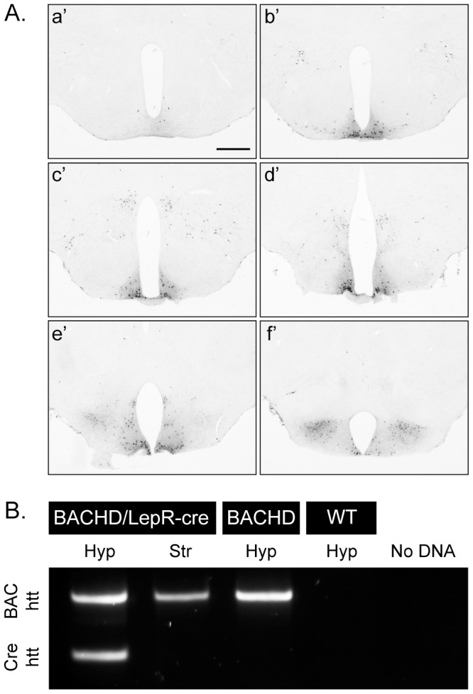
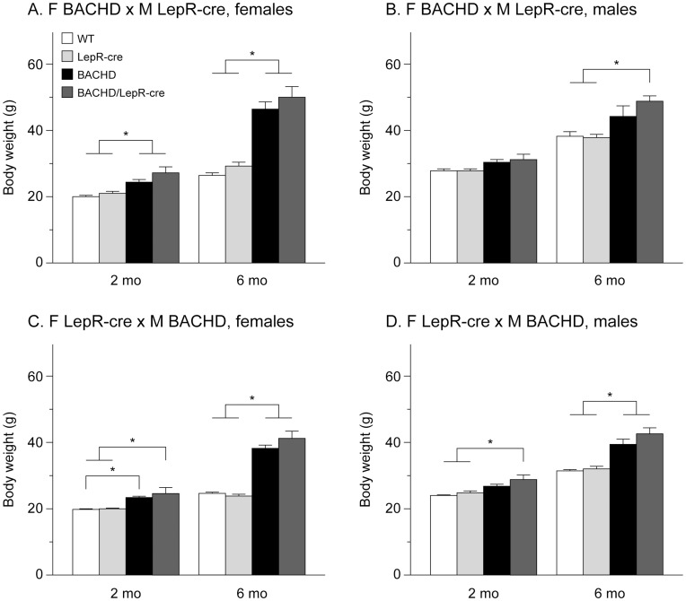
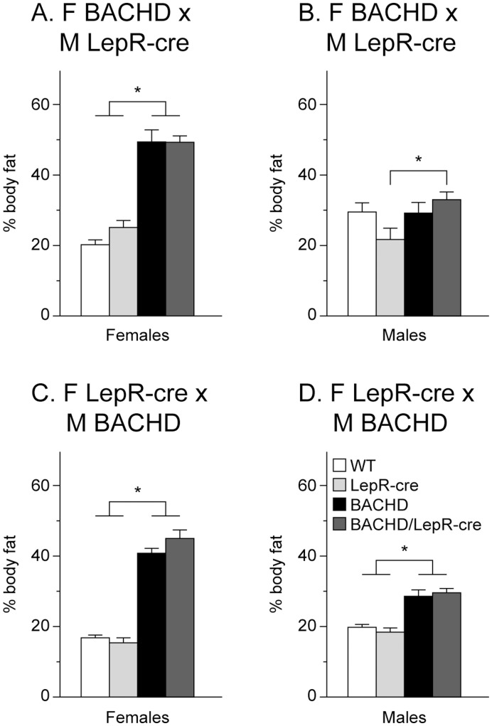
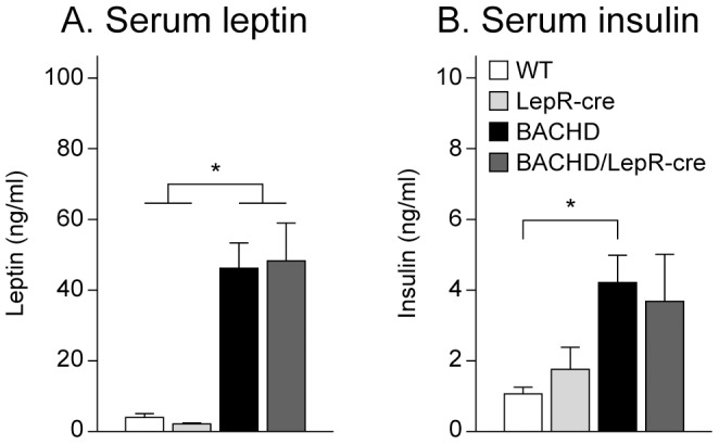
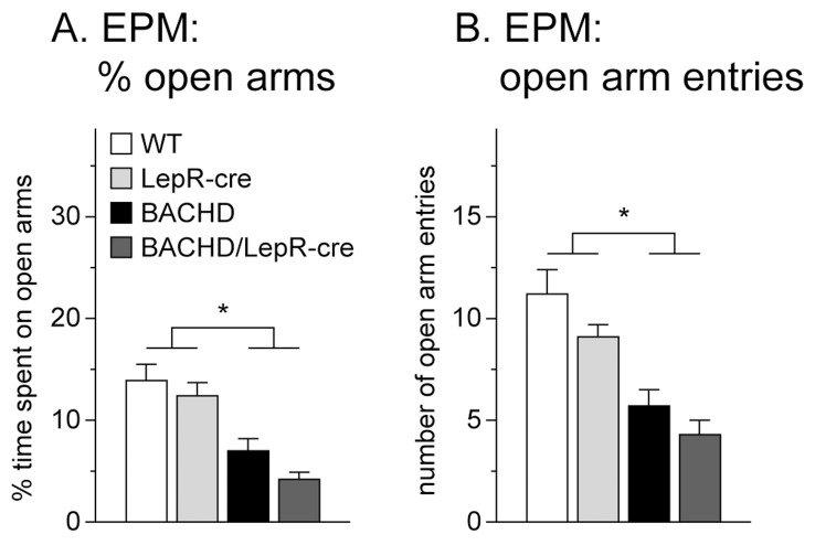
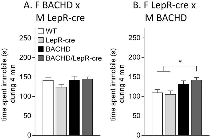
Metabolic and psychiatric disturbances occur early on in the clinical manifestation of Huntington's disease (HD), a neurodegenerative disorder caused by an expanded CAG repeat in the huntingtin (HTT) gene. Hypothalamus has emerged as an important site of pathology and alterations in this area and its neuroendocrine circuits may play a role in causing early non-motor symptoms and signs in HD. Leptin is a hormone that controls energy homeostasis by signaling through leptin receptors in the hypothalamus. Disturbed leptin action is implicated in both obesity and depression and altered circulating levels of leptin have been reported in both clinical HD and rodent models of the disease. Pathological leptin signaling may therefore be involved in causing the metabolic and psychiatric disturbances of HD. Here we tested the hypothesis that expression of mutant HTT in leptin receptor carrying neurons plays a role in the development of the non-motor phenotype in the BACHD mouse model. Our results show that inactivation of mutant HTT in leptin receptor-expressing neurons in the BACHD mouse using cross-breeding based on a cre-loxP system did not have an effect on the metabolic phenotype or anxiety-like behavior. The data suggest that mutant HTT disrupts critical hypothalamic pathways by other mechanisms than interfering with intracellular leptin signaling.


Conflict of interest statement

Competing Interests: The authors have declared that no competing interests exist.

Figures

Figure 1
Figure 1. Demonstration and validation of Cre-excision.
(A) GFP staining of hypothalamic sections from crossbred LepR-cre mice×ROSA-eYFP mice illustrates the cre-excision pattern in the hypothalamus, a’f’ represents rostral (bregma −1) to caudal (bregma −2.5) hypothalamic sections, scale bar = 500 µm. (B) PCR analysis confirmed successful excision of human mutant htt exon1 in leptin receptor-expressing neurons in the hypothalamus, but not in a region (striatum), which lacks leptin receptor-expressing neurons.
Figure 2
Figure 2. Body weight in BACHD×LepR-cre offspring.
Body weight at 2- and 6-months in males and females from the two different breedings between BACHD and BACHD/LepR-cre (n = 4–10/genotype/sex/breeding). Both BACHD and BACHD/LepR-cre developed early onset obesity but no significant differences between BACHD and BACHD/LepR-cre could be detected in any of the breedings or sexes. (A) F BACHD×M LepR-cre, females, (B) F BACHD×M LepR-cre, males, (C) F LepR-cre×M BACHD, females, and (D) F LepR-cre×M BACHD, males. All data are expressed as means ± SEM. Significant differences from WT and/or LepR-cre mice: * p<0.05 (see Statistical Results S1).
Figure 3
Figure 3. Body composition in BACHD×LepR-cre offspring.
The graphs show percentage body fat as assessed with dual energy x-ray absorptiometry (DEXA) at 6-months in males and females from the two different breedings (n = 4–10/genotype/sex/breeding). Both BACHD and BACHD/LepR-cre showed increased percentage of body fat but there were no significant differences between BACHD and BACHD/LepR-cre in the two breedings and sexes. (A) F BACHD×M LepR-cre, females, (B) F BACHD×M LepR-cre, males, (C) F LepR-cre×M BACHD, females, and (D) F LepR-cre×M BACHD, males. All data are expressed as means ± SEM. Significant differences from WT and/or LepR-cre mice: * p<0.05 (see Statistical Results S1).
Figure 4
Figure 4. Endocrine measurements in BACHD×LepR-cre offspring.
Circulating levels of leptin and insulin were measured in 6-month old female mice from the second breeding (n = 9–10/genotype). (A) Both BACHD and BACHD/LepR-cre showed elevated leptin levels but no significant differences between BACHD and BACHD/LepR-cre could be detected. (B) Only BACHD mice displayed significantly higher insulin levels than wt mice. However, no significant differences could be detected between BACHD and BACHD/LepR-cre. All data are expressed as means ± SEM. Significant differences from WT and/or LepR-cre mice: * p<0.05 (see Statistical Results S1).
Figure 5
Figure 5. Assessment of anxiety-like behavior in BACHD×LepR-cre offspring.
Both BACHD and BACHD/LepR-Cre displayed anxiety-like behavior at 6 months of age, with reduced time spent on open arms (A) as well as reduced number of entries onto the open arms (B) in the EPM at 6-months (n = 4–10/genotype/sex/breeding). However, no difference could be detected between BACHD and BACHD/LepR-cre mice. All data are expressed as means ± SEM. Significant differences from WT and LepR-cre mice: * p<0.05 (see Statistical Results S1).
Figure 6
Figure 6. Assessment of depressive-like behavior in BACHD×LepR-cre offspring.
No differences in immobility could be detected in the FST between any of the genotypes in the breeding between F BACHD and M LepR-cre (A). For the F LepR-cre×M BACHD breeding (B), LepR-cre mice spent significantly more time immobile than WT and LepR-cre mice. (n = 4–10/genotype/sex/breeding). All data are expressed as means ± SEM. significant differences from WT and LepR-cre mice: * p<0.05 (see Statistical Results S1).

References

    1. HDCRG (1993) A novel gene containing a trinucleotide repeat that is expanded and unstable on Huntington’s disease chromosomes. The Huntington’s Disease Collaborative Research Group. Cell 72: 971–983. - PubMed
    1. Ross CA, Tabrizi SJ (2011) Huntington’s disease: from molecular pathogenesis to clinical treatment. Lancet Neurol 10: 83–98. - PubMed
    1. Sturrock A, Leavitt BR (2010) The clinical and genetic features of Huntington disease. J Geriatr Psychiatry Neurol 23: 243–259. - PubMed
    1. Aziz NA, Swaab DF, Pijl H, Roos RA (2007) Hypothalamic dysfunction and neuroendocrine and metabolic alterations in Huntington’s disease: clinical consequences and therapeutic implications. Rev Neurosci 18: 223–251. - PubMed
    1. Lalic NM, Maric J, Svetel M, Jotic A, Stefanova E, et al. (2008) Glucose homeostasis in Huntington disease: abnormalities in insulin sensitivity and early-phase insulin secretion. Arch Neurol 65: 476–480. - PubMed

Publication types