. 2012;7(12):e51242.
doi: 10.1371/journal.pone.0051242. Epub 2012 Dec 14.

Phosphorylation of SET protein at Ser171 by protein kinase D2 diminishes its inhibitory effect on protein phosphatase 2A

Affiliations

Phosphorylation of SET protein at Ser171 by protein kinase D2 diminishes its inhibitory effect on protein phosphatase 2A

Atsushi Irie et al. PLoS One. 2012.

Abstract

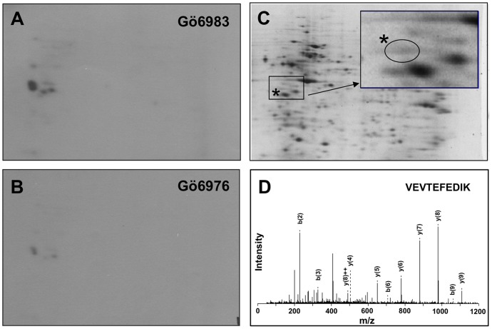
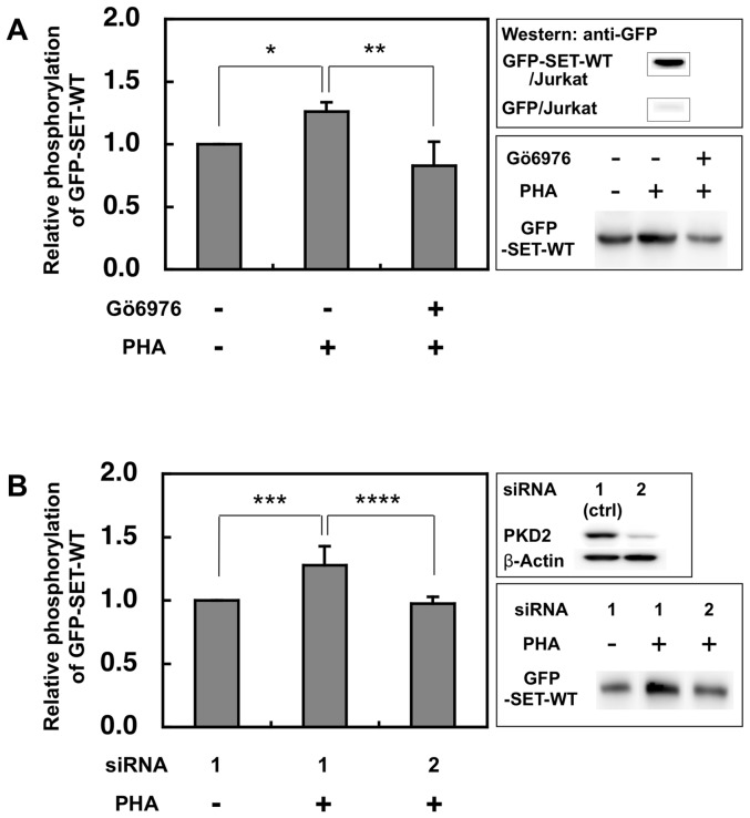
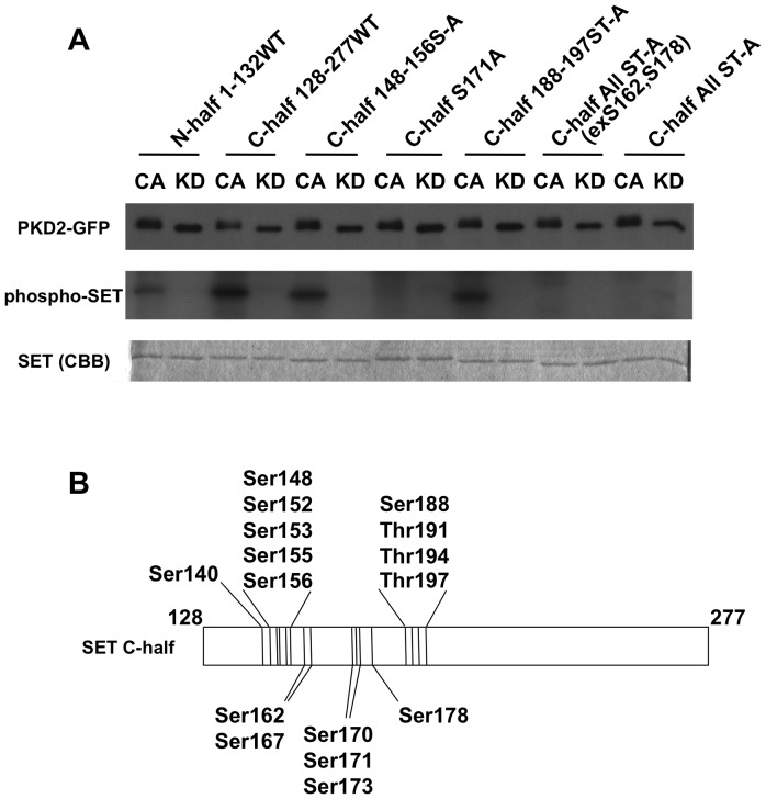
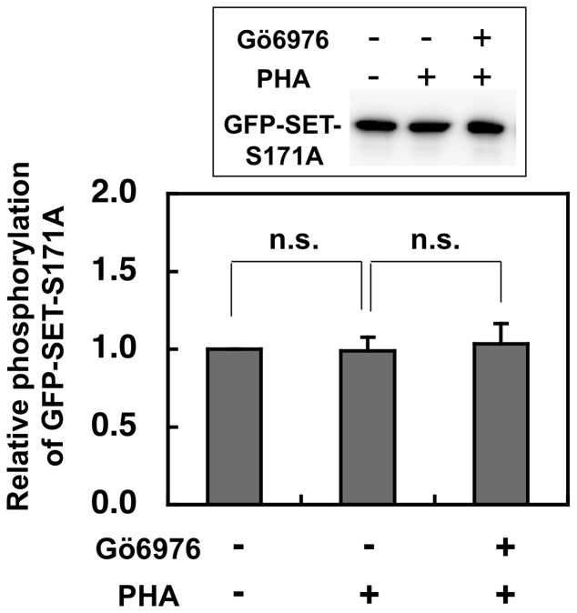
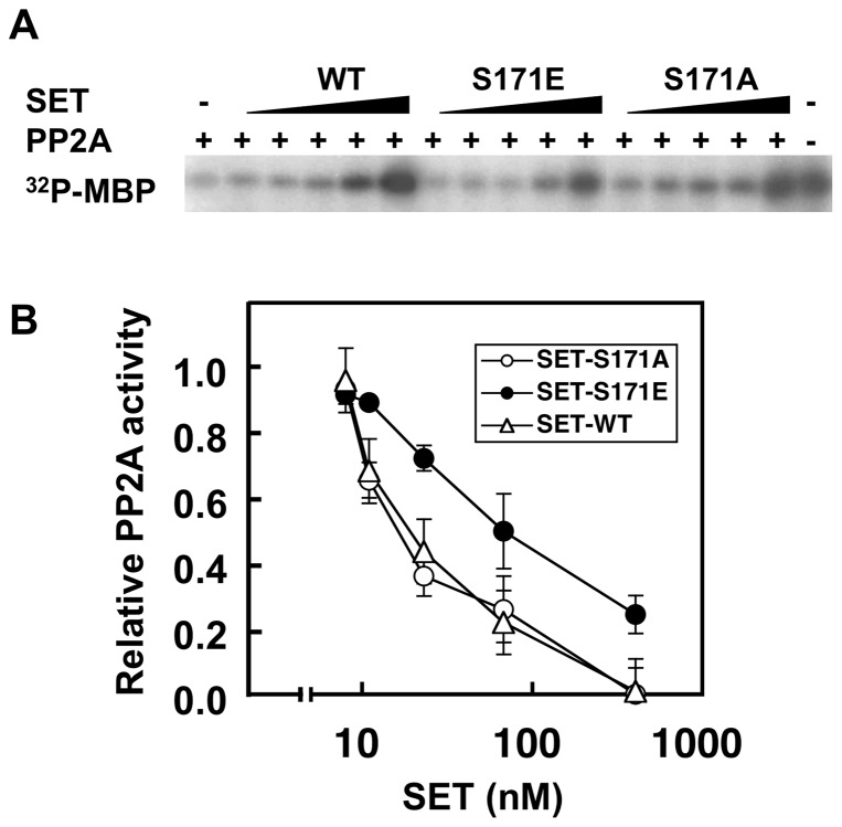
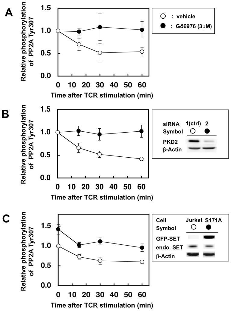
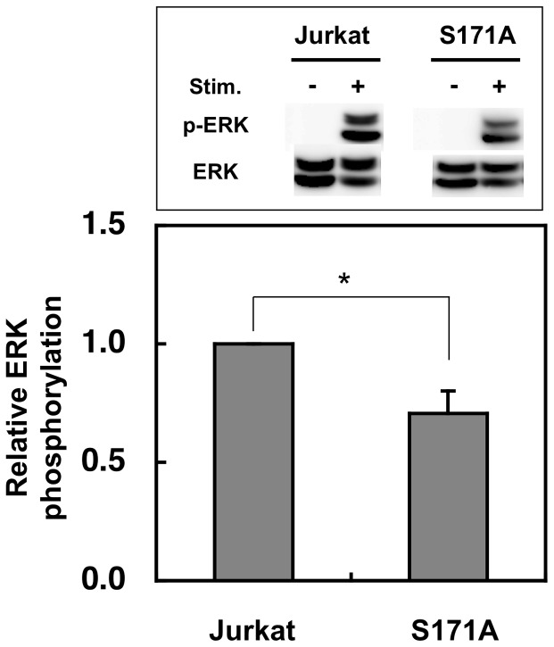
We previously reported that protein kinase D2 (PKD2) in T cells is promptly activated after T-cell receptor (TCR) stimulation and involved in the activation of interleukin-2 promoter and T cell death, and that one of its candidate substrate is SET protein, a natural inhibitor for protein phosphatase 2A (PP2A). In this study, we investigated the target amino acid residues of SET phosphorylated by PKD2 and the effects of phosphorylation of SET on PP2A phosphatase activity. In vitro kinase assay using various recombinant SET mutants having Ser/Thr to Ala substitutions revealed that Ser171 of SET is one of the sites phosphorylated by PKD2. Recombinant SET with phosphorylation-mimic Ser171 to Glu substitution reduced its inhibitory effects on PP2A phosphatase activity compared with Ser171 to Ala substituted or wild-type SET. In addition, knockdown of PKD2 in Jurkat cells by RNAi or treatment of human CD4(+) T cell clone with the PKD2 inhibitor Gö6976 resulted in reduced PP2A activity after TCR-stimulation judged from phosphorylation status of Tyr307 of the catalytic subunit of PP2A. These results suggest that PKD2 is involved in the regulation of PP2A activity in activated T cells through phosphorylation of Ser171 of SET.


Conflict of interest statement

Competing Interests: The authors have declared that no competing interests exist.

Figures

Figure 1
Figure 1. SET protein in Jurkat nuclear extract is phosphorylated by PKD2.
Activated PKD2 was immunoprecipitated from lysate of Jurkat cells stimulated with anti-CD3ε antibody. The heat-inactivated (50°C, 5 min) Jurkat nuclear extract was used as a substrate for PKD2 and analyzed by 2D-gel electrophoresis. (A) Activated PKD2 was incubated with the nuclear extracts in the presence of [γ-32P]-ATP and Gö6983 (an irrelevant PKC inhibitor) and the radioactivity of 2D-electrophregram was detected. (B) Activated PKD2 was incubated with the nuclear extracts in the presence of [γ-32P]-ATP and Gö6976 (PKD2 inhibitor) and the radioactivity of 2D-electrophoregram was detected. (C) The proteins in the Jurkat nuclear extract treated with non-radioactive ATP and PKD2 were stained with syproruby. Inset; the enlarged view of the boxed area. A protein spot indicated by the asterisks, which overlaps to the radioactive spots indicated in A or in B was analyzed by mass spectrometry. (D) One of the MS/MS spectra assigned to be SET protein is shown (m/z = 604, 110VEVTEFEDIK119).
Figure 2
Figure 2. PKD2 is involved in the up-regulation of SET phoshorylation in activated Jurkat cells.
(A) After Jurkat cells expressing GFP-SET were stimulated by PHA (2 µg/ml) with or without Gö6976 (3 µM), the phosphorylated GFP-tagged SET was collected by PhosphoProtein Purification kit and separated by SDS-PAGE. The phosphoprotein was transferred onto the PVDF membrane and quantified by ECL using anti-GFP antibody and the secondary antibody. The typical blot was shown in the lower-right panel. The luminescence of samples from cells without both stimulation and the inhibitor was assigned to be 1. The results were expressed as mean +SD for three independent experiments. *, p<0.01; **, p<0.05. As shown in the upper-right panel, phosphorylated GFP was not detected from the eluate of the PHA stimulated Jurkat cells expressing GFP only. (B) Jurkat cells expressing GFP-SET treated with control (ctrl) siRNA1 had no effect on the PKD2 expression, while siRNA2 markedly reduced the PKD2 expression (upper-right panel). The typical blot was shown in the lower-right panel. The luminescence of the samples from non-stimulated and siRNA1-treated cells was assigned to be 1. The results were expressed as mean +SD for three independent experiments. ***, p<0.05; ****, p<0.05, as determined by Student's t test.
Figure 3
Figure 3. Ser171 of SET is phosphorylated by PKD2.
(A) The indicated 6×His-tag SET mutant proteins were treated with immunoprecipitated constitutively active-PKD2-GFP (CA) or kinase dead-PKD2-GFP (KD) in the presence of [γ-32P]-ATP. The kinase assay mixture was analyzed with SDS-PAGE. Immunoprecipitated PKD2-GFP was blotted with anti-GFP antibody (upper panel) to monitor equal loading of the PKD2 mutants. Phosphorylation of the SET mutants was monitored by incorporated radioactivity (middle panel). Equal loading of each SET mutant protein was confirmed by staining with Coomassie Brilliant Blue (CBB, lower panel). The data shown are representative results from three independent experiments with similar results. (B) A diagram of Ser/Thr residues present in C-terminal half (128–277) of SET protein.
Figure 4
Figure 4. Phospho-GFP-SET-S171A levels in Jurkat were unchanged by PHA stimulation with or without PKD2 inhibitor.
The Jurkat cells expressing GFP-SET-S171A were stimulated and phosphorylation status of GFP-SET-S171A was investigated by the same method as described in Fig. 2. The results were indicated as mean +SD for three independent experiments. There was no significant up- or down-regulation of recovered phospho-GFP-SET-S171A by PHA stimulation with or without PKD2 inhibitor, suggesting that Ser171 of SET is the major target site of TCR-activated PKD2.
Figure 5
Figure 5. Substitution of Ser171 to Glu (SET-S171E) in SET compromised its inhibitory effect on PP2A.
Substitution of Ser171 to Glu (SET-S171E), which mimics phosphorylated SET at Ser171 residue, reduced its inhibitory effect on PP2A compared with Ser171 to Ala substitution (SET-S171A) or wild-type SET did. (A) The phosphatase activity of PP2A (37 µU) under the presence of indicated concentration of recombinant SET was monitored by using 32P-labeled MBP as a substrate. After SET proteins and PP2A were preincubated for 15 min at 4°C, 32P-MBP was added and incubated for 10 min at 37°C. The reaction mixture was separated with SDS-PAGE. The inhibitory effect of SET proteins on PP2A activity was visualized by exposing the SDS-PAGE gel with PP2A-treated 32P-MBP to X-ray film. The data shown are representative results from three independent experiments with similar results. (B) The relative PP2A activity to the radioactivity of 32P-MBP without SET (assigned to be 1) was calculated from the radioactivity of MBP bands in the dried gel shown in (A). Values represent the means ±SD from three independent experiments.
Figure 6
Figure 6. Suppression of PKD2 up-regulates Tyr307 phosphorylation of PP2A catalytic subunit after TCR stimulation.
(A) Human CD4+ T cell clone was stimulated by L cells expressing its cognate ligands in the presence (closed circles) or absence (open circlse) of PKD2 inhibitor Gö6976 (3 µM). (B) Jurkat cells treated with siRNA were stimulated by anti-CD3ε antibody and anti-mouse IgG antibody. Treatment with siRNA2 (closed circles) significantly reduced the PKD2 expression, while siRNA1 (open circles) had almost no effect and hence used as negative controls (right panel). (C) Normal Jurkat cells (open circles) and Jurkat cells expressing GFP-SET-S171A (closed circles) were stimulated by anti-CD3ε antibody and anti-mouse IgG antibody. The latter cells expressed 4.5-fold higher GFP-SET-S171A than the endogenous SET as judged by the intensity of the bands using anti-SET antibody on the Western blot (upper box). Cell lysate were subjected to SDS-PAGE followed by western blot using anti-PP2A phospho-Tyr307 antibody and horseradish peroxidase-conjugated second antibody. The amount of each band was quantified by using ECL and LAS-4000. The same membrane was stripped and reprobed with the anti-PP2A C-subunit antibody to monitor relative abundance of total PP2A protein. Values represent the means ±SD from three independent experiments.
Figure 7
Figure 7. Overexpression of GFP-SET-S171A reduced the ERK activation after stimulation.
Normal and GFP-SET-S171A-overexpressing Jurkat cells were stimulated by anti-CD3e antibody and anti-mouse IgG antibody for 7 min were lyzed and subjected to SDS-PAGE followed by the western blot analyses. The phospho-ERK (p-ERK) was first detected and quantified and after stripping off the antibodies, ERK1 was detected and quantified to monitor p-ERK/ERK1 ratio. The p-ERK/ERK1 ratio of Jurkat cell was assigned to be 1. The results were indicated as mean +SD for three independent experiments. *, p<0.01, as determined by Student's t test.

References

    1. Seo SB, McNamara P, Heo S, Turner A, Lane WS, et al. (2001) Regulation of histone acetylation and transcription by INHAT, a human cellular complex containing the set oncoprotein. Cell 104: 119–130. - PubMed
    1. Li M, Makkinje A, Damuni Z (1996) The myeloid leukemia-associated protein SET is a potent inhibitor of protein phosphatase 2A. J Biol Chem 271: 11059–11062. - PubMed
    1. Fan Z, Beresford PJ, Oh DY, Zhang D, Lieberman J (2003) Tumor suppressor NM23-H1 is a granzyme A-activated DNase during CTL-mediated apoptosis, and the nucleosome assembly protein SET is its inhibitor. Cell 112: 659–672. - PubMed
    1. Millward TA, Zolnierowicz S, Hemmings BA (1999) Regulation of protein kinase cascades by protein phosphatase 2A. Trends Biochem Sci 24: 186–191. - PubMed
    1. Zolnierowicz S (2000) Type 2A protein phosphatase, the complex regulator of numerous signaling pathways. Biochem Pharmacol 60: 1225–1235. - PubMed

Publication types