. 2012;7(12):e51270.
doi: 10.1371/journal.pone.0051270. Epub 2012 Dec 12.

Two types of Tet-On transgenic lines for doxycycline-inducible gene expression in zebrafish rod photoreceptors and a gateway-based tet-on toolkit

Affiliations

Two types of Tet-On transgenic lines for doxycycline-inducible gene expression in zebrafish rod photoreceptors and a gateway-based tet-on toolkit

Leah J Campbell et al. PLoS One. 2012.

Abstract

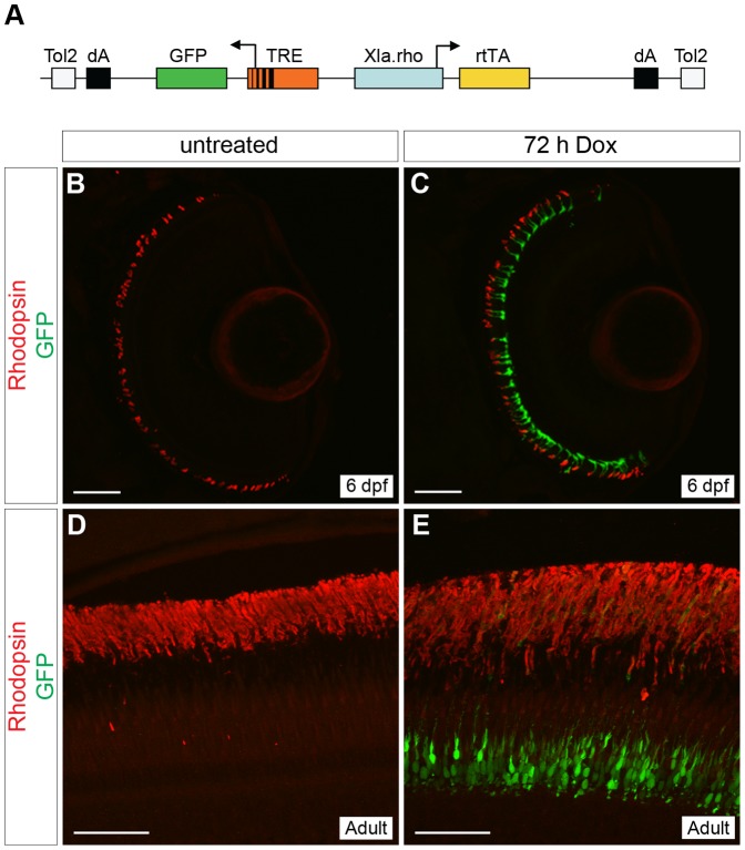
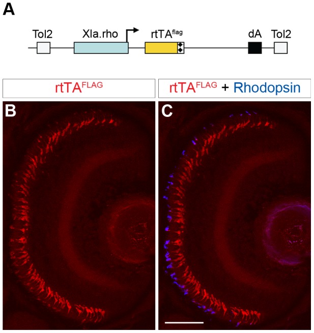
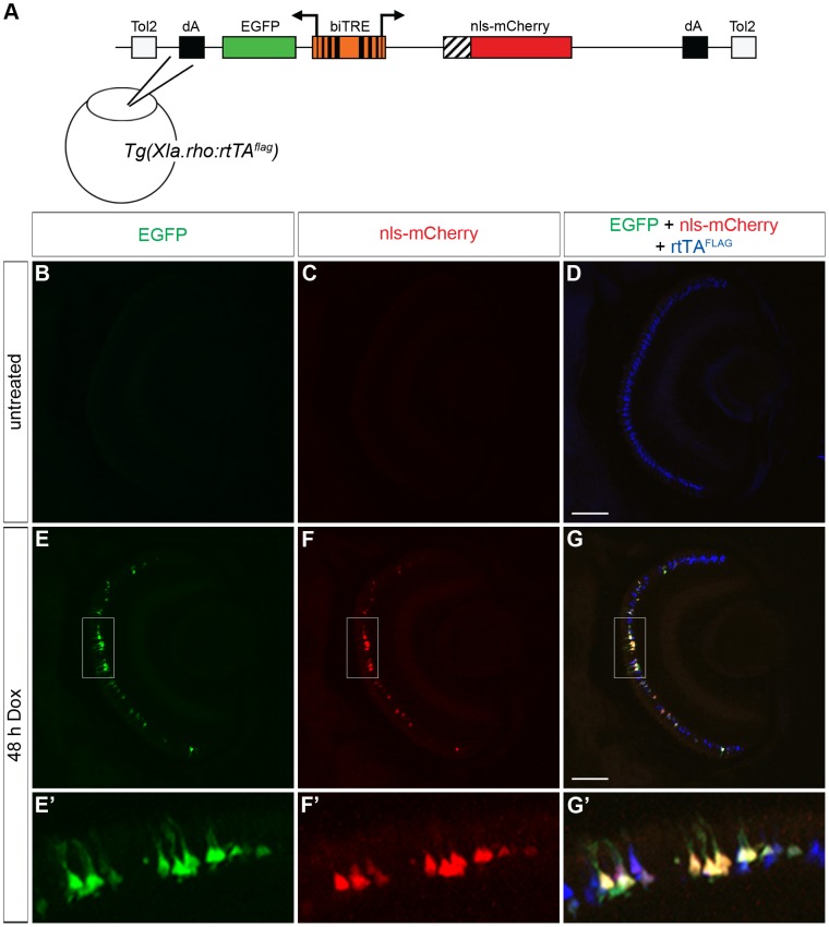
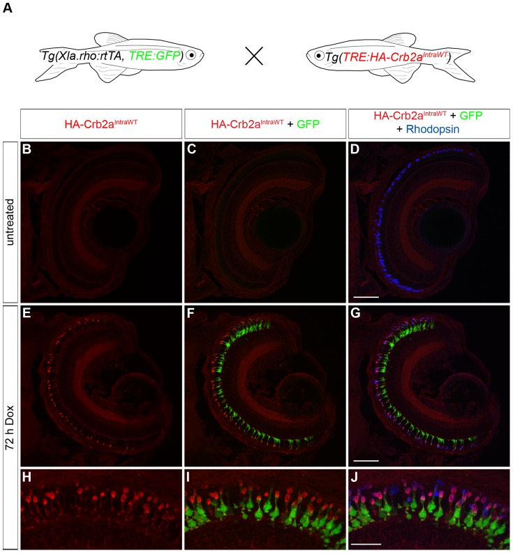
The ability to control transgene expression within specific tissues is an important tool for studying the molecular and cellular mechanisms of development, physiology, and disease. We developed a Tet-On system for spatial and temporal control of transgene expression in zebrafish rod photoreceptors. We generated two transgenic lines using the Xenopus rhodopsin promoter to drive the reverse tetracycline-controlled transcriptional transactivator (rtTA), one with self-reporting GFP activity and one with an epitope tagged rtTA. The self-reporting line includes a tetracycline response element (TRE)-driven GFP and, in the presence of doxycycline, expresses GFP in larval and adult rods. A time-course of doxycycline treatment demonstrates that maximal induction of GFP expression, as determined by the number of GFP-positive rods, is reached within approximately 24 hours of drug treatment. The epitope-tagged transgenic line eliminates the need for the self-reporting GFP activity by expressing a FLAG-tagged rtTA protein. Both lines demonstrate strong induction of TRE-driven transgenes from plasmids microinjected into one-cell embryos. These results show that spatial and temporal control of transgene expression can be achieved in rod photoreceptors. Additionally, system components are constructed in Gateway compatible vectors for the rapid cloning of doxycycline-inducible transgenes and use in other areas of zebrafish research.


Conflict of interest statement

Competing Interests: The authors have declared that no competing interests exist.

Figures

Figure 1
Figure 1. Generation of a rod-specific, doxycycline-inducible, self-reporting gene expression system.
(A) Diagram of the construct used to generate a stable transgenic zebrafish line that expresses doxycycline (Dox)-inducible GFP specifically in rod photoreceptors. The Xenopus rhodopsin promoter (Xla.rho) drives expression of the reverse tetracycline-controlled transcriptional activator (rtTA) gene while the tetracycline responsive element (TRE) drives expression of GFP in the converse direction. (B, C) Confocal z-projections of retinal sections from 6 dpf Tg(Xla.rho:rtTA, TRE:GFP) larvae labeled with anti-Rhodopsin antibody (red). (B) No GFP fluorescence (green) is visible in the absence of Dox. (C) Rod photoreceptors show strong GFP fluorescence (green) when transgenic larvae are treated for 72 h with Dox (3–6 dpf). (D, E) Confocal z-projections of the photoreceptor layer of retinas from Tg(Xop:rtTA, TRE:GFP) adult fish labeled with anti-Rhodopsin (red) and anti-GFP (green) antibodies. (D) No anti-GFP immunofluorescence (green) is visible in the untreated adult photoreceptors, whereas strong anti-GFP immunofluorescence (green) is visible in the adult photoreceptors after treatment with Dox for 72 h (E). dA, polyadenylation signal; Tol2, pTol integration site. Scale bars, 50 µm.
Figure 2
Figure 2. Time course of doxycycline-induced GFP expression in Tg(Xla.rho:rtTA, TRE:GFP) larvae.
Confocal z-projections of retinal sections from 6 dpf Tg(Xla.rho:rtTA, TRE:GFP); alb−/− larvae that were (A) untreated or treated with doxycycline (Dox) for (B) 1, (C) 2, (D) 4, (E) 8, (F) 16, (G) 24, (H) 48, or (I) 72 h. Anti-Rhodopsin immunofluorescence (red) labels rod photoreceptor outer segments and is visible for all treatment conditions. Anti-GFP immunofluorescence (green) is not visible in the untreated retinal section (A). The number of rods showing anti-GFP immunofluorescence (green) noticeably increases with an increase in the Dox exposure time (B–I). Scale bar (A), 50 µm.
Figure 3
Figure 3. Quantitative analysis of the time course of doxycycline-induced GFP expression in Tg(Xla.rho:rtTA, TRE:GFP) larvae.
Tg(Xla.rho:rtTA, TRE:GFP); alb−/− embryos were reared to 6 dpf. Larvae were treated with 10 µg/mL doxycycline (Dox) for the durations indicated prior to fixation at 6 dpf. Fixed larval tissues were processed for cryosectioning and immunofluorescence with antibodies against GFP and Rhodopsin. GFP-positive and Rhodopsin-positive rod outer segments were counted and averaged amongst 3 confocal z-projections for each of the 2–72 h treatment groups. The ratio of GFP-positive to Rhodopsin-positive cells is plotted for each treatment group; error bars represent standard deviation. (formula image
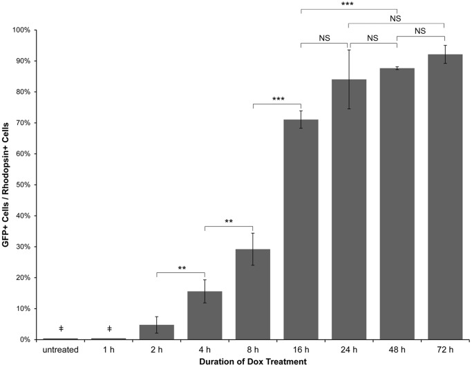
) The untreated and 1 h Dox-treated samples were quantified by counting the number of GFP-positive cells in all retinal sections from 10 eyes and dividing by an estimation of the total number of rods per eye. Statistical comparisons are indicated by brackets. ***, p<0.001; **, p<0.01; NS, not significant.
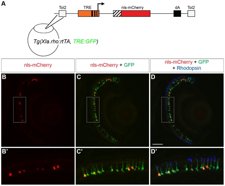
Figure 4
Figure 4. Transactivation of an injected plasmid into Tg(Xop:rtTA, TRE:GFP).
(A) Diagram of the tetracycline response element (TRE) construct injected into Tg(Xla.rho:rtTA, TRE:GFP) one-cell embryos. The TRE drives the mCherry gene with a nuclear localization sequence (nls-mCherry). (B–D) Confocal z-projections of retinal sections from injected Tg(Xla.rho:rtTA, TRE:GFP) larvae at 6 dpf that were treated for the final 48 h with doxycycline (Dox) and were labeled with anti-Rhodopsin antibody. (B, B′) nls-mCherry fluorescence (red) is visible in the photoreceptor layer. (C, C′) nls-mCherry fluorescence (red) co-localizes with green fluorescent protein (GFP) fluorescence (green) in the photoreceptor layer. (D, D′) Anti-Rhodopsin label (blue) shows that GFP and nls-mCherry are expressed in rod photoreceptors. Boxed regions in B, C, and D correspond to B′, C′, and D′. dA, polyadenylation sequence; Tol2, pTol integration site. Scale bar, 50 µm.
Figure 5
Figure 5. Generation of a rod-specific, epitope-tagged, rtTA-expressing transgenic line for doxycycline-inducible gene expression.
(A) Diagram of the construct used to generate a stable transgenic zebrafish line that expresses a FLAG-tagged rtTA protein specifically in rod photoreceptors. The Xenopus rhodopsin promoter (Xla.rho) drives the flag epitope-tagged reverse tetracycline-controlled transcriptional activator (rtTAflag) gene. (B, C) Confocal z-projection of a retinal section from a 6 dpf Tg(Xla.rho:rtTAflag) larva labeled with anti-FLAG and anti-Rhodopsin antibodies. (B) Anti-FLAG immunofluorescence (red) is visible in the photoreceptor layer. (C) The anti-FLAG immunofluorescence (red) colocalizes with the anti-Rhodopsin immunofluorescence (blue) in the rod photoreceptors. dA, polyadenylation sequence; Tol2, pTol integration site. Scale bar, 50 µm.
Figure 6
Figure 6. Bidirectional transactivation of an injected biTRE-containing plasmid into Tg(Xla.rho:rtTAflag).
(A) Diagram of the bidirectional tetracycline response element (biTRE)-containing construct injected into Tg(Xla.rho:rtTAflag) one-cell embryos. EGFP and mCherry with a nuclear localization sequence (nls-mCherry) flank the biTRE. (B–G) Confocal z-projections of retinal sections from injected Tg(Xla.rho:rtTAflag) larvae at 6 dpf labeled with anti-FLAG antibody (blue). (B–D) GFP fluorescence (B, green) and nls-mCherry fluorescence (C, red) are undetectable in the absence of doxycycline (Dox) treatment, while anti-FLAG labeling (D, blue) is visible in rod photoreceptors. (E–G) GFP fluorescence (E, E′, green) and nls-mCherry fluorescence (F, F′, red) are visible in the photoreceptor layer and co-localize with anti-FLAG labeling (G, G′, blue) in the rod photoreceptors after 48 h Dox treatment. Boxed regions in E, F, and G correspond to E′, F′, and G′. dA, polyadenylation sequence; Tol2, pTol integration site. Scale bar (G), 50 µm.
Figure 7
Figure 7. Transactivation of the transgenic TRE:HA-Crb2aIntraWT.
(A) Larvae from an outcross of Tg(Xla.rho:rtTA, TRE:GFP) to Tg(TRE:HA-Crb2aIntraWT) were untreated or doxycycline (Dox)-treated for 72 hours and genotyped for the transgenes before immunofluorescence analysis. (B–J) Confocal z-projections of retinal sections from 6 dpf Tg(Xla.rho:rtTA, TRE:GFP; TRE:HA-Crb2aIntraWT) larvae labeled with anti-HA (red) and anti-Rhodopsin (blue) antibodies. (B–D) Anti-HA-Crb2aIntraWT immunofluorescence (B, red) and GFP fluorescence (C, green) are undetectable in the untreated rod photoreceptors with anti-Rhodopsin immunofluorescence (D, blue). (E–J) Anti-HA-Crb2aIntraWT immunofluorescence (E, H, red) and GFP fluorescence (F, I, green) are visible in the photoreceptor layer and co-localize with the anti-Rhodopsin immunofluorescence in the rod photoreceptors after 72 h of Dox treatment. Scale bar (D, G), 50 µm and (J), 20 µm.

References

    1. Ekker SC (2000) Morphants: a new systematic vertebrate functional genomics approach. Yeast 17: 302–306. - PMC - PubMed
    1. Nasevicius A, Ekker SC (2000) Effective targeted gene ‘knockdown’ in zebrafish. Nat Genet 26: 216–220. - PubMed
    1. Hesselson D, Anderson RM, Beinat M, Stainier DYR (2009) Distinct populations of quiescent and proliferative pancreatic β-cells identified by HOTcre mediated labeling. Proc Natl Acad Sci U S A 106: 14896–14901. - PMC - PubMed
    1. Hans S, Freudenreich D, Geffarth M, Kaslin J, Machate A, et al. (2011) Generation of a non-leaky heat shock–inducible Cre line for conditional Cre/lox strategies in zebrafish. Dev Dyn 240: 108–115. - PubMed
    1. Hans S, Kaslin J, Freudenreich D, Brand M (2009) Temporally-controlled site-specific recombination in zebrafish. PLoS ONE 4: e4640. - PMC - PubMed

Publication types

LinkOut - more resources