. 2012;7(12):e51368.
doi: 10.1371/journal.pone.0051368. Epub 2012 Dec 12.

Cubozoan venom-induced cardiovascular collapse is caused by hyperkalemia and prevented by zinc gluconate in mice

Affiliations

Cubozoan venom-induced cardiovascular collapse is caused by hyperkalemia and prevented by zinc gluconate in mice

Angel A Yanagihara et al. PLoS One. 2012.

Abstract

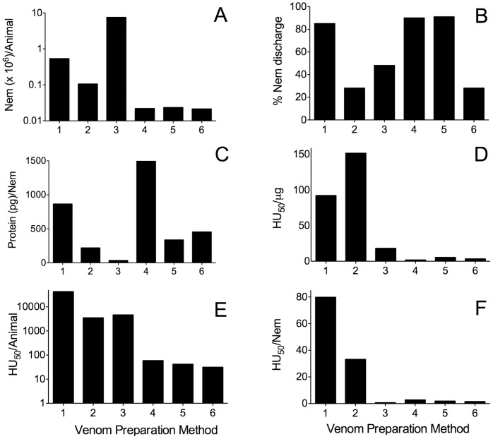
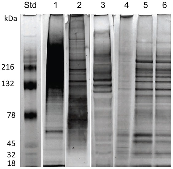
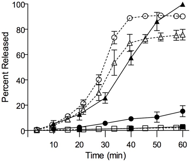
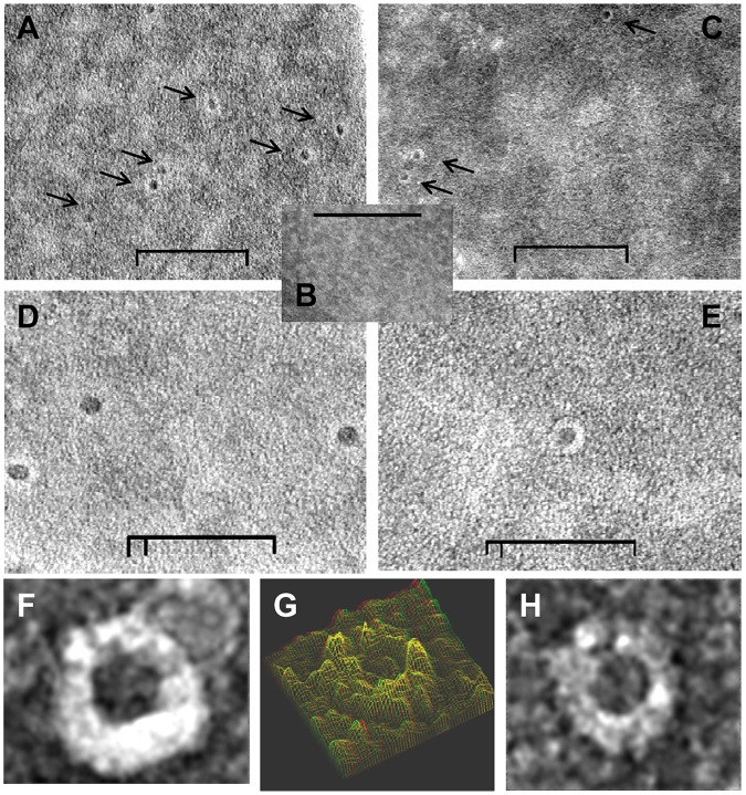
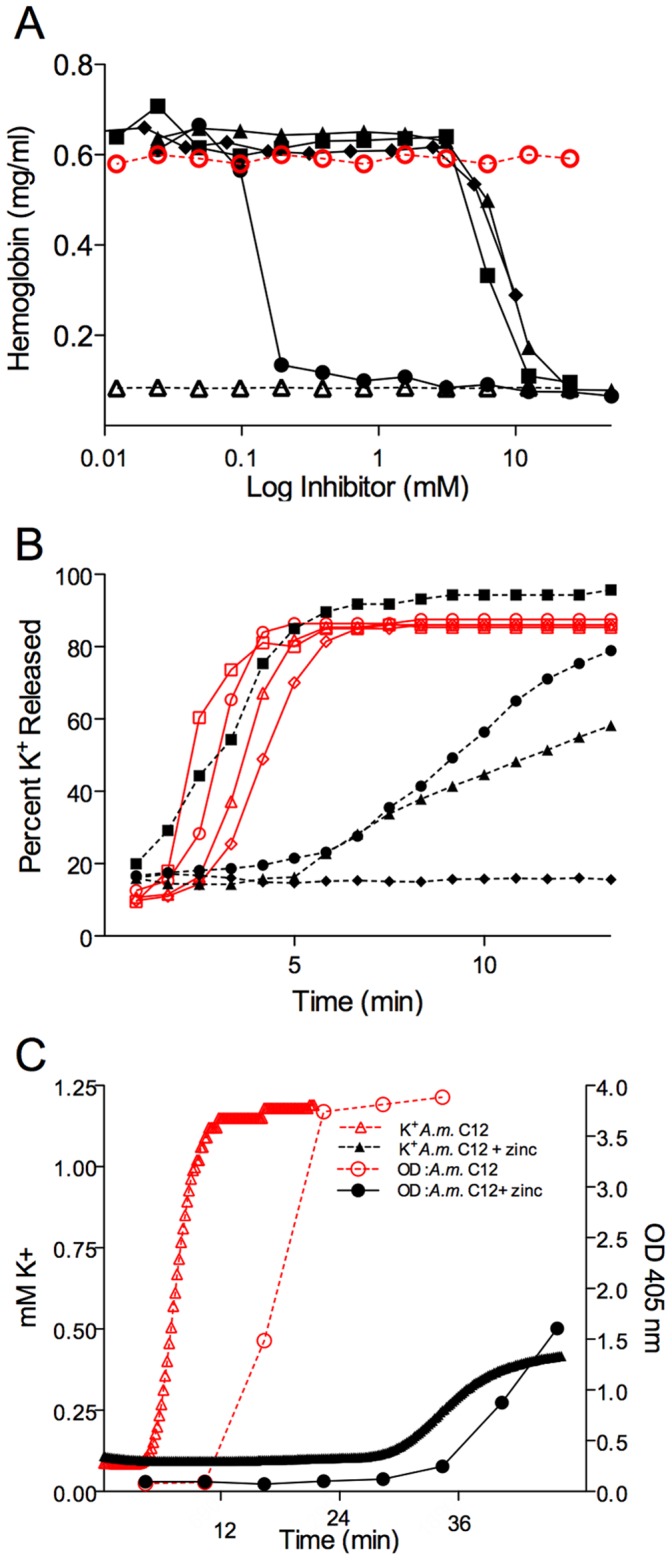
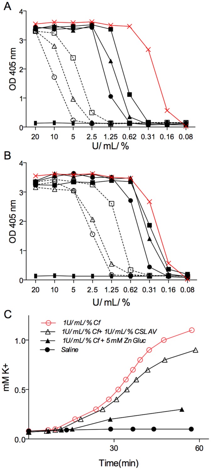
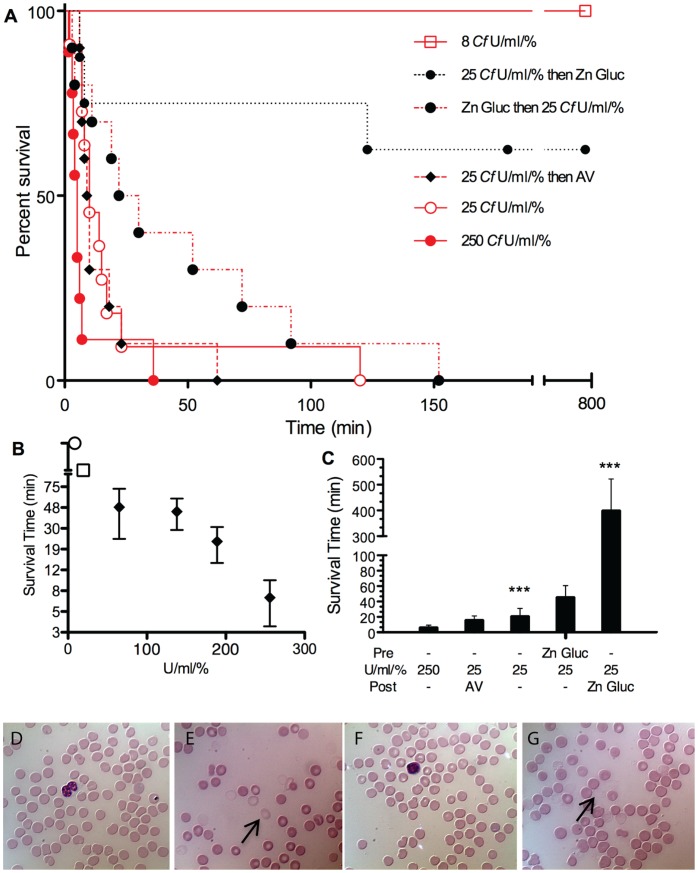
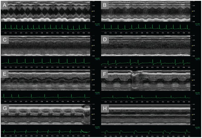
Chironex fleckeri (Australian box jellyfish) stings can cause acute cardiovascular collapse and death. We developed methods to recover venom with high specific activity, and evaluated the effects of both total venom and constituent porins at doses equivalent to lethal envenomation. Marked potassium release occurred within 5 min and hemolysis within 20 min in human red blood cells (RBC) exposed to venom or purified venom porin. Electron microscopy revealed abundant ~12-nm transmembrane pores in RBC exposed to purified venom porins. C57BL/6 mice injected with venom showed rapid decline in ejection fraction with progression to electromechanical dissociation and electrocardiographic findings consistent with acute hyperkalemia. Recognizing that porin assembly can be inhibited by zinc, we found that zinc gluconate inhibited potassium efflux from RBC exposed to total venom or purified porin, and prolonged survival time in mice following venom injection. These findings suggest that hyperkalemia is the critical event following Chironex fleckeri envenomation and that rapid administration of zinc could be life saving in human sting victims.


Conflict of interest statement

Competing Interests: AAY has a provisional patent for treatment of syndromes associated with pore-forming toxins, U.S. Patent Office #61245238. RVS is listed as a contributing party to this document. The filing of a provisional patent application for treatment of syndromes associated with pore-forming toxins, U.S. Patent Office #61245238 by the University of Hawaii Office of Technology Transfer and Economic Development with RVS is listed as a contributing party does not alter the adherence of the authors (AAY and RVS) to all the PLOS ONE policies on sharing data and materials.

Figures

Figure 1
Figure 1. Comparison of venom preparation methods.
Venom was prepared in parallel as described in the Materials and Methods section: Lane 1, Yanagihara; Lane 2, Winkel et al.; Lane 3, Mustafa et al.; Lane 4 Bloom et al.; Lane 5 Bailey et al.; Lane 6, Carrette and Seymour. Histogram plots show the comparative metrics of Alatina moseri venom recovery and activity in terms of (A) nematocysts (Nem) recovered per animal, (B) percentage ruptured nematocysts, (C) protein yield in picogram per nematocyst, and relative toxicity in terms of (D) hemolytic units (HU50) recovered per microgram of protein, (E) HU50 per animal and (F) HU50 per nematocyst.
Figure 2
Figure 2. Comparative polyacrylamide gel electrophoresis (PAGE) profiles of proteins comprising various venom preparations.
Venom prepared using various methods was electrophoresed on SDS-PAGE gels and silver stained to compare the recovered size range and distribution of Alatina moseri venom. Lane 1: Yanagihara method, Lane 2: Winkel et al., Lane 3: Mustafa et al., Lane 4: Bloom et al., Lane 5: Bailey et al., Lane 6: Carrette and Seymour, and Std: Molecular weight standards.
Figure 3
Figure 3. Cubozoan (Chironex fleckeri or Alatina moseri) venom elicits potassium and hemoglobin release.
1 U/mL/% Chironex fleckeri venom-exposed whole blood time course of plasma potassium (open circles), 1 U/mL% Alatina moseri venom (open triangles) and hemoglobin release in whole blood exposed to 1 U/mL/% Chironex fleckeri venom (closed circles), or 1 U/mL/% Alatina moseri venom (closed triangles) expressed as a percentage of total with respective potassium and hemoglobin controls (shown as open and closed squares).
Figure 4
Figure 4. Ultrastructure of negatively stained RBC after exposure to cubozoan porins (Chironex fleckeri or Alatina moseri).
Transmission electron micrographs of 2% ammonium molybdate negative stained purified venom porin pretreated RBC showed the presence of distinct ring shaped pores. (A) Chironex fleckeri porin (A+B isoforms) treated human RBC membrane; (B) control mock treated RBC; (C) Alatina moseri porin exposed human RBC membrane; (D) higher magnification of A; (E) higher magnification of B; (F) and (H) highest magnification of individual exemplar pores from panel B; (G) 3-D modeling using analySIS EsiVision 3.2.0. Inner and outer diameter diameter of pores measured approximately 12 nm and 25 nm (size bars: 200 nm in panels A–C, 100 nm in panels D and E).
Figure 5
Figure 5. Inhibition of both venom and purified porin action.
(A) Comparative dose-dependent inhibition of hemolysis was examined after 1 U/mL/% cubozoan venom alone (positive control, open red circles) in the presence of zinc gluconate (closed black circles), calcium gluconate (closed black squares), magnesium sulfate (closed black triangles), strontium chloride (closed black diamonds) or sodium chloride alone (negative control, open black triangles). (B) The potency of 5 mM zinc gluconate to inhibit time course of potassium release at various doses of venom from 100 U/mL/% (open square without and closed square with zinc gluconate), 40 U/mL/% (open circle without and closed circle with zinc gluconate), 20 U/mL/% (open triangle without and closed triangle with zinc gluconate) and 10 U/mL/% (open diamond without and closed diamond with zinc gluconate) was examined. (C) The effect of 5 mM zinc gluconate was examined on purified porin (1 U/mL/% Alatina moseri porin) exposed washed RBC. Time course levels of released potassium (red open triangles) and hemoglobin (red open circles) are shown.
Figure 6
Figure 6. Inhibitory effects of zinc gluconate compared to CSL antivenom in Chironex fleckeri exposed human RBC.
Hemoglobin release was measured over (A) a concentration range of venom in the presence of high concentration potential inhibitors: 50 mM zinc gluconate (open circle), 25 mM zinc gluconate (open triangle), 12.5 mM zinc gluconate (open square), and 250 U/mL/% CSL antivenom (closed circle), 125 U/mL/% CSL antivenom (closed triangle), 62.5 U/mL/% CSL antivenom (closed square) or saline (red×marks). (B) Hemoglobin release was measured over a concentration range of venom in the presence of therapeutically relevant concentration range of zinc gluconate and antivenom 6.25 mM zinc gluconate (open circle), 3.25 mM zinc gluconate (open triangle), 1.56 mM zinc gluconate (open square), and 31.2 U/mL/% CSL antivenom (closed circle), 15.6 U/mL/% CSL antivenom (closed triangle), 7.8 U/mL/% CSL antivenom (closed square) or saline (red×marks). (C) Time course release of potassium from RBC was measured in the presence of saline (closed circle), 1 U/mL/% Chironex fleckeri venom (open red circle), venom together with 1 U/mL/% CSL antivenom (open black triangle), or venom with 5 mM zinc gluconate (closed black triangle).
Figure 7
Figure 7. Kaplan-Meier survival plots of Chironex fleckeri venom-injected mice.
(A) Survival data of C57BL/6 mice administered Chironex fleckeri venom by tail-vein injection at doses of 250 U/mL/% (solid red line and closed red circles), 25 U/mL/% (solid fine red line and open red circles), 8 U/mL/% (dashed red line and open red box), 25 U/mL/% venom followed by CSL antivenom 60 sec later (black diamond and dashed red line), 25 U/mL/% venom preceded by 100 mM zinc gluconate to achieve plasma concentration of 5 mM (solid black circles with red dot-dash line), and 25 U/mL/% venom followed by 100 mM zinc gluconate to achieve plasma concentration of 5 mM 60 sec later dashed black line (in 4 out of 8 animals survived but were sacrificed at 12 hr per to protocol). A Log-rank (Mantel-Cox) Test analysis yielded Chi square value of 33.44, df of 5 and P value of <0.0001. The mean survival times were 6.45 min for 250 U/mL/% (SEM 3.0, n = 11), 21.2 min for 25 U/mL/% (SEM 10.0, n = 11), 16 min for 25 U/mL/% then 100 µL of 1∶10 saline diluted CSL antivenom (SEM 5.39, n = 10), 45.7 min for 25 U/mL/% preceded by 60 sec by weight derived blood volume calculation of 100 mM zinc gluconate to reach 5 mM (SEM 15.1, n = 10), and 399 min for 25 U/mL/% followed 60 sec later by weight derived blood volume calculation of 100 mM zinc gluconate to reach 5 mM (SEM 122.7, n = 8). One way ANOVA demonstrates a P value of <0.0001 for comparison of 8 U/mL/% with all other groups examined as well as for 25 U/mL/% followed by zinc gluconate to reach 5 mM plasma levels as compared to all other 25 U/mL/% venom injected groups. No animals died during the 18-hr observation period after injection of 8 U/mL/% Chironex fleckeri venom (n = 5) or after 100 mM zinc gluconate to reach circulating plasma levels of 5 mM (n = 4). All PBS-injected control mice survived. (B) Dose response. Mouse survival time means with SEM error bars shown as a function of venom dose injected at 250, 190, 140, 65 (black solid diamonds), 12 (open square) and 8 (open circle) U/mL/%. Mice injected with below 15 U/mL/% exhibited 1–4 hr of lethargic or anxious behaviors but survived (represented by open square). Mice injected at doses below 8 U/mL/% survived and showed some transient 10–60 min unusual behaviors, hyperactivity, grooming or stillness (represented by open circle). (C) Histogram plot of survival study data from Figure 7A. Means with SEM error bars are shown. The treatment of 25 U/mL/% venom-injected mice with 100 mM zinc gluconate to achieve 5 mM circulating concentration 60 sec after (Post) venom injection resulted in a highly significant (p<0.0001) enhancement of survival time as compared with the 25 U/mL/% venom-injected mice. (D through G) Blood smear images. Tail-vein control (D and F) and immediate postmortem cardiac-puncture blood droplet smears (E and G) were performed and stained with a modified Wright-Giemsa stain (Accustain, Sigma Aldrich) from mice injected with 25 U/mL/% Alatina moseri (E, for comparison) and Chironex fleckeri (G) venom.
Figure 8
Figure 8. Representative ECG and ECHO recording of Chironex fleckeri venom-injected mouse.
Preinjection (A) recording is shown together with 90 sec (B), 135 sec (C), 225 sec (D), 390 sec (E), 525 sec (F), 660 sec (G) and 840 sec (H) recording after injection with 25 U/mL/% Chironex fleckeri venom. Left ventricular function was markedly impaired, progressing to electromechanical dissociation, 90 sec after Chironex fleckeri venom injection. Contractility briefly recovered slightly, but not sufficiently to maintain perfusion. ECG showed second-degree block at 225 sec, with progression to nodal- and ventricular-escape arrhythmia before death.

References

    1. Bentlage B, Cartwright P, Yanagihara AA, Lewis C, Richards GS, et al. (2010) Evolution of box jellyfishes (Cnidaria: Cubozoa), a group of highly toxic invertebrates. Proc Biol Sci 277: 493–501. - PMC - PubMed
    1. Tibballs J (2006) Australian venomous jellyfish, envenomation syndromes, toxins and therapy. Toxicon 48: 830–859. - PubMed
    1. Flecker H (1945) Injuries by unknown agents to bathers in North Queensland. Med J Aust 20: 128–129.
    1. Barnes JH (1960) Observations on jellyfish stingings in North Queensland Med J Aust. 2: 993–999. - PubMed
    1. Richardson AJ, Bakum A, Hays GC, Gibbons MJ (2009) The jellyfish joyride: causes, consequences and management responses to a more gelatinous future. Trends Ecol Evol 24: 312–322. - PubMed

Publication types

MeSH terms