. 2012;7(12):e51431.
doi: 10.1371/journal.pone.0051431. Epub 2012 Dec 12.

Isoflurane induces learning impairment that is mediated by interleukin 1β in rodents

Affiliations

Isoflurane induces learning impairment that is mediated by interleukin 1β in rodents

Lin Cao et al. PLoS One. 2012.

Abstract

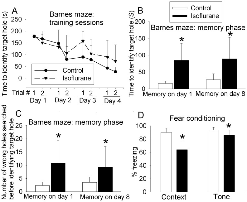
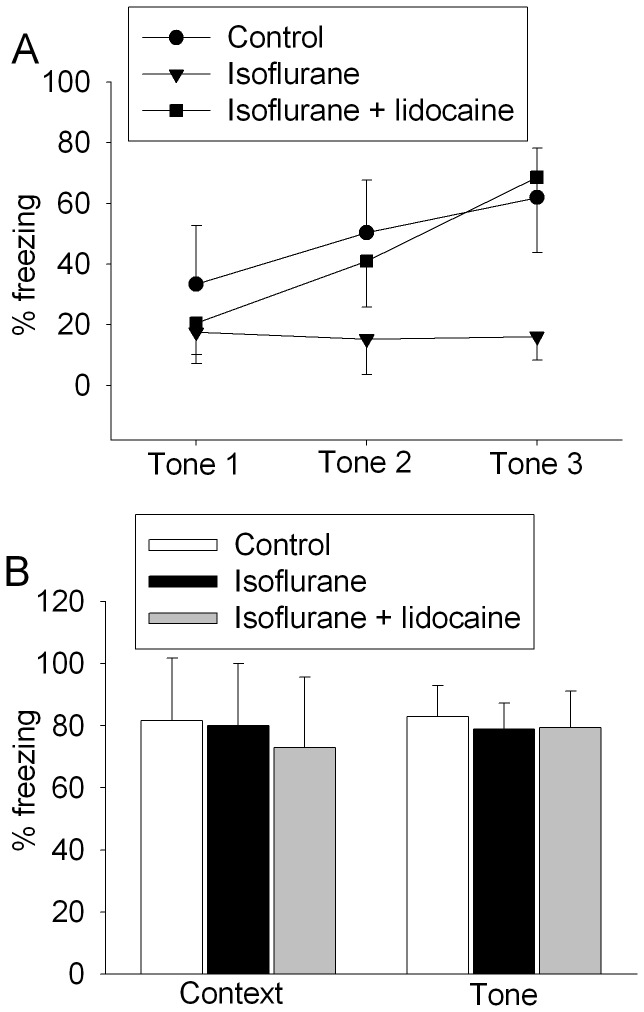
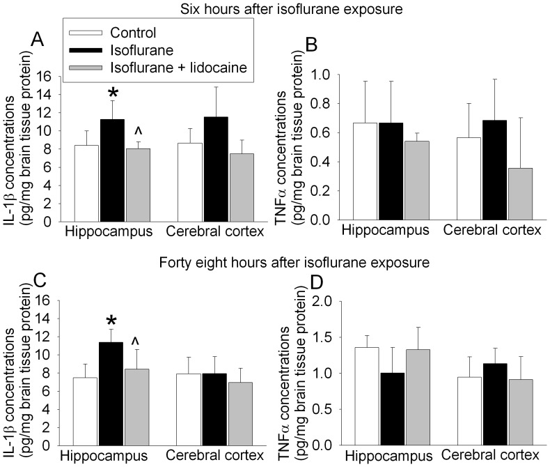
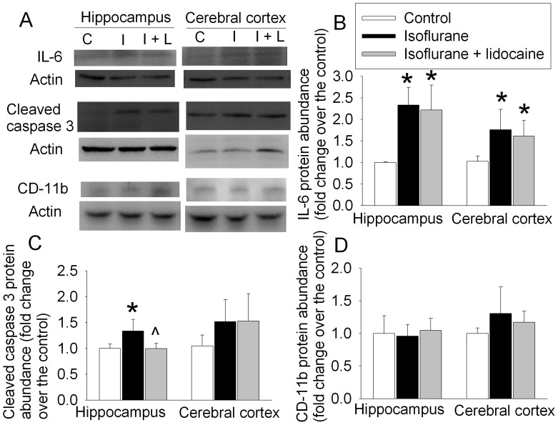
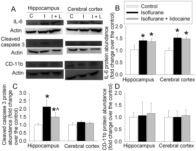
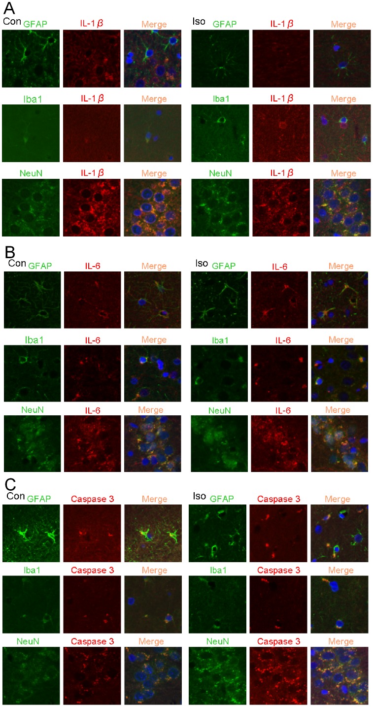
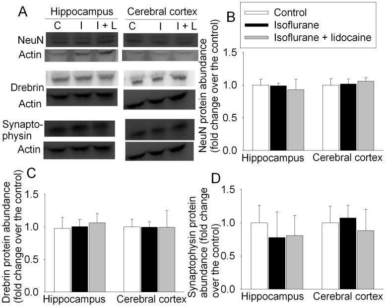
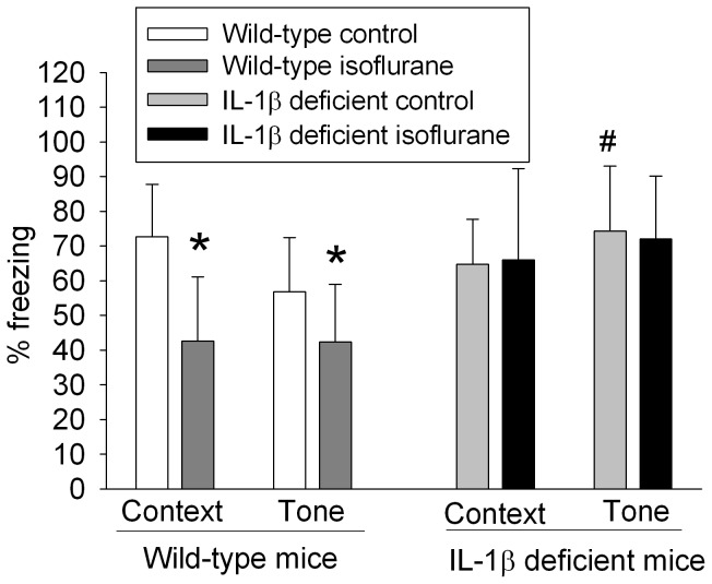
Postoperative cognitive decline is a clinical syndrome. Volatile anesthetics are commonly used during surgery. It is conceivable that volatile anesthetics may contribute to postoperative cognitive decline. Isoflurane can impair cognitive functions of animals under certain conditions. However, the mechanisms for this impairment are not clear. Here, male 18-month old Fisher 344 rats or 10-week old mice were exposed to 1.2 or 1.4% isoflurane for 2 h. Our studies showed that isoflurane impaired the cognitive functions of the rats in Barnes maze. Isoflurane-exposed rats had reduced freezing behavior during the training sessions in the fear conditioning test. This isoflurane effect was attenuated by lidocaine, a local anesthetic with anti-inflammatory property. Rats that had training sessions and were exposed to isoflurane 30 min later had freezing behavior similar to that of control animals. Isoflurane increased the expression of interleukin 1β (IL-1β), interleukin-6 and activated caspase 3 in the hippocampus of the 18-month old rats. IL-1β positive staining was co-localized with that of NeuN, a neuronal marker. The increase of IL-1β and activated caspase 3 but not interleukin-6 was attenuated by lidocaine. Isoflurane also impaired the cognitive functions of 10-week old C57BL/6J mice and increased IL-1β in their hippocampi. However, isoflurane did not affect the cognitive functions of IL-1β deficient mice. Our results suggest that isoflurane impairs the learning but may not affect the recall of the aged rats. IL-1β may play an important role in this isoflurane effect.


Conflict of interest statement

Competing Interests: The authors have declared that no competing interests exist.

Figures

Figure 1
Figure 1. Isoflurane-induced cognitive impairment measured by Barnes maze and fear conditioning.
Eighteen-month-old Fisher 344 rats were exposed to or were not exposed to 1.2% isoflurane for 2 h. They were subjected to Barnes maze 2 weeks later or fear conditioning 27 days later. A: performance in the training sessions of Barnes maze test. There is a significant effect of training sessions on the latency to identify the target hole (P<0.001 by two-way repeated measures analysis of variance). B: latency to identify the target hole at 1 day or 8 days after the training sessions. C: number of other hole searched before identifying the target hole at 1 day or 8 days after the training sessions. D: performance in fear conditioning test. Results are mean±S.D. (n = 6). *P<0.05 compared with the corresponding control in panels B, C and D by t-test.
Figure 2
Figure 2. Isoflurane-induced learning impairment measured by fear conditioning test.
A: freezing behavior during training sessions for 18-month old Fisher 344 rats that were exposed to isoflurane in the presence or absence of lidocaine 27 days ago. Results are means±S.D. (n = 6 for control and isoflurane only groups and  = 7 for isoflurane plus lidocaine group). There is a significant effect of training trial (P<0.001, among the control animals and animals exposed to isoflurane plus lidocaine), isoflurane use (P = 0.002, control animals vs. animals exposed to isoflurane only) and lidocaine use (P = 0.003, animals exposed to isoflurane only vs. animals exposed to isoflurane plus lidocaine) on the freezing behavior. Statistical analysis was performed by two-way analysis of variance. B: freezing behavior during the context- and tone-related fear conditioning tests for 18-month old Fisher 344 rats that first had the training sessions and were exposed to isoflurane in the presence or absence of lidocaine at 30 min after the training sessions. Results are means±S.D. (n = 4 for control group and  = 5 for the isoflurane only and isoflurane plus lidocaine groups).
Figure 3
Figure 3. Isoflurane effects on interleukin 1β (IL-1β) and tumor necrosis factor α (TNFα) contents in rat brain tissues.
A and B: eighteen-month-old Fisher 344 rats were exposed to or were not exposed to 1.2% isoflurane in the presence or absence of lidocaine for 2 h. Hippocampus and cerebral cortex were harvested at 6 h after anesthetic exposure for ELISA of IL-1β or TNFα content. C and D: eighteen-month-old Fisher 344 rats had training sessions of fear conditioning test and 30 min later were exposed to or were not exposed to 1.2% isoflurane in the presence or absence of lidocaine for 2 h. Hippocampus and cerebral cortex were harvested at 48 h after anesthetic exposure for ELISA of IL-1β or TNFα content. Results are means±S.D. (n = 4). *P<0.05 compared to control. ? P<0.05 compared to isoflurane alone. Statistical analysis was performed by one-way analysis of variance.
Figure 4
Figure 4. Isoflurane effects on the expression of interleukin 6 (IL-6), activated/cleaved caspase 3 and cluster of differentiation 11b (CD-11b) in rat brain tissues.
Eighteen-month-old Fisher 344 rats were exposed to or were not exposed to 1.2% isoflurane in the presence or absence of lidocaine for 2 h. Hippocampus and cerebral cortex were harvested at 6 h after anesthetic exposure for Western blotting. A: representative Western blot images. B, C and D: graphic presentation of the IL-6, cleaved caspase 3 and CD-11b protein abundance quantified by integrating the volume of autoradiograms from 4 rats for each experimental condition. Values in graphs are expressed as fold changes over the mean values of control animals and are presented as the means±S.D. *P<0.05 compared with the control group. ? P<0.05 compared to isoflurane alone. Statistical analysis was performed by one-way analysis of variance. C: control, I: isoflurane, I+L: isoflurane plus lidocaine.
Figure 5
Figure 5. Isoflurane effects on the expression of interleukin 6 (IL-6), activated/cleaved caspase 3 and cluster of differentiation 11b (CD-11b) in rat brain tissues.
Eighteen-month-old Fisher 344 rats had the training sessions of fear conditioning and 30 min later were exposed to or were not exposed to 1.2% isoflurane in the presence or absence of lidocaine for 2 h. Hippocampus and cerebral cortex were harvested at 48 h after anesthetic exposure for Western blotting. A: representative Western blot images. B, C and D: graphic presentation of the IL-6, cleaved caspase 3 and CD-11b protein abundance quantified by integrating the volume of autoradiograms from 4 rats for each experimental condition. Values in graphs are expressed as fold changes over the mean values of control animals and are presented as the means±S.D. *P<0.05 compared with the control group. ? P<0.05 compared to isoflurane alone. Statistical analysis was performed by one-way analysis of variance. C: control, I: isoflurane, I+L: isoflurane plus lidocaine.
Figure 6
Figure 6. Expression of interleukin 1β (IL-1β), IL-6 and activated/cleaved caspase 3 in rat brain tissues.
Eighteen-month-old Fisher 344 rats were exposed to or were not exposed to 1.2% isoflurane for 2 h. Hippocampus was harvested at 48 h after isoflurane exposure for immunofluorescent staining of IL-1β (red), IL-6 (red), cleaved caspase 3 (red), NeuN (green), glial fibrillary acidic protein (GFAP, green) and ionized calcium binding adaptor molecule 1 (Iba1, green). The merged panels also include Hoechst staining (blue) to show cell nuclei. A: co-staining of IL-1β with GFAP, Iba1 and NeuN. B: co-staining of IL-6 with GFAP, Iba1 and NeuN. C: co-staining of cleaved caspase 3 with GFAP, Iba1 and NeuN. Con: control, Iso: isoflurane.
Figure 7
Figure 7. Isoflurane effects on the expression of NeuN, drebrin and synaptophysin in rat brain tissues.
Eighteen-month-old Fisher 344 rats had the training sessions of fear conditioning and 30 min later were exposed to or were not exposed to 1.2% isoflurane in the presence or absence of lidocaine for 2 h. Hippocampus and cerebral cortex were harvested at 48 h after anesthetic exposure for Western blotting. A: representative Western blot images. B, C and D: graphic presentation of the NeuN, drebrin and synaptophysin protein abundance quantified by integrating the volume of autoradiograms from 4 rats for each experimental condition. Values in graphs are expressed as fold changes over the mean values of control animals and are presented as the means±S.D. C: control, I: isoflurane, I+L: isoflurane plus lidocaine.
Figure 8
Figure 8. Isoflurane effects on cognitive functions assessed by fear conditioning.
Ten-week old male C57BL/6J (wild-type) mice or interleukin 1β (IL-1β) deficient mice were exposed to 1.4% isoflurane for 2 h and then subjected to the fear conditioning test 48 h later. Results are means±S.D. (n = 15 for the wild-type mice and 6 for the IL-1β deficient mice). *P<0.05 compared with the corresponding control group. # P<0.05 compared to the wild-type control mice. Statistical analysis was performed by t-test.

References

    1. Silva AC, ORyan F, Poor DB (2006) Postoperative nausea and vomiting (PONV) after orthognathic surgery: a retrospective study and literature review. J Oral Maxillofac Surg 64: 1385–1397. - PubMed
    1. Zheng S, Zuo Z (2004) Isoflurane preconditioning induces neuroprotection against ischemia via activation of p38 mitogen-activated protein kinase. Mol Pharmacol 65: 1172–1180. - PubMed
    1. Lin D, Zuo Z (2011) Isoflurane induces hippocampal cell injury and cognitive impairments in adult rats. Neuropharmacology 61: 1354–1359. - PMC - PubMed
    1. Culley DJ, Baxter M, Yukhananov R, Crosby G (2003) The memory effects of general anesthesia persist for weeks in young and aged rats. Anesth Analg 96: 1004–1009. - PubMed
    1. Baranov D, Bickler PE, Crosby GJ, Culley DJ, Eckenhoff MF, et al. (2009) Consensus statement: First International Workshop on Anesthetics and Alzheimer’s disease. Anesth Analg 108: 1627–1630. - PMC - PubMed

Publication types

MeSH terms