. 2012;7(12):e51566.
doi: 10.1371/journal.pone.0051566. Epub 2012 Dec 12.

Exome sequencing identifies a founder frameshift mutation in an alternative exon of USH1C as the cause of autosomal recessive retinitis pigmentosa with late-onset hearing loss

Affiliations

Exome sequencing identifies a founder frameshift mutation in an alternative exon of USH1C as the cause of autosomal recessive retinitis pigmentosa with late-onset hearing loss

Samer Khateb et al. PLoS One. 2012.

Abstract

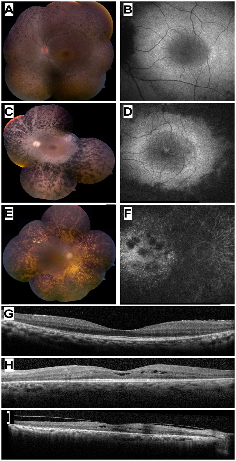
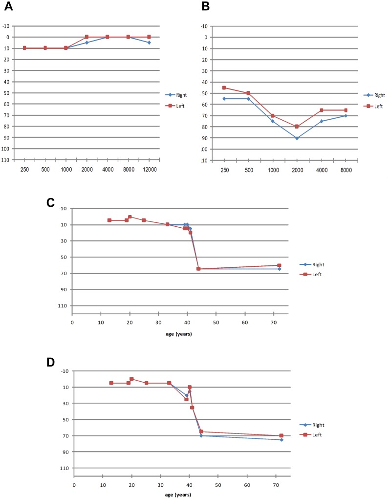
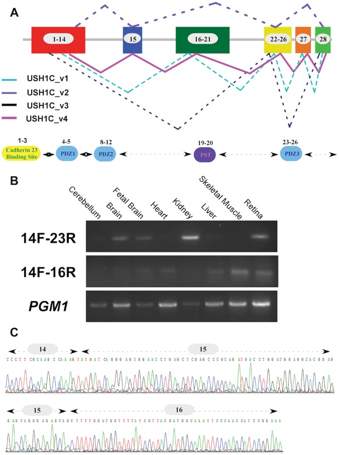
We used a combined approach of homozygosity mapping and whole exome sequencing (WES) to search for the genetic cause of autosomal recessive retinitis pigmentosa (arRP) in families of Yemenite Jewish origin. Homozygosity mapping of two arRP Yemenite Jewish families revealed a few homozygous regions. A subsequent WES analysis of the two index cases revealed a shared homozygous novel nucleotide deletion (c.1220delG) leading to a frameshift (p.Gly407Glufs*56) in an alternative exon (#15) of USH1C. Screening of additional Yemenite Jewish patients revealed a total of 16 homozygous RP patients (with a carrier frequency of 0.008 in controls). Funduscopic and electroretinography findings were within the spectrum of typical RP. While other USH1C mutations usually cause Usher type I (including RP, vestibular dysfunction and congenital deafness), audiometric screening of 10 patients who are homozygous for c.1220delG revealed that patients under 40 years of age had normal hearing while older patients showed mild to severe high tone sensorineural hearing loss. This is the first report of a mutation in a known USH1 gene that causes late onset rather than congenital sensorineural hearing loss. The c.1220delG mutation of USH1C accounts for 23% of RP among Yemenite Jewish patients in our cohort.


Conflict of interest statement

Competing Interests: The authors have declared that no competing interests exist.

Figures

Figure 1
Figure 1. Family trees and segregation analysis of the USH1C c.1220delG mutation.
A. Structure of the two families that were selected for whole exome sequencing. The family number is indicated above the corresponding family tree. Filled symbols represent affected individuals, whereas clear symbols represent unaffected individuals. Generation numbers are depicted on the left and individual numbers below each symbol. The USH1C genotype is presented as: M/M, homozygous for the c.1220delG mutation; M/+, heterozygous; +/+, homozygous for the wildtype allele. Index cases are marked with an arrow. B. Sequence chromatograms of part of USH1C exon 15 of a control individual (top), a heterozygote (middle), and a patient who is homozygous for the mutation (bottom). The c.1220delG mutation (boxed) results in a frameshift (p.Gly407Glufs*56).
Figure 2
Figure 2. Ocular Phenotype of patients who are homozygous for the USH1C c.1220delG mutation.
(A, C, E) Color fundus photographs of three patients aged 13 (MOL0486 II:3), 33 (TB16-R12), and 72 (MOL1023-1) years old, respectively. Note the increasing severity of fundus changes with age, and the presence of dense bone spicule-like pigmentation. (B, D, F) Corresponding fundus autofluorescence imaging of the macular area in the three patients shown in A,C,E. Note hyeprfluorescent rings around the foveas in the younger patients, and hypofluorescent areas of atrophy that encroach upon the macula in the 33 year-old patient and invade the macula in the 72 year-old. (G, H, I) Horizontal optical coherence tomography (OCT) cross-sections through the fovea in the 13 yo (panel G), 33 yo (H) and 72 yo (I) USH1C patients showing progressive loss of retinal and particularly photoreceptor layer thickness with age. Intra-retinal cysts of fluid (cystoid macular edema) are evident in two of the cases (H, I).
Figure 3
Figure 3. Audiometric results of patients who are homozygous for the USH1C c.1220delG mutation.
A. Patient MOL0486 II:3 (13 yo) with normal hearing function. B. Patient MOL0887-3 (44 yo) with severe SNHL, particularly at higher frequencies (descending pattern). C,D. Hearing threshold versus age at a frequency of 4,000 Hz (C) and 8,000 Hz (D). At younger ages, normal hearing function is evident, but after the age of 40 years, sensitivity is markedly reduced. For each of the 10 patients, the data from both the right and left ears is presented, showing symmetry between both ears.
Figure 4
Figure 4. USH1C alternatively-spliced transcripts.
A. USH1C can produce four different transcripts through alternative splicing. The scheme represents the full-length open reading frame (ORF) of USH1C and the alternatively-spliced transcripts that can be produced. Exon numbers are depicted within boxes. Previously reported transcripts are dashed and the novel transcript (USH1C_v4) is presented with a solid line. The bottom panel shows the USH1C functional domains and the corresponding coding exons. B. RT-PCR analysis of USH1C splicing variants. A PCR product of 249 bp (top panel) amplified by the 14F and 23R primers (Table S1), representing the most common USH1C splicing isoform, USH1C_v2, is depicted in different human tissues. The newly discovered transcript (USH1C_v4) is depicted in the middle panel (a PCR band of 189 bp amplified by primers 14F and 16R; Table S1) showing a relatively low expression level. Both PCR products were sequenced and verified in a few tissues, including the retina. PGM1 RT-PCR was used as a control (bottom panel). C. Sequence chromatogram of the newly-identified USH1C splicing isoform. The splicing variant (USH1C_v4) contains all USH1C coding exons and the new splicing event (exon14-exon15-exon16) is depicted.

References

    1. Haim M (2002) Epidemiology of retinitis pigmentosa in Denmark. Acta Ophthalmol Scand Suppl: 1–34. - PubMed
    1. Bunker CH, Berson EL, Bromley WC, Hayes RP, Roderick TH (1984) Prevalence of retinitis pigmentosa in Maine. Am J Ophthalmol 97: 357–365. - PubMed
    1. Hartong DT, Berson EL, Dryja TP (2006) Retinitis pigmentosa. Lancet 368: 1795–1809. - PubMed
    1. Rivolta C, Sweklo EA, Berson EL, Dryja TP (2000) Missense Mutation in the USH2A Gene: Association with Recessive Retinitis Pigmentosa without Hearing Loss. Am J Hum Genet 66: 1975–1978. - PMC - PubMed
    1. Kaiserman N, Obolensky A, Banin E, Sharon D (2007) Novel USH2A mutations in Israeli patients with retinitis pigmentosa and Usher syndrome type 2. Arch Ophthalmol 125: 219–224. - PubMed

Publication types

MeSH terms

Substances