. 2012;7(12):e51777.
doi: 10.1371/journal.pone.0051777. Epub 2012 Dec 14.

Cryopreserved dental pulp tissues of exfoliated deciduous teeth is a feasible stem cell resource for regenerative medicine

Affiliations

Cryopreserved dental pulp tissues of exfoliated deciduous teeth is a feasible stem cell resource for regenerative medicine

Lan Ma et al. PLoS One. 2012.

Abstract

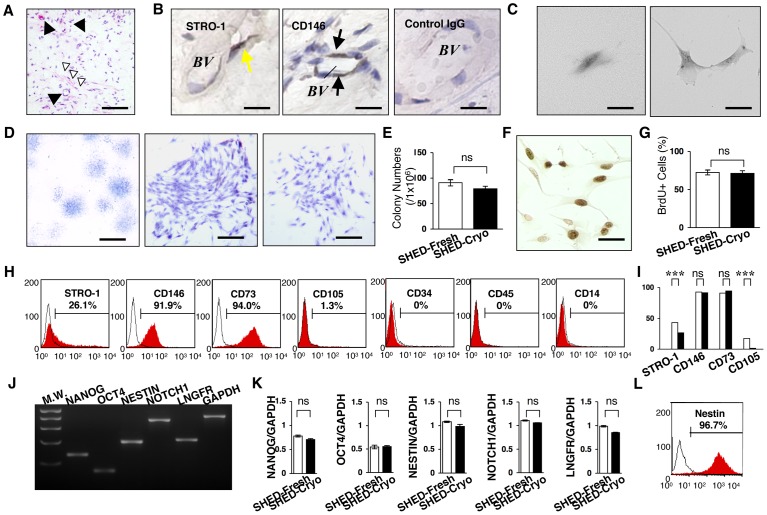
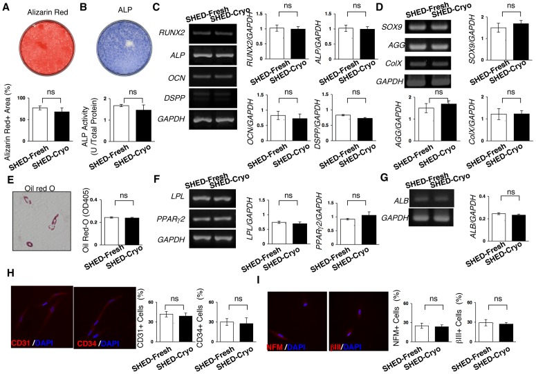
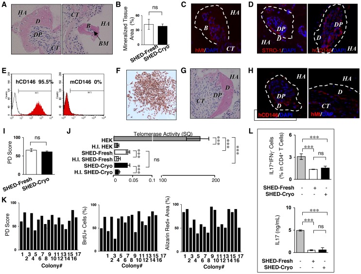
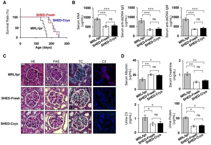
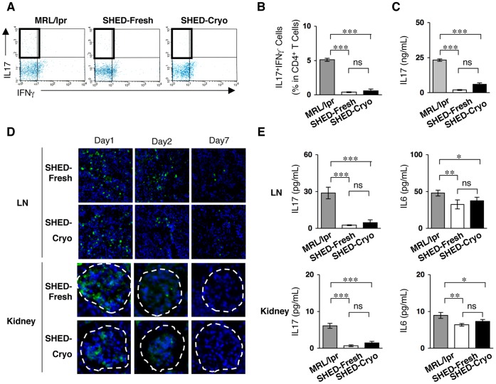
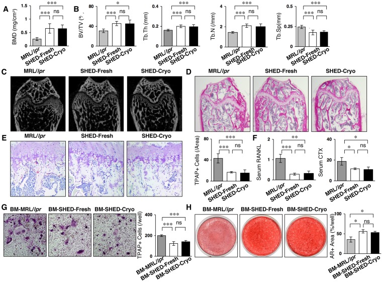
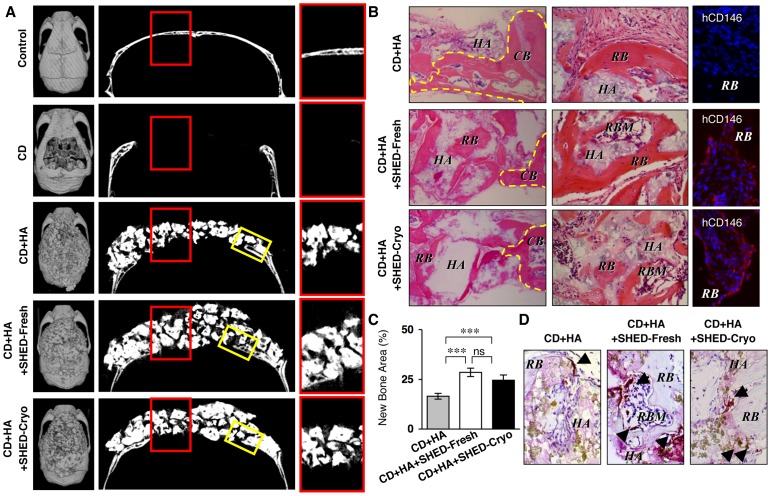
Human exfoliated deciduous teeth have been considered to be a promising source for regenerative therapy because they contain unique postnatal stem cells from human exfoliated deciduous teeth (SHED) with self-renewal capacity, multipotency and immunomodulatory function. However preservation technique of deciduous teeth has not been developed. This study aimed to evaluate that cryopreserved dental pulp tissues of human exfoliated deciduous teeth is a retrievable and practical SHED source for cell-based therapy. SHED isolated from the cryopreserved deciduous pulp tissues for over 2 years (25-30 months) (SHED-Cryo) owned similar stem cell properties including clonogenicity, self-renew, stem cell marker expression, multipotency, in vivo tissue regenerative capacity and in vitro immunomodulatory function to SHED isolated from the fresh tissues (SHED-Fresh). To examine the therapeutic efficacy of SHED-Cryo on immune diseases, SHED-Cryo were intravenously transplanted into systemic lupus erythematosus (SLE) model MRL/lpr mice. Systemic SHED-Cryo-transplantation improved SLE-like disorders including short lifespan, elevated autoantibody levels and nephritis-like renal dysfunction. SHED-Cryo amended increased interleukin 17-secreting helper T cells in MRL/lpr mice systemically and locally. SHED-Cryo-transplantation was also able to recover osteoporosis bone reduction in long bones of MRL/lpr mice. Furthermore, SHED-Cryo-mediated tissue engineering induced bone regeneration in critical calvarial bone-defect sites of immunocompromised mice. The therapeutic efficacy of SHED-Cryo transplantation on immune and skeletal disorders was similar to that of SHED-Fresh. These data suggest that cryopreservation of dental pulp tissues of deciduous teeth provide a suitable and desirable approach for stem cell-based immune therapy and tissue engineering in regenerative medicine.


Conflict of interest statement

Competing Interests: The authors have declared that no competing interests exist.

Figures

Figure 1
Figure 1. Clonogenicity, cell proliferation capacity and stem cell marker expression of SHED-Cryo.
(A) Histology of cryopreserved dental pulp tissue of exfoliated deciduous teeth. Black arrowheads: blood vessel, white arrowheads: nerve fibers. H&E staining. (B) Localization of MSC markers in the cryopreserved deciduous pulp tissues. Yellow arrows: STRO-1-positive cells, black arrows: CD146-positive cells. BV: blood vessel, Control: subclass-matched antibody staining. (C–E) CFU-F assay. Formation of a clonogenic cell cluster from a single attached cell (C). Images of attached colonies of SHED-Cryo. Toluidine blue staining (D). Comparison of CFU-F number (E). (F, G) Cell proliferation assay. Immunostaining of BrdU-positive nuclei. (F). Comparison of cell proliferation (G). (H, I) Flow cytometry of MSC markers in SHED-Cryo. Representative histograms (H). Comparison of STRO-1, CD146, CD73 and CD105. Black columns: SHED-Cryo, white columns: SHED-Fresh (I). (J, K) Gene expression of embryonic stem and neural crest cell markers. MW: molecular weight markers (J). Comparative analysis of NANOG, octamer 4 (OCT4), NESTIN, NOTCH1 and low affinity nerve growth factor receptor (LNGFR) (K). (L) Flow cytometry of Nestin in SHED-Cryo. A, B: n = 3. C–L: n = 5 for all group. A–E: Bar = 30 µm (A), 5 µm (B, C, F), 1 mm (D, left) 25 µm (D, middle and right). G, I, K: ***P<0.005. ns: no significance. The graph bars represent mean±SD.
Figure 2
Figure 2. Multipotency of SHED-Cryo.
(A–C) Dentinogenic/osteogenic differentiation capacity. Images of Alizarin Red staining (A) and alkaline phosphatase (ALP) activity (B) of SHED-Cryo. Comparison of Alizarin Red-positive (Alizarin Red+) area (A), ALP activity (B) and odontoblast/osteoblast-specific genes, runt-related gene 2 (RUNX2), ALP, osteocalcin (OCN), and dentin sialophosphoprotein (DSPP) (C). (D) Chondrogenic differentiation capacity. Comparison of chondrocyte-specific genes, SOX9, aggrecan (AGG) and type X collagen (ColX). (E, F) Adipogenic differentiation assay. A representative image of Oil Red-O staining and comparison of Oil Red-O accumulation (E). Comparison of adipocyte-specific genes lipoprotein lipase (LPL) and peroxisome proliferator activated receptor-gamma2 (PPARgamma2) (F). (G) Hepatogenic differentiation capacity. Comparison of hepatocyte-specific gene albumin (ALB). (H) Endothelial cell differentiation assay. Comparison of endothelial cell markers CD31 and CD34. (H) Neural cell differentiation assay. Comparison of neural cell markers neurofilament M (NFM) and tubulin betaIII (betaIII). A–I: n = 5 for all group. ns: no significance. The graph bars represent mean±SD.
Figure 3
Figure 3. Tissue regeneration capability, self-renewal potency, heterogeneity and in vitro immunomodulatory functions of SHED-Cryo.
(A–D) Images of primary transplant tissues of SHED-Cryo. H&E staining (A). Comparison of newly formed-mineralized tissue (B). Immunofluorescence with anti-human specific mitochondria (hMt) (C) and anti-STRO-1/human CD146 (hCD146) (D) antibodies. (E, F) Purity of hCD146 antibody-sorted cells from primary transplants. Flow cytometry with hCD146 and mouse CD146 (mCD146). (E). Immunocytochemistry with hCD146 antibody of sorted cell-derived CFU-F (F). (G, H) in vivo self-renewal assay. Images of secondary transplant tissues. H&E staining (G). Immunofluorescence with anti-hCD146/anti-Mt antibodies. (H). (I) Comparison of population doubling (PD) scores. (J) Comparison of telomerase activity. (K) Single-colony-derived cell assay with 17 single cell colonies from a cryopreserved deciduous pulp tissues. (L) In vitro direct immunosuppressive effects of SHED-Cryo on human Th17 cells. A–J, L: n = 5 for all group. A, C, D, F, H: B: bone, BM: bone marrow, CT: connective tissue, D: dentin, DP: dental pulp, HA: HA/TCP, I: HEK: HEK293 cells, H.I. HEK: heat inactivated HEK, H.I. SHED-Cryo: heat inactivated SHED-Cryo, H.I. SHED-Fresh: heat inactivated SHED-Fresh. C, D, H: Dot lined areas: mineralized tissue. Nuclei are counterstained with DAPI. B, H, I, L: ***P<0.005, ns: no significance. The graph bars represent mean±SD.
Figure 4
Figure 4. Systemic SHED-Cryo-transplantation improves lifespan and SLE-like disorders in MRL/lpr mice.
(A) Kaplan-Meier survival curve of MRL/lpr mice. (B) ELISA of serum levels of autoantibodies ANA and anti-dsDNA IgG and IgM antibodies. (C) Histopathology of kidneys. G and dot-circled area: glomerular. HE: H&E staining, TC: Gomori trichrome staining, PAS: Periodic acid-Schiff staining, C3: Immunofluorescence of Complement C3. DAPI staining. (D) Levels of serum albumin and creatinine and urine C3 and protein. AD: MRL/lpr: control group, SHED-Cryo: SHED-Cryo-transplant group, SHED-Fresh: SHED-Fresh-transplant group. A: n = 7, BD: n = 5 for all group. B, D: *P<0.05, **P<0.01, ***P<0.005, ns: no significance. The graph bars represent mean±SD.
Figure 5
Figure 5. SHED transplantation suppresses circulating and local levels of Th17 cells in MRL/lpr mice.
(A, B) Flow cytometry of peripheral CD4+IL17+IFNgamma Th17 cells. (C) Serum levels of IL-17. (D) Homing of systemically infused CFSE-labeled SHED-Cryo and SHED-Fresh to lymph node (LN) and kidney of MRL/lpr Mice after 1- (Day 1), 2- (Day 2) or 7- (Day 7) day transplantation. Dot-circled area: glomerular. (E) ELISA of IL-17 an IL-6 levels in lymph node and kidney. AE: n = 5 for all group. MRL/lpr: control group, SHED-Cryo: SHED-Cryo-transplant group, SHED-Fresh: SHED-Fresh-transplant group. B, C, E: *P<0.05, **P<0.01, ***P<0.005, ns: no significance. The graph bars represent mean±SD.
Figure 6
Figure 6. SHED-Cryo transplantation ameliorates osteoporotic bone disorder in MRL/lpr mice. (A, B)
MicroCT analysis of tibiae. BMD (A). Trabecular parameters, bone volume ratio to tissue volume (BV/TV), trabecular thickness (Tb.Th), and trabecular number (Tb.N) along with increased trabecular separation (Tb.Sp) (B). (C, D) MicroCT (C) and histological (D) images of trabecular bone structures of tibiae. H&E staining (D). (E, F) In vivo osteoclast activity. TRAP staining (E). ELISA of serum sRANKL and C-terminal telopeptides of type I collagen (CTX) (F). (G) Ex vivo sRANKL-induced osteoclastogenesis. TRAP+ cells: TRAP-positive osteoclast-like cells. (H) Ex vivo osteogenic capacity. Alizarin red-positive (AR+) area after four-week induction. A–H: n = 5 for all groups. A–F: MRL/lpr: control group, SHED-Cryo: SHED-Cryo-transplant group, SHED-Fresh: SHED-Fresh-transplant group, G, H: BM-MRL/lpr: control MRL/lpr mice-derived bone marrow cells, BM-SHED-Cryo: SHED-Cryo-transplanted mice-derived bone marrow cells, BM-SHED-Fresh: SHED-Fresh-transplanted mice-derived bone marrow cells. A, B, E, F: *P<0.05, ***P<0.005 (vs. MRL/lpr). G, H: *P<0.05, **P<0.01, ***P<0.005 (vs. BM-MRL/lpr), ns: no significance. A, B, E–H: The graph bars represent mean±SD.
Figure 7
Figure 7. SHED-Cryo are capable of repairing critical calvarial bone defects in immunocompromised mice.
(A) MicroCT images of mouse calvariae. Left panels: cranial images, middle panels: saggital images, right panels: images of red-bowed area in riddle panels. (B) Histology of bone regeneration in mouse calvariae. Left panels: edge parts of the defect area (yellow-boxed area in Figure 7A). H&E staining; middle panels: middle parts of the defect area (red red-bowed area in Figure 7A). H&E staining; right panels: immunofluorescence with anti-human CD146 antibody (hCD146). DAPI staining. CB, yellow dot-circled area: calvarial bone, HA: HA/TCP, RB: regenerated bone, RBM: regenerated bone marrow. (C) Regenerated bone area in the defect area. (D) Distribution of osteoclasts. Arrowheads: TRAP-positive cells. TRAP staining. A–D: n = 5 for all groups. Control: control (non-defect) group, CD: calvarial defect group, CD+HA: HA/TCP-implanted group, CD+HA+SHED-Fresh: SHED-Fresh-implanted group, CD+HA+SHED-Cryo: SHED-Cryo-implanted group. C: ***P<0.005, ns: no significance. The graph bars represent mean±SD.

References

    1. Porada CD, Almeida-Porada G (2010) Mesenchymal stem cells as therapeutics and vehicles for gene and drug delivery. Adv Drug Deliv Rev 62: 1156–1166. - PMC - PubMed
    1. Kwan MD, Slater BJ, Wan DC, Longaker MT (2008) Cell-based therapies for skeletal regenerative medicine. Hum Mol Genet 17: R93–98. - PubMed
    1. Panetta NJ, Gupta DM, Quarto N, Longaker MT (2009) Mesenchymal cells for skeletal tissue engineering. Panminerva Med 51: 25–41. - PubMed
    1. Aggarwal S, Pittenger MF (2005) Human mesenchymal stem cells modulate allogeneic immune cell responses. Blood 105: 1815–1822. - PubMed
    1. Nauta AJ, Fibbe WE (2007) Immunomodulatory properties of mesenchymal stromal cells. Blood 110: 3499–3506. - PubMed

Publication types

MeSH terms