. 2012;7(12):e51964.
doi: 10.1371/journal.pone.0051964. Epub 2012 Dec 14.

A potent anti-HB-EGF monoclonal antibody inhibits cancer cell proliferation and multiple angiogenic activities of HB-EGF

Affiliations

A potent anti-HB-EGF monoclonal antibody inhibits cancer cell proliferation and multiple angiogenic activities of HB-EGF

Shuji Sato et al. PLoS One. 2012.

Abstract

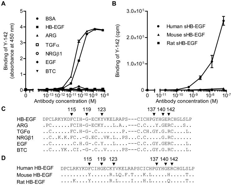
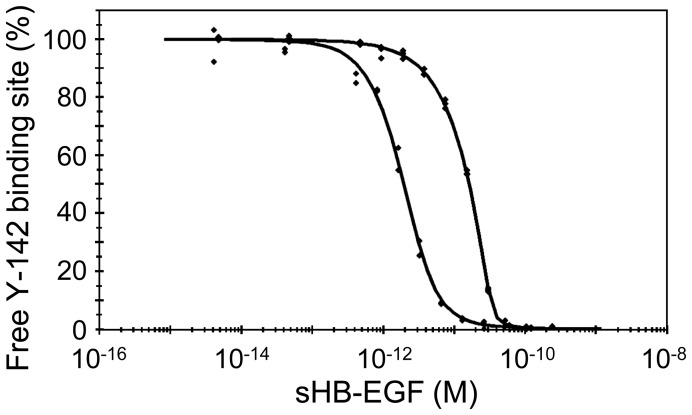
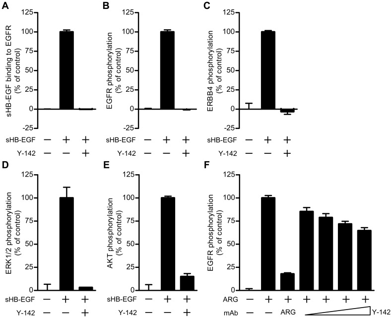
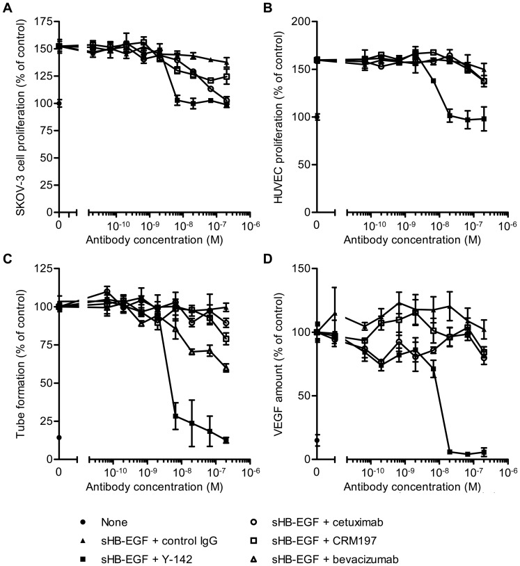
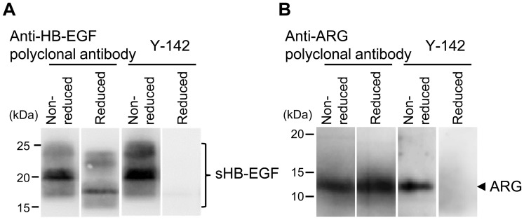
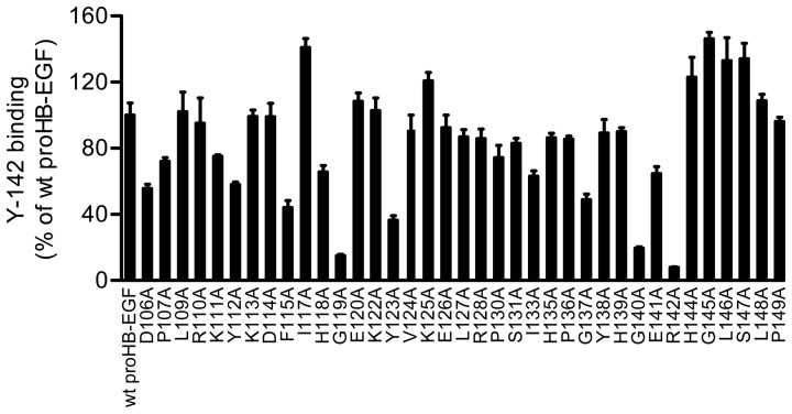
Heparin-binding epidermal growth factor-like growth factor (HB-EGF) is a member of the epidermal growth factor family and has a variety of physiological and pathological functions. Modulation of HB-EGF activity might have a therapeutic potential in the oncology area. We explored the therapeutic possibilities by characterizing the in vitro biological activity of anti-HB-EGF monoclonal antibody Y-142. EGF receptor (EGFR) ligand and species specificities of Y-142 were tested. Neutralizing activities of Y-142 against HB-EGF were evaluated in EGFR and ERBB4 signaling. Biological activities of Y-142 were assessed in cancer cell proliferation and angiogenesis assays and compared with the anti-EGFR antibody cetuximab, the HB-EGF inhibitor CRM197, and the anti-vascular endothelial growth factor (VEGF) antibody bevacizumab. The binding epitope was determined with alanine scanning. Y-142 recognized HB-EGF as well as the EGFR ligand amphiregulin, and bound specifically to human HB-EGF, but not to rodent HB-EGF. In addition, Y-142 neutralized HB-EGF-induced phosphorylation of EGFR and ERBB4, and blocked their downstream ERK1/2 and AKT signaling. We also found that Y-142 inhibited HB-EGF-induced cancer cell proliferation, endothelial cell proliferation, tube formation, and VEGF production more effectively than cetuximab and CRM197 and that Y-142 was superior to bevacizumab in the inhibition of HB-EGF-induced tube formation. Six amino acids in the EGF-like domain were identified as the Y-142 binding epitope. Among the six amino acids, the combination of F115 and Y123 determined the amphiregulin cross-reactivity and that F115 accounted for the species selectivity. Furthermore, it was suggested that the potent neutralizing activity of Y-142 was derived from its recognition of R142 and Y123 and its high affinity to HB-EGF. Y-142 has a potent HB-EGF neutralizing activity that modulates multiple biological activities of HB-EGF including cancer cell proliferation and angiogenic activities. Y-142 may have a potential to be developed into a therapeutic agent for the treatment of HB-EGF-dependent cancers.


Conflict of interest statement

Competing Interests: The authors have the following interests. This study was funded by Takeda San Francisco, Inc. and Takeda Pharmaceutical Company Limited. All authors were employees of Takeda San Francisco, Inc. or Takeda Pharmaceutical Company Limited and were paid as such. There are no patents, products in development or marketed products to declare. This does not alter the authors’ adherence to all the PLOS ONE policies on sharing data and materials, as detailed online in the guide for authors.

Figures

Figure 1
Figure 1. Binding specificity of Y-142 to EGFR ligands and to different species of sHB-EGF.
(A) The binding activity of Y-142 to EGFR ligands by ELISA. The various concentrations of Y-142 were incubated in an EGFR ligand-immobilized plate. The binding was then detected with HRP-labeled anti-mouse IgG antibody. Data points represent the mean ± standard deviation (SD) of values acquired in duplicate. (B) The binding activity of Y-142 to human, mouse, and rat HB-EGF by ELISA. The binding activity to different species HB-EGF was measured by ELISA using an electroluminescence-based technology. The various concentrations of Y-142 were incubated in a sHB-EGF-immobilized plate. The binding was detected with sulfo-tagged anti-mouse IgG antibody. Data points represent the mean ± SD of values acquired in duplicate. (C) Amino acid alignment of the EGF-like domain of EGFR ligands. A dot indicates an amino acid different than HB-EGF. A dash represents a gap. Arrowheads labeled with a number indicate the Y-142 binding epitopes identified in Fig. 6. (D) Amino acid alignment of the EGF-like domain of human, mouse, and rat HB-EGF. A dot indicates an amino acid identical to human HB-EGF. Arrowheads labeled with a number indicate the Y-142 binding epitopes identified in Fig. 6.
Figure 2
Figure 2. Measuring the KD of the Y-142/HB-EGF complex.
Dual-curve KinExA equilibrium titration of sHB-EGF binding to Y-142. KD-controlled data (bottom fitted curve) were acquired by equilibrating sHB-EGF at a concentration range of 4.04 fM–207 pM with 1.03 pM Y-142 binding sites. Antibody-controlled data (top fitted curve) were acquired by equilibrating sHB-EGF at a concentration range of 4.67 fM–239 pM with 35.6 pM Y-142 binding sites. All data points were acquired in duplicate. Both curves were simultaneously fit to a standard positive cooperativity equilibrium model, yielding an effective KD = 1.50 pM (0.31) where the number in parentheses is the 95% confidence interval of the fit, and a Hill coefficient n = 1.68.
Figure 3
Figure 3. Neutralizing activities of Y-142 against sHB-EGF and ARG signaling.
(A) Inhibitory activity of Y-142 to sHB-EGF binding to EGFR. EGFR-hFc was incubated in an anti-human IgG Fc antibody-coated plate. Y-142 was then incubated at a concentration of 6.7 nM in the presence of 0.63 nM biotinylated sHB-EGF for 1 hour at 37°C. sHB-EGF bound to EGFR-hFc was detected by HRP-labeled streptavidin. sHB-EGF binding to EGFR-hFc in the presence of Y-142 was calculated as a percentage of the “control” sHB-EGF binding to EGFR which occurred without Y-142. Data points represent the mean + SD of values acquired in triplicate. (B) Neutralizing activity of Y-142 against EGFR phosphorylation. SK-OV-3 cells were treated with 10 nM sHB-EGF and 67 nM Y-142. Cell lysates were incubated in an anti-EGFR antibody-coated plate, followed by an incubation with HRP-labeled anti-phosphorytosine antibody. EGFR phosphorylation in the presence of Y-142 was calculated as a percentage of the “control” EGFR phosphorylation which occurred without Y-142. Data points represent the mean + SD of values acquired in triplicate. (C) Neutralizing activity of Y-142 against ERBB4 phosphorylation. Cell lysates of T47D cells as prepared in Fig. 3A were incubated on an anti-ERBB4 antibody-coated plate. The phosphorylation of ERBB4 was detected by a sulfo-tagged anti-phosphotyrosine antibody. ERBB4 phosphorylation in the presence of Y-142 was calculated as a percentage of the “control” ERBB4 phosphorylation which occurred without Y-142. Data points represent the mean + SD of values acquired in duplicate. (D) and (E) Neutralizing activity of Y-142 against (D) ERK1/2 phosphorylation and (E) AKT phosphorylation. In (D) and (E) SK-OV-3 cells treated with 10 nM sHB-EGF and 200 nM Y-142 were stained with an anti-phosphorylated ERK1/2 antibody or an anti-phosphorylated AKT antibody, respectively, followed by an Alexa488-labeled anti-rabbit IgG antibody. Phosphorylated ERK1/2 and phosphorylated AKT were both detected with an ImageXpress Micro instrument and calculated as a percentage of the “control” phosphorylation levels which occurred without Y-142. Data points represent the mean + SD of values acquired in duplicate. (F) Neutralizing activity of Y-142 to ARG. SK-OV-3 cells were treated with 10 nM ARG plus various concentrations of Y-142 (2 nM, 6.7 nM, 20 nM, and 67 nM). Anti-ARG monoclonal antibody (67 nM) was used as a positive control. Cell lysates were incubated in an anti-EGFR antibody-coated plate followed by an incubation with an HRP-labeled anti-phosphorytosine antibody. EGFR phosphorylation was calculated as a percentage of the “control” EGFR phosphorylation which occurred without Y-142. Data points represent the mean + SD of values acquired in triplicate.
Figure 4
Figure 4. Inhibitory activity of Y-142 against sHB-EGF functions.
(A) and (B) Neutralizing activities of Y-142 against (A) sHB-EGF-induced SK-OV-3 cell proliferation and (B) HUVEC proliferation. SK-OV-3 cells or HUVEC were cultured for 3 days in the presence of sHB-EGF and the indicated concentrations of Y142, cetuximab, or CRM197. Cell proliferation was detected with CellTiter-Glo and calculated as a percentage of the “control” cell proliferation without sHB-EGF. Data points represent the mean ± SD of values acquired in triplicate. (C) Inhibition of HUVEC tube formation by Y-142. HUVEC were cultured on a monolayer of NHDF in the presence of 50 nM sHB-EGF and the indicated concentrations of Y142, cetuximab, CRM197, or bevacizumab for 4 days. HUVEC were then stained with FITC-labeled anti-CD31 antibody. Tube formation (CD31-positive area) was calculated as a percentage of the “control” amount of tube formation in the presence of sHB-EGF. Data points represent the mean ± SD of values acquired in triplicate. (D) Inhibition of VEGF production by Y-142. HUVEC were prepared as in Figure 4C and treated with 50 nM sHB-EGF and the indicated concentrations of Y142, cetuximab, or CRM197 for 4 days. VEGF concentration in the supernatant of co-culture was measured in an electrochemiluminescence-based method. VEGF production was calculated as a percentage of the “control” amount of VEGF produced in the presence of sHB-EGF. Data points represent the mean ± SD of values acquired in triplicate.
Figure 5
Figure 5. Recognition of a conformational epitope by Y-142.
The binding of Y-142 to a linear or conformational epitope was tested using Western blot. sHB-EGF (A) or ARG (B) was prepared in reducing or non-reducing conditions with or without dithiothreitol, respectively. The sHB-EGF was probed with an anti-HB-EGF polyclonal antibody and with Y-142. The ARG was probed with anti-ARG polyclonal antibody and with Y-142.
Figure 6
Figure 6. Recognition of F115, G119, Y123, G137, G140, and R142 by Y-142.
Epitope mapping of Y-142 was performed using alanine scanning. Each mutant proHB-EGF expression plasmid was transfected into SW480 cells. The binding activity of Y-142 to the cells was measured in a cell ELISA. The expression level of mutant proHB-EGF was normalized with the binding of anti-HB-EGF polyclonal antibody by each mutant. The binding of Y-142 to mutant proHB-EGF was calculated as a percentage of the “control” binding of Y-142 to wild-type proHB-EGF. Data points represent the mean + SD of values acquired in triplicate.

References

    1. Higashiyama S, Abraham JA, Miller J, Fiddes JC, Klagsbrun M (1991) A heparin-binding growth factor secreted by macrophage-like cells that is related to EGF. Science 251: 936–939. - PubMed
    1. Elenius K, Paul S, Allison G, Sun J, Klagsbrun M (1997) Activation of HER4 by heparin-binding EGF-like growth factor stimulates chemotaxis but not proliferation. EMBO J 16: 1268–1278. - PMC - PubMed
    1. Higashiyama S, Lau K, Besner GE, Abraham JA, Klagsbrun M (1992) Structure of heparin-binding EGF-like growth factor. Multiple forms, primary structure, and glycosylation of the mature protein. J Biol Chem 267: 6205–6212. - PubMed
    1. Higashiyama S, Iwamoto R, Goishi K, Raab G, Taniguchi N, et al. (1995) The membrane protein CD9/DRAP 27 potentiates the juxtacrine growth factor activity of the membrane-anchored heparin-binding EGF-like growth factor. J Cell Biol 128: 929–938. - PMC - PubMed
    1. Shiomi T, Lemaître V, D’Armiento J, Okada Y (2010) Matrix metalloproteinases, a disintegrin and metalloproteinases, and a disintegrin and metalloproteinases with thrombospondin motifs in non-neoplastic diseases. Pathol Int 60: 477–496. - PMC - PubMed

MeSH terms