. 2012;7(12):e51975.
doi: 10.1371/journal.pone.0051975. Epub 2012 Dec 14.

Regional brain atrophy and functional disconnection in Broca's area in individuals at ultra-high risk for psychosis and schizophrenia

Affiliations

Regional brain atrophy and functional disconnection in Broca's area in individuals at ultra-high risk for psychosis and schizophrenia

Wi Hoon Jung et al. PLoS One. 2012.

Abstract

Background: Abnormalities in cognitive abilities such as verbal fluency and in cognitive-related brain regions, particularly Broca's area, have been reported in patients with schizophrenia. Additionally, previous studies have demonstrated that structural and functional abnormalities in Broca's area were associated with clinical symptoms and cognitive deficits in patients with schizophrenia, suggesting that deficits in this area may reflect the core pathology of schizophrenia. Thus, it is important to understand how the structural volume and functional connectivity in this area changes at rest according to the course of the illness.

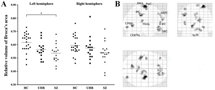
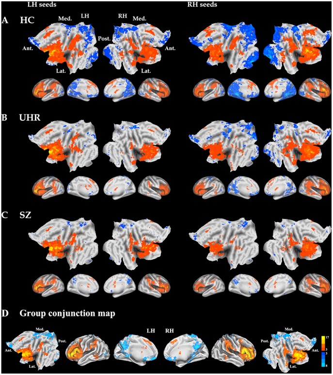
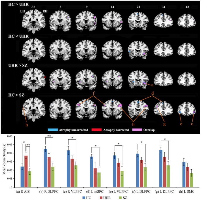
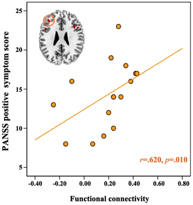
Methods/principal findings: We used magnetic resonance imaging (MRI) to measure the structural volume of Broca's area as a region of interest in 16 schizophrenia, 16 ultra-high risk (UHR), and 23 healthy matched controls. We also assessed verbal fluency and analyzed differences across groups in the functional connectivity patterns using resting-state functional MRI. The UHR group showed significantly reduced structural volume in Broca's area and significantly reduced functional connectivity between Broca's area and the lateral and medial frontal cortex as well as decreased cognitive performance. Altered functional connectivity in patients was correlated with their positive symptoms.

Conclusions/significance: Our results suggest the existence of functional disconnections in Broca's area, even during resting-states, among those with schizophrenia as well as those at UHR for this disorder. These alterations may contribute to their clinical symptoms, suggesting that this is one of the key regions involved in the pathophysiology of schizophrenia.


Conflict of interest statement

Competing Interests: The authors have declared that no competing interests exist.

Figures

Figure 1
Figure 1. Gray matter volume differences in Broca’s area using Freesurfer (A) and in whole brain using voxel-based morphometry (B) across groups using ANOVA.
HC, healthy controls; UHR, ultra-high risk; SZ, schizophrenia; SMA, supplementary motor area; PreC, precentral cortex; mSFC, medial superior frontal cortex; IFC, inferior frontal cortex; OFC, orbitofrontal cortex; IPC, inferior parietal cortex; THAL, thalamus; LING, lingual gyrus; CER, cerebellum.
Figure 2
Figure 2. Functional connectivity maps for each group of healthy controls (A), individuals at ultra-high risk for developing psychosis (B), and schizophrenia patients (C), and common neural network across groups generated by performing a conjunction analysis (D).
Warm and cold color indicate positive and negative connectivity with each seed region, respectively. HC, healthy controls, UHR, ultra-high risk; SZ, schizophrenia; LH, left hemisphere; RH, right hemisphere; Ant, anterior; Post, posterior; Med, medial; Lat, lateral.
Figure 3
Figure 3. Brain regions showing group differences in functional connectivity.
Blue and red color show significant regions before and after atrophy correction, respectively. Pink color shows overlap regions between significant regions before and after atrophy correction. Graphs depicted below represent average functional connectivity in each region of interest. An ROI (a) defined as the regions (5 mm radius sphere centered at the peak coordinate) showing quadratic trend in FC across three groups in voxel-wise manner and ROIs (b-h) are defined as the regions (5 mm radius spheres centered at the peak coordinates) showing significant differences in FC between HC and SZ in voxel-wise manner. We extracted the value in each ROI using MarsBaR software (http://marsbar.sourceforge.net/) and then performed ANOVA and post-hoc t-tests with Bonferroni correction as well as trend analyses using SPSS (refer to Table S1). LH, left hemisphere; RH, right hemisphere; HC, healthy controls; UHR, ultra-high risk; SZ, schizophrenia; AIS, anterior insula; DLPFC, dorsolateral prefrontal cortex; VLPFC, ventrolateral prefrontal cortex; mSFC, medial superior frontal cortex; SMC, supramarginal cortex.
Figure 4
Figure 4. Correlation of functional connectivity with cognitive performance and positive symptom.
Combined group data (black line), collapsed across groups, and SZ group data (orange line) showed significant correlation between functional connectivity and cognitive performance. LH, left hemisphere; RH, right hemisphere; HC, healthy controls(blue circle); UHR, ultra-high risk(green circle); SZ, schizophrenia(orange circle); PANSS, Positive and Negative Syndrome Scale.

References

    1. Simon AE, Cattapan-Ludewig K, Zmilacher S, Arbach D, Gruber K, et al. (2007) Cognitive functioning in the schizophrenia prodrome. Schizophr Bull 33: 761–771. - PMC - PubMed
    1. Fusar-Poli P, Perez J, Broome M, Borgwardt S, Placentino A, et al. (2007) Neurofunctional correlates of vulnerability to psychosis: a systematic review and meta-analysis. Neurosci Biobehav Rev 31: 465–484. - PubMed
    1. Jung WH, Jang JH, Byun MS, An SK, Kwon JS (2010) Structural brain alterations in individuals at ultra-high risk for psychosis: a review of magnetic resonance imaging studies and future directions. J Korean Med Sci 25: 1700–1709. - PMC - PubMed
    1. Choi JS, Park JY, Jung MH, Jang JH, Kang DH, et al... (2011) Phase-specific brain change of spatial working memory processing in genetic and ultra-high risk groups of schizophrenia. Schizophr Bull In press. - PMC - PubMed
    1. Liu Y, Liang M, Zhou Y, He Y, Hao Y, et al. (2008) Disrupted small-world networks in schizophrenia. Brain 131(Pt 4): 945–961. - PubMed

Publication types

MeSH terms