. 2012:2:994.
doi: 10.1038/srep00994. Epub 2012 Dec 18.

Helicobacter pylori colonization critically depends on postprandial gastric conditions

Affiliations

Helicobacter pylori colonization critically depends on postprandial gastric conditions

Roland Bücker et al. Sci Rep. 2012.

Abstract

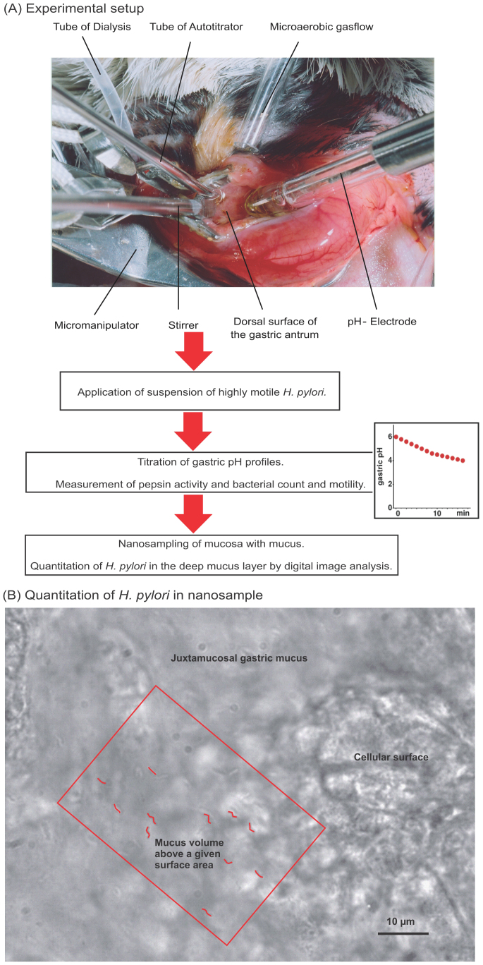
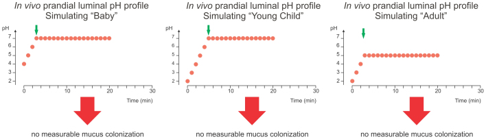
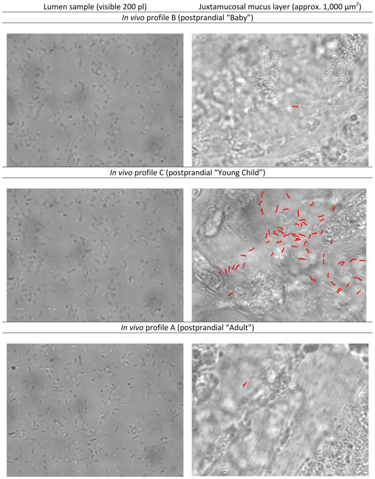
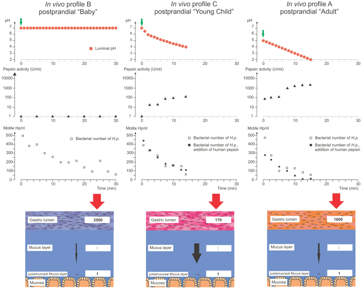
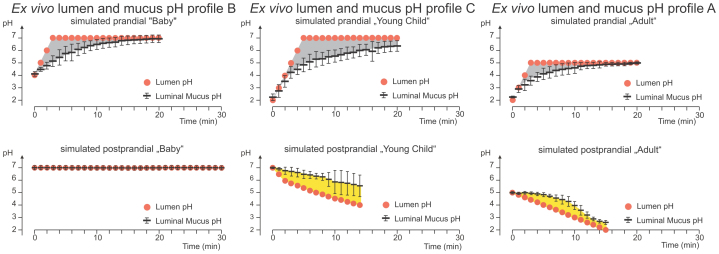
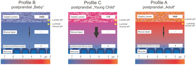
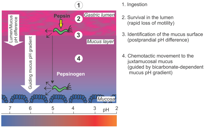
The risk of Helicobacter pylori infection is highest in childhood, but the colonization process of the stomach mucosa is poorly understood. We used anesthetized Mongolian gerbils to study the initial stages of H. pylori colonization. Prandial and postprandial gastric conditions characteristic of humans of different ages were simulated. The fraction of bacteria that reached the deep mucus layer varied strongly with the modelled postprandial conditions. Colonization success was weak with fast gastric reacidification typical of adults. The efficiency of deep mucus entry was also low with a slow pH decrease as seen in pH profiles simulating the situation in babies. Initial colonization was most efficient under conditions simulating the postprandial reacidification and pepsin activation profiles in young children. In conclusion, initial H. pylori colonization depends on age-related gastric physiology, providing evidence from an in vivo infection model that suggests an explanation why the bacterium is predominantly acquired in early childhood.


Figures

Figure 1
Figure 1. Flow diagram of the key steps of the Mongolian gerbil in vivo model system.
(A) Photograph of opened gerbil stomach and setup for the simulation of prandial and postprandial conditions. Luminal gastric conditions can be manipulated through a small incision within the wall of the Mongolian gerbil stomach. A micro-pH-electrode measured the luminal pH value continuously, while the small stirrer ensured efficient mixing of the gastric content. Defined pH profiles were generated by an autotitrator, which applies 0.25 M HCl and 100 mM urea or 0.15 M NaOH. A gas flow of 5% O2, 10% CO2 and 85% N2 ensured a microaerobic atmosphere. 50 µl bacterial suspension was applied into the gastric lumen, which contained approx. 0.5 ml of gastric juice. Each animal received the same number of highly motile Helicobacter pylori. After simulation of defined pH profiles representing different gastric conditions of certain age groups, bacterial numbers in the mucosa can be sampled by the nanosampling method. (B) Micrograph of H. pylori in the juxtamucosal mucus layer. Microscopical detection of H. pylori in the mucus volume above a given surface area. The volume of gastric juice in the lumen above the gastric surface in the setup constellation is known. Spatial distribution of H. pylori can be identified in the mucus layer near the epithelium. Colonized H. pylori cells were coloured red (see also supplemental Figure S2 (B) for the original image).
Figure 2
Figure 2. In vivo colonization under simulated prandial gastric conditions.
The three panels show the luminal pH profiles (red bullets) that were titrated to simulate the prandial phase in breast-fed babies (left), young children (middle), and adults (right). The H. pylori inoculum is given during the simulation of the meal, as administration at the beginning of the profile would lead to rapid loss of bacterial motility due to the acidic fasting pH in profiles A and C. In profile B, the pH is remaining at 4 between the meals. The time points when an inoculum of 108 motile H. pylori was added in the in vivo experiments in the Mongolian gerbil are marked by green arrows.
Figure 3
Figure 3. Luminal and juxtamucosal densities of H. pylori after application of three age-dependent postprandial gastric pH profiles.
Left panels: Luminal samples analyzed in a heated chamber. The visible volume is 200 picoliters. Right panels: Nanosamples from the anesthetized gerbil, with different numbers of H. pylori that have entered the juxtamucosal mucus. The profile simulating the postprandial gastric conditions in young children causes a remarkably higher colonization. To improve contrast and readability, all sharply visible bacteria (± 2,5 µm from focus plane) were coloured in red. A composite with the original (uncoloured) micrographs is included as supplemental figure S2 (A). Five nanosamples of juxtamucosal mucus were averaged to determine the colonization density of H. pylori in each animal.
Figure 4
Figure 4. Postprandial gastric conditions strongly affect H. pylori colonization.
The figure shows the effects of three different postprandial pH profiles on pepsin activity, H. pylori motility, and H. pylori mucus entry in anesthetized Mongolian gerbils. pH profiles simulate postprandial conditions in breast-fed babies (profile B, left column), young children (profile C, middle column), and adults (profile A, right column). The three top panels show the titrated profiles (red bullets). The time points of H. pylori inoculation are marked by green arrows. The next two rows of graphs depict pepsin activity in logarithmic scale (triangles) and the number of motile H. pylori during the in vivo experiment (Mongolian gerbil), open squares indicate values measured during experiments performed without addition of exogenous pepsin (i.e. the gastric juice contains native gerbil pepsin only), black squares are values determined in experiments where human pepsin was added. The values depicted here are mean values of at least three independent experiments for each experimental group. The lowest panel visualizes the number of motile H. pylori in the lumen required for one bacterium to reach the juxtamucosal mucus.
Figure 5
Figure 5. Luminal pH profiles and the pH values within the mucus layer.
The figure shows the effects of three different prandial and postprandial pH profiles on mucus pH as measured by ultrafine tipped double-barrelled microelectrodes in explanted guinea pig mucosa. pH profiles simulate prandial (top panel) and postprandial (bottom panel) conditions in breast-fed babies (left column), young children (middle column), and adults (right column). Black horizontal lines represent measurements of the resulting pH within the mucus layer (mean ± SD, n = 3 each) in the antral guinea pig mucosa. The pH difference between lumen and mucus is indicated by grey shading following prandial pH profiles. Under all three prandial profiles, the mucus pH was more acidic than the lumen pH. The three bottom panels show the titrated postprandial profiles and the resulting mucus pH values following postprandial profiles (B, C, A). The yellow-shaded area represents the difference between lumen pH and the pH in the mucus. Note that, by contrast to the prandial conditions, the pH is lower in the lumen than in the mucus in postprandial profiles A and C.
Figure 6
Figure 6. Schematical model summarizing the results from both experimental setups.
Depicted is the pH gradient within the mucus layer and the luminal pH by coloration gradient (as measured in guinea pig experiments), as well as the colonization ratio following postprandial conditions (from Mongolian gerbil experiments).
Figure 7
Figure 7. A hypothetical model of the steps required for a successful Helicobacter pylori colonization of the stomach.
-1- Oral ingestion -2- H. pylori has to remain motile in the gastric lumen, where active pepsins, particularly pepsin C, can cause a rapid loss of motility and could therefore prevent an acute colonization.-3- As deduced from the presented experimental series, a postprandial pH difference between lumen and mucus caused by a dynamic decrease of the gastric lumen pH might be essential for H. pylori to be guided toward the mucus surface for colonization. -4- In the gastric mucus, chemotactic movement guided by the bicarbonate-dependent mucus pH-gradient appeared to be one condition required for H. pylori orientation to reach the mucus adjacent to the tissue (juxtamucosal mucus). Within this deep mucus layer, the secreted pepsinogen is not activated and the surrounding pH is less acidic.

References

    1. Suerbaum S. & Michetti P. Helicobacter pylori infection. .N Engl J Med. 347, 1175–1186 (2002). - PubMed
    1. Dooley C. P. et al. Prevalence of Helicobacter pylori infection and histologic gastritis in asymptomatic persons. N Engl J Med. 321, 1562–1566 (1989). - PubMed
    1. Kuipers E. J., Thijs J. C. & Festen H. P. The prevalence of Helicobacter pylori in peptic ulcer disease. Aliment Pharmacol Ther. 9 Suppl 2, 59–69 (1995). - PubMed
    1. Eck M. et al. MALT-type lymphoma of the stomach is associated with Helicobacter pylori strains expressing the CagA protein. Gastroenterology 112, 1482–1486 (1997). - PubMed
    1. Brenner H. et al. Is Helicobacter pylori infection a necessary condition for noncardia gastric cancer? Am J Epidemiol. 159, 252–258 (2004). - PubMed

Publication types