. 2013 Feb 14;56(3):1084-97.
doi: 10.1021/jm301490d. Epub 2013 Feb 4.

Design and exploration of novel boronic acid inhibitors reveals important interactions with a clavulanic acid-resistant sulfhydryl-variable (SHV) β-lactamase

Affiliations

Design and exploration of novel boronic acid inhibitors reveals important interactions with a clavulanic acid-resistant sulfhydryl-variable (SHV) β-lactamase

Marisa L Winkler et al. J Med Chem. .

Abstract

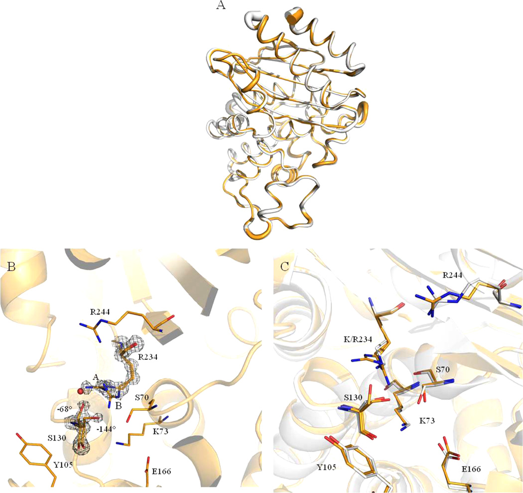
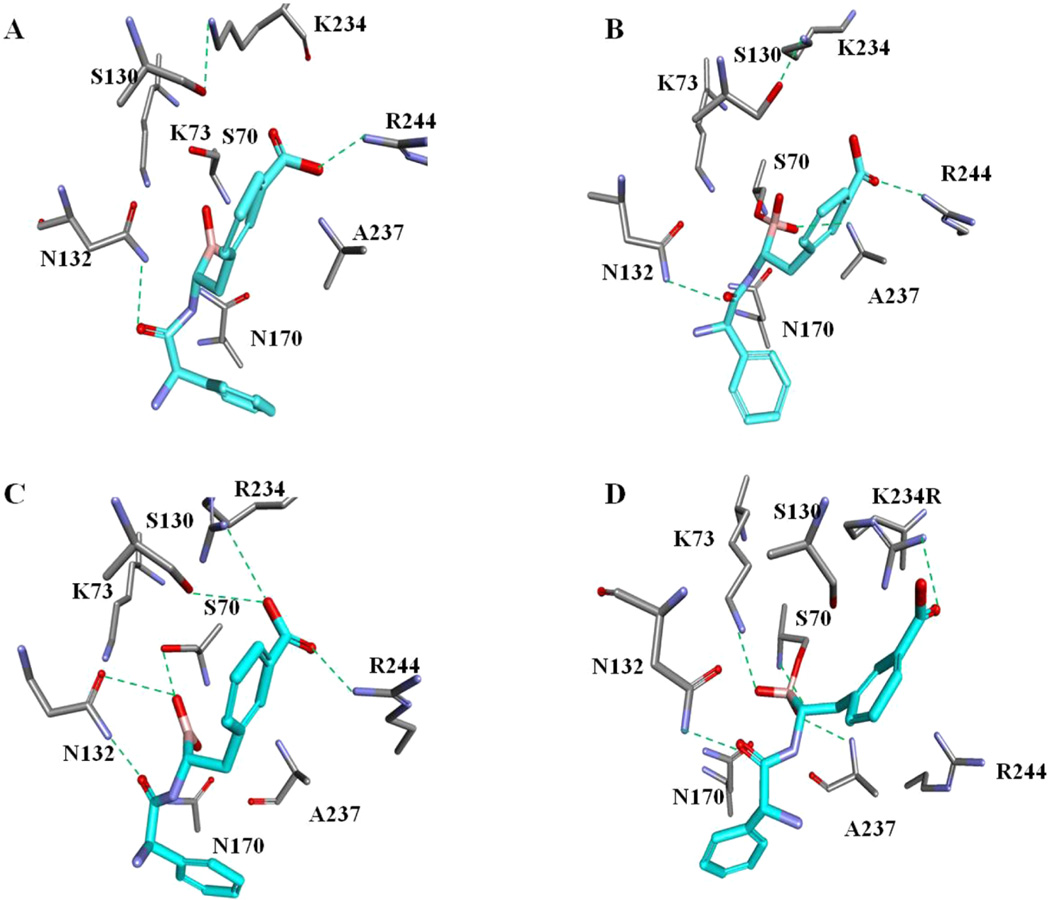
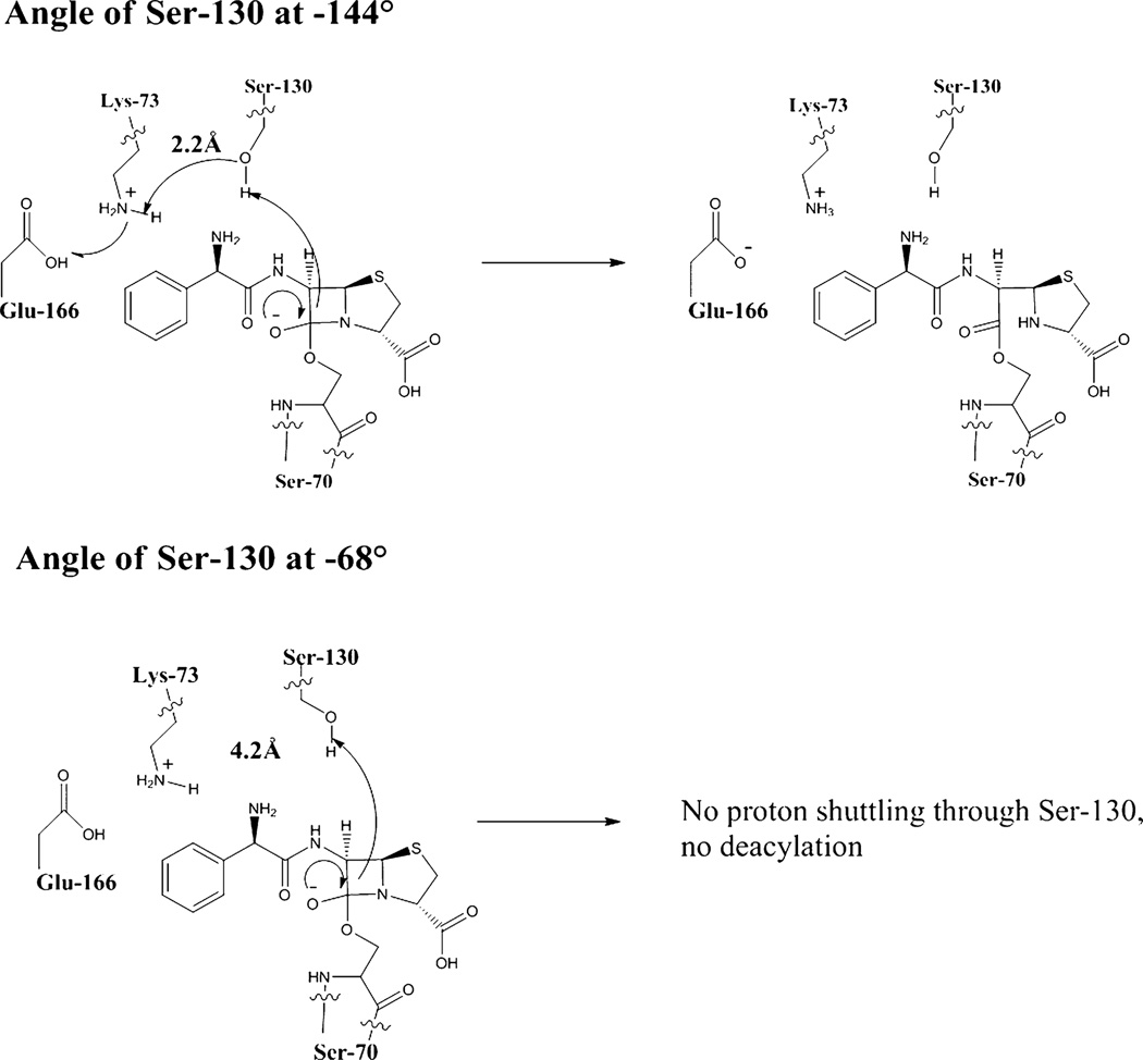
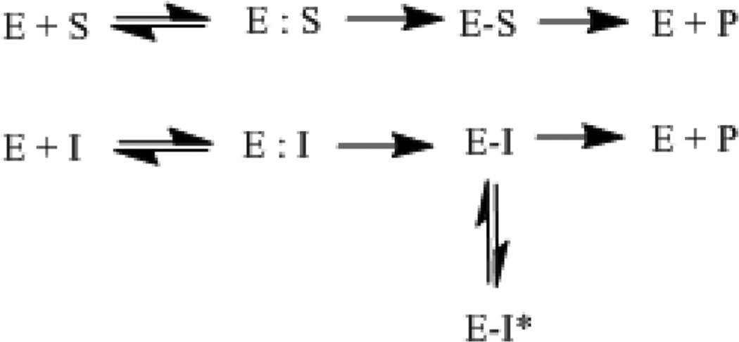
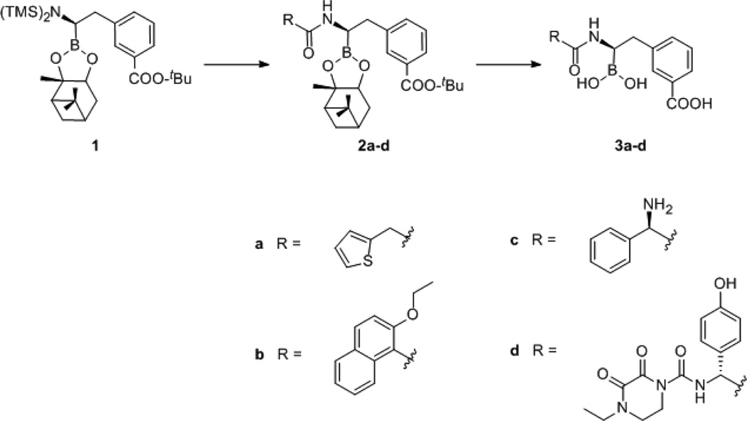
Inhibitor resistant (IR) class A β-lactamases pose a significant threat to many current antibiotic combinations. The K234R substitution in the SHV β-lactamase, from Klebsiella pneumoniae , results in resistance to ampicillin/clavulanate. After site-saturation mutagenesis of Lys-234 in SHV, microbiological and biochemical characterization of the resulting β-lactamases revealed that only -Arg conferred resistance to ampicillin/clavulanate. X-ray crystallography revealed two conformations of Arg-234 and Ser-130 in SHV K234R. The movement of Ser-130 is the principal cause of the observed clavulanate resistance. A panel of boronic acid inhibitors was designed and tested against SHV-1 and SHV K234R. A chiral ampicillin analogue was discovered to have a 2.4 ± 0.2 nM K(i) for SHV K234R; the chiral ampicillin analogue formed a more complex hydrogen-bonding network in SHV K234R vs SHV-1. Consideration of the spatial position of Ser-130 and Lys-234 and this hydrogen-bonding network will be important in the design of novel antibiotics targeting IR β-lactamases.


Figures

Figure 1
Figure 1
Chemical structures of the compounds tested in this paper.
Figure 2
Figure 2
Deconvoluted ESI-MS spectra of (A) SHV-1 and (B) K234R β-lactamases. All measurements have an error of ±3amu.
Figure 3
Figure 3
(A) Far UV spectra of SHV-1 (dashed) and K234R (solid black) measured at 10 µM enzyme concentration. (B) Fraction of folded protein was calculated as described and is plotted here relative to the temperature in order to compute the Tm of each enzyme SHV-1 (dashed) has a Tm of 54.2 °C and K234R (solid black) has a melting temperature of 48.2 °C.
Figure 4
Figure 4
(A) SHV-1 at 10 µM enzyme concentration in the presence of 50 µM of various inhibitors monitored by CD showing changes in the secondary structure upon the enzyme binding to the inhibitor. (B) Changes in the thermal denaturation curve of SHV-1 at 10 µM enzyme concentration in the presence of 50 µM of various inhibitors (the chiral ampicillin BATSI is the only compound to significantly shift the curve). (C) K234R at 10 µM enzyme concentration in the presence of 50 µM of various inhibitors monitored by CD showing changes in the secondary structure upon the enzyme binding to the inhibitor (large changes are seen with tazobactam and sulbactam, thin solid line and dashed black line, respectively). Additionally, clavulanate and the achiral cephalothin BATSI seem to increase the flexibility and movement of the protein (dotted and dashed-dotted black lines, respectively). (D) Changes in the thermal denaturation cruve of K234R at 10 µM enzyme concentration in the presence of 50 µM of various inhibitors (the chiral ampicillin BATSI is the only compound to greatly shift the curve). Sulbactam seems to cause an initial destabilization of the K234R variant, with stability recovered after the first few temperature determinations are made.
Figure 5
Figure 5
(A) Ribbon cartoon overlay of K234R (gold) and SHV-1 (PDB ID: 1SHV, white). Overall rmsd is 0.14 Å, all Cα superposition performed using SSM Superpose in COOT. Ribbon width is proportionate to B-factor, i.e., larger ribbon radius indicates higher B-factor. (B) SHVK234R active site. 2Fo–Fc density contoured at 1.5σ is shown for both R234 orientations. Two S130 orientations are also observed, with χ1 angles of −144 and −68°. χ1 angle defined as the angle between the N, CA, CB and CA, CB, Oγ planes as in Mendonca et. al. The red sphere represents the water molecule found in SHV-1. This space is filled by the A orientation of Arg-234 in the K234R structure, which leads the water molecule to be absent in the crystal structure of the K234R protein. (C) Active site overlay of SHV-1 (PDB ID: 1SHV, white) and K234R (gold). The change in position of K/R234, S130, and S70 is evident in this image.
Figure 6
Figure 6
Molecular docking of the chiral ampicillin BATSI (light blue) and SHV-1 in the preacylation state (before dative bond formation) (A) and after dative bond formation (B) and K234R before dative bond formation (C) and after dative bond formation (D).
Figure 7
Figure 7
The top figure shows the wild-type enzyme with Ser-130 at a χ angle of −144° where proton shuffling can occur to allow breakdown of ampicillin by the β-lactamase. The bottom figure shows the movement of the hydroxyl group of Ser-130 away from Lys-73 when Ser-130 is at χ = −68°, which prevents the proton shuffling and ampicillin breakdown.
Scheme 1
Scheme 1
Scheme Showing the Breakdown of a β-Lactam (S) by the Enzyme (E) to Form the Inactive β-Lactam (P) and the Interaction of an Inhibitor (I) with the Enzyme to Either Form an Inactivated Inhibitor (P) and Active Enzyme or Inactivation of the Enzyme through a Long-Lived Inhibitor Complex (E–I*)
Scheme 2
Scheme 2
Synthesis Scheme Leading to the Creation of the Various BATSI Compounds (a = Chiral Cephalothin, b = Chiral Nafcillin, c = Chiral Ampicillin, d = Chiral Cefoperazone)a aSynthesis provided in Supporting Information.

References

    1. Klevens RM, Edwards JR, Richards CL, Horan TC, Gaynes RP, Pollock DA, Cardo DM. Estimating Health Care-Associated Infections and Deaths in U.S. Hospitals, 2002. Public Health Rep. 2007;122:160–166. - PMC - PubMed
    1. Drawz S, Bonomo R. Three Decades of β-Lactamase Inhibitors. Clin. Microbiol. Rev. 2010;23:160–201. - PMC - PubMed
    1. Buynak JD. Cutting and Stitching: The Cross-linking of Peptidoglycan in the Assembly of the Bacterial Cell Wall. ACS Chem. Biol. 2007;2:602–605. - PubMed
    1. Meroueh SO, Bencze KZ, Hesek D, Lee M, Fisher JF, Stemmler TL, Mobashery S. Three-Dmensional Structure of the Bacterial Cell Wall Peptidoglycan. Proc. Natl. Acad. Sci. U.S.A. 2006;103:4404–4409. - PMC - PubMed
    1. Lakaye B, Dubus A, Lepage S, Groslambert S, Frere JM. When Drug Inactivation Renders the Target Irrelevant to Antibiotic Resistance: A Case Story with β-Lactams. Mol. Microbiol. 1999;31:89–101. - PubMed

Publication types

Associated data