

Myasthenogenicity of the main immunogenic region
- PMID: 23252892
- PMCID: PMC3531903
- DOI: 10.1111/j.1749-6632.2012.06766.x
Myasthenogenicity of the main immunogenic region
Abstract
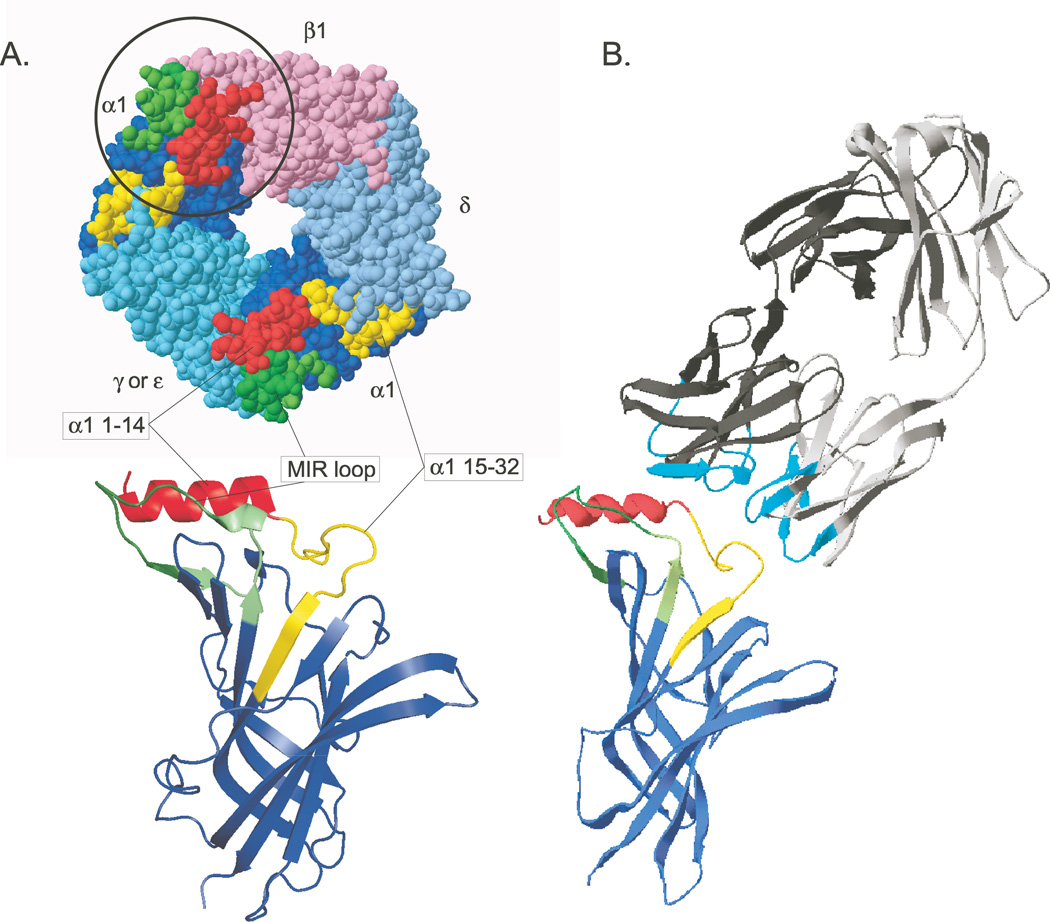
In myasthenia gravis (MG) and experimental autoimmune MG (EAMG), many pathologically significant autoantibodies are directed to the main immunogenic region (MIR) of muscle nicotinic acetylcholine receptors (AChRs), a conformation-dependent region at the extracellular tip of α1 subunits of AChRs. Human muscle AChR α1 MIR sequences were integrated into Aplesia ACh-binding protein (AChBP). The chimera potently induced EAMG, while AChBP induced EAMG much less potently. AChBP is a water-soluble protein resembling the extracellular domain of AChRs; yet, rats immunized with chimeras developed autoantibodies to both extracellular and cytoplasmic domains of muscle AChRs. We propose that an initial autoimmune response directed at the MIR leads to an autoimmune response sustained by muscle AChRs. Autoimmune stimulation sustained by endogenous muscle AChR may be a target for specific immunosuppression. These studies show that the α1 MIR is highly myasthenogenic, and that AChR-like proteins distantly related to muscle AChR can induce EAMG and, potentially, MG.
© 2012 New York Academy of Sciences.
Conflict of interest statement
The authors declare no conflicts of interest.
Figures
References
-
- Lindstrom J. Acetylcholine receptors and myasthenia. Muscle and Nerve. 2000;23:453–477. - PubMed
-
- Luo J, Taylor P, Losen M, de Baets M, Shelton G, Lindstrom J. Main immunogenic region structure promotes binding of conformation-dependent myasthenia gravis autoantibodies, nicotinic acetylcholine receptor conformation maturation, and agonist sensitivity. J. Neuroscience. 2009;29:13898–13908. - PMC - PubMed
-
- Unwin N. Refined structure of the nicotinic acetylcholine receptor at 4Å resolution. J. Mol. Biol. 2005;346:967–989. - PubMed
-
- Brejc K, van Dyk W, Klassen R, Schuurmans M, vanderOost J, Smit A, Sixma T. Crystal structure of an ACh-binding protein reveals the ligand-binding domain of nicotinic receptors. Nature. 2001;411:269–276. - PubMed
Publication types
MeSH terms
Substances
Grants and funding
LinkOut - more resources
Full Text Sources
Other Literature Sources
Medical
