Review
. 2012 Dec;1274(1):68-76.
doi: 10.1111/j.1749-6632.2012.06840.x.

Functional defect in regulatory T cells in myasthenia gravis

Affiliations
Review

Functional defect in regulatory T cells in myasthenia gravis

Muthusamy Thiruppathi et al. Ann N Y Acad Sci. 2012 Dec.

Abstract

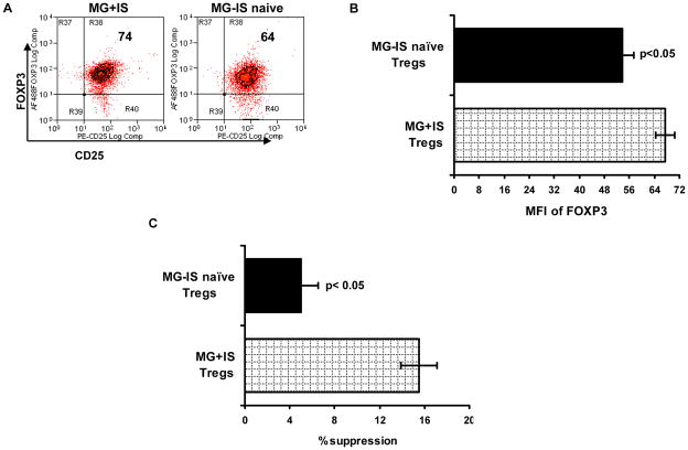
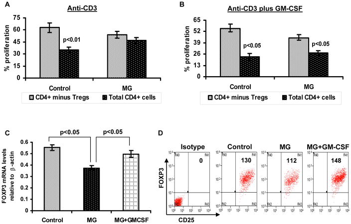
Forkhead box P3 (FOXP3) is a transcription factor necessary for the function of regulatory T cells (T(reg) cells). T(reg) cells maintain immune homeostasis and self-tolerance and play an important role in the prevention of autoimmune disease. Here, we discuss the role of T(reg) cells in the pathogenesis of myasthenia gravis (MG) and review evidence indicating that a significant defect in T(reg) cell in vitro suppressive function exists in MG patients, without an alteration in circulating frequency. This functional defect is associated with a reduced expression of key functional molecules, such as FOXP3 on isolated T(reg) cells, and appears to be more pronounced in immunosuppression-naive MG patients. In vitro administration of granulocyte macrophage-colony-stimulating factor (GM-CSF) enhanced the suppressive function of T(reg) cells and upregulated FOXP3 expression. These findings indicate a clinically relevant T(reg) cell-intrinsic defect in immune regulation in MG that may reveal a novel therapeutic target.


Conflict of interest statement

Conflict of Interest

The authors declare no competing financial interests.

Figures

Figure 1
Figure 1
FOXP3 expression and suppressive function of Treg cells in treated versus untreated MG patients. Intensity of FOXP3 expression (MFI = mean fluorescent intensity) in CD4+ CD25high CD127low/− Treg cells. (A) Representative data for immunosuppressive naive (MG - IS naive) and immunosuppressed (MG + IS) subjects. (B) MFI of FOXP3 expression in isolated CD4+ CD25high CD127low/− cells. (C) Percent suppression of Tresp cell proliferation by Treg cells. Data in (B) and (C) represent n = 5 for MG-IS naive subjects and n = 12 for MG-IS subjects. Results are expressed as mean ± SEM.
Figure 2
Figure 2
In vitro GM-CSF treatment to lymphocytes enhances FOXP3 expression and suppressive function of Treg cells in MG. CFSE-labeled total CD4+ T cells and Treg cell–depleted CD4+ T cells were stimulated with human anti-CD3 alone or anti-CD3 plus GM-CSF in the presence of autologus irradiated APCs. Cultures consisting of total CD4+ cells (black bars) and CD4+ cells after removal of CD4+ CD25high CD127low/− cells (grey bars), were compared to evaluate suppressive function of Treg cells. After 5 days of culture, percentage of T cell proliferation was analyzed based on CFSE dilution. The bar diagram represents percentage Tresp cell proliferation in response to anti-CD3 alone (A) and anit-CD3 plus GM-CSF (B) for four MG patients and four healthy controls.(C) FOXP3 mRNA expression was determined by multiplex PCR using isolated CD4+ CD25high CD127low/−Treg cells obtained from a control subject, MG patient and an MG patient’s PBMCs treated with GM-CSF. Results are expressed as relative FOXP3 mRNA expression. Results are expressed as mean ± SEM. (D) Representative flow cytometry plots illustrate mean florescent intensity of FOXP3 expression within isolated CD4+ CD25high CD127low/− Treg cells.
Figure 3
Figure 3
Hypothetical model linking Treg cell suppressive function to FOXP3 expression level. In this model, high expression of FOXP3 in T cells endows them with enhanced suppressive capacity and a characteristic phenotype. Absence of FOXP3 expression is associated with no suppressive capacity, but reduced FOXP3 expression may result in an intermediate phenotype without suppressive function, and possibly a tendency for enhanced functional plasticity.

References

    1. Meriggioli MN, Sanders DB. Autoimmune myasthenia gravis: emerging clinical and biological heterogeneity. Lancet Neurol. 2009;8:475–490. - PMC - PubMed
    1. Berrih S, Morel E, Gaud C, et al. Anti-AChR antibodies, thymic histology, and T cell subsets in myasthenia gravis. Neurology. 1984;34:66–71. - PubMed
    1. Roxanis I, Micklem K, Willcox N. True epithelial hyperplasia in the thymus of early-onset myasthenia gravis: implications for immunopathogenesis. J Neuroimmunol. 2001;112:163–173. - PubMed
    1. Giraud M, Vandiedonck C, Garchon HJ. Genetic factors in autoimmune myasthenia gravis. Ann NY Acad Sci. 2008;1132:180–192. - PubMed
    1. Landoure G, Knight MA, Stanescu H, et al. A candidate gene for autoimmune myasthenia gravis. Neurology. 2012;79:342–347. - PMC - PubMed

Publication types

MeSH terms

Substances