

Case Reports
doi: 10.1016/j.ihj.2012.07.016.
Epub 2012 Jul 27.
Ruptured cardiac angiosarcoma with pulmonary metastases: a rare disease with a common (mis)diagnosis!
Affiliations
- PMID: 23253417
- PMCID: PMC3860970
- DOI: 10.1016/j.ihj.2012.07.016
Item in Clipboard
Case Reports
Ruptured cardiac angiosarcoma with pulmonary metastases: a rare disease with a common (mis)diagnosis!
Indian Heart J.
2012 Nov-Dec.
Abstract
In Indian settings pulmonary tuberculosis remains the most common diagnosis in a patient presenting with constitutional symptoms, hemoptysis and lung opacities. We describe a case report of a fifty-year-old woman who was receiving empirical anti-tubercular drugs for a metastatic illness to lungs arising from a primary angiosarcoma in the right atrium. This rare entity was misdiagnosed and typical echocardiographic findings suggested this diagnosis.
Copyright © 2012 Cardiological Society of India. Published by Elsevier B.V. All rights reserved.
Figures
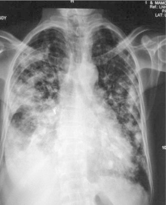
Chest X-ray reveals right-sided pleural effusion with multiple nodular opacities in both lung fields. Consolidation of right middle lobe also noted silhouetting right heart border.
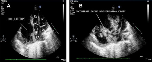
(A) Apical 4c view showing friable looking sessile, mobile mass (✰) arising from right atrial wall and a large loculated effusion around right atria and ventricle. (B) Apical 4c view showing leakage of saline bubble contrast (white arrow) from right atrial free wall to surrounding pericardial effusion thus confirming atrio-pericardial fistula.
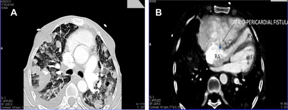
(A) Axial CT reveals multiple non cavitating nodular lesions of varying sizes scattered in bilateral lung parenchyma in angiocentric distribution suggesting metastatic deposits. Bilateral pleural effusion (R > L) is also seen. (B) Contrast enhanced axial CT reveals pericardial collection communicating with right atria and causing attenuation of the right ventricle. Associated papillary like projection (*) noted in the free wall of the right atria.
Similar articles
-
Cardiac angiosarcoma.Intern Med. 1996 Oct;35(10):795-8. doi: 10.2169/internalmedicine.35.795. Intern Med. 1996. PMID: 8933189 Review.
-
Cardiac angiosarcoma presenting as diffuse pulmonary disease.Eur Heart J. 1995 Dec;16(12):2005-6. doi: 10.1093/oxfordjournals.eurheartj.a060863. Eur Heart J. 1995. PMID: 8682042 No abstract available.
-
FDG PET/CT of Cardiac Angiosarcoma With Pulmonary Metastases.Clin Nucl Med. 2018 Oct;43(10):744-746. doi: 10.1097/RLU.0000000000002215. Clin Nucl Med. 2018. PMID: 30036249
-
Rapidly progressive respiratory failure with multiple halo signs on computed tomography in a patient with primary cardiac angiosarcoma derived from the right atrium: a case report.BMC Pulm Med. 2020 Dec 9;20(1):321. doi: 10.1186/s12890-020-01366-6. BMC Pulm Med. 2020. PMID: 33297995 Free PMC article.
-
Angiosarcomas of the interatrial septum mimicking atrial myxomas.J Am Soc Echocardiogr. 1996 Mar-Apr;9(2):209-12. doi: 10.1016/s0894-7317(96)90033-4. J Am Soc Echocardiogr. 1996. PMID: 8849621 Review.
Cited by
-
Cardiac angiosarcoma with multiple pulmonary metastases: A case report and literature review.Radiol Case Rep. 2023 Feb 1;18(4):1446-1451. doi: 10.1016/j.radcr.2022.11.081. eCollection 2023 Apr. Radiol Case Rep. 2023. PMID: 36798066 Free PMC article.
-
Clinical Detection of Primary Pulmonary Angiosarcoma.Cureus. 2021 Aug 10;13(8):e17059. doi: 10.7759/cureus.17059. eCollection 2021 Aug. Cureus. 2021. PMID: 34522537 Free PMC article.
-
A surgical case of right atrial wall perforation caused by an invasive angiosarcoma.Interdiscip Cardiovasc Thorac Surg. 2024 Dec 25;40(1):ivae222. doi: 10.1093/icvts/ivae222. Interdiscip Cardiovasc Thorac Surg. 2024. PMID: 39774640 Free PMC article.
-
Primary cardiac angiosarcoma with right atrial wall rupture: A case report.Medicine (Baltimore). 2019 Apr;98(14):e15020. doi: 10.1097/MD.0000000000015020. Medicine (Baltimore). 2019. PMID: 30946333 Free PMC article.
-
Clinical and diagnostic features of angiosarcoma with pulmonary metastases: A retrospective observational study.Medicine (Baltimore). 2017 Sep;96(36):e8033. doi: 10.1097/MD.0000000000008033. Medicine (Baltimore). 2017. PMID: 28885371 Free PMC article.
References
-
- Burke A.P., Cowan D., Virmani R. Primary sarcomas of the heart. Cancer. 1992;69:387–395. - PubMed
-
- Herrmann M.A., Shankerman R.A., Edwards W.D. Primary cardiac angiosarcoma: a clinicopathologic study of six cases. J. Thorac Cardiovasc Surg. 1992;103(4):655–664. - PubMed
-
- Chul H.K., Jane Y.D., Donna C. Clinicopathologic study of 24 patients with primary cardiac sarcomas: a 10-year single institution experience. Hum Pathol. 2008;39:933–938. - PubMed
-
- Corso R.B., Kraychete N., Nardeli S. Spontaneous rupture of a right atrial angiosarcoma and cardiac tamponade. Arq Bras Cardiol. 2003;81:611–613. - PubMed
-
- Sakaguchi M., Minato N., Katayama Y. Cardiac angiosarcoma with right atrial perforation and cardiac tamponade. Ann Thorac Cardiovasc Surg. 2006 Apr;12(2):145–148. - PubMed
Publication types
MeSH terms
LinkOut - more resources
Full Text Sources
Medical
