Review
. 2013 Apr;83(4):759-69.
doi: 10.1124/mol.112.083758. Epub 2012 Dec 19.

P2X receptors as drug targets

Affiliations
Review

P2X receptors as drug targets

R Alan North et al. Mol Pharmacol. 2013 Apr.

Abstract

The study of P2X receptors has long been handicapped by a poverty of small-molecule tools that serve as selective agonists and antagonists. There has been progress, particularly in the past 10 years, as cell-based high-throughput screening methods were applied, together with large chemical libraries. This has delivered some drug-like molecules in several chemical classes that selectively target P2X1, P2X3, or P2X7 receptors. Some of these are, or have been, in clinical trials for rheumatoid arthritis, pain, and cough. Current preclinical research programs are studying P2X receptor involvement in pain, inflammation, osteoporosis, multiple sclerosis, spinal cord injury, and bladder dysfunction. The determination of the atomic structure of P2X receptors in closed and open (ATP-bound) states by X-ray crystallography is now allowing new approaches by molecular modeling. This is supported by a large body of previous work using mutagenesis and functional expression, and is now being supplemented by molecular dynamic simulations and in silico ligand docking. These approaches should lead to P2X receptors soon taking their place alongside other ion channel proteins as therapeutically important drug targets.


Figures

Fig. 1.
Fig. 1.
Trimeric P2X receptor compared with tetrameric glutamate and pentameric nicotinic (Cys-loop) receptor. Zebrafish P2X4 receptor (modified from PDB ID 4DW1) (left). Rat GluA2 receptor (modified from PDB ID 3KG2) (center). Caenorhabditis elegans glutamate-gated chloride channel (modified from PDB ID 3RHW) (center). Each is shown from the outside of the cell.
Fig. 2.
Fig. 2.
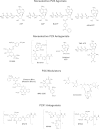
Chemical structures of nonselective P2X receptor agonists, antagonists, and modulators, and antagonists at P2X1 receptors. Nucleotide-based agonists are active at multiple P2X receptor subunits. For example, 2′,3′-O-(benzoyl-4-benzoyl)-5′-ATP is a nanomolar agonist at P2X1 and P2X3 receptors, and has an approximately 10,000 lower affinity for P2X7 receptors. Suramin and PPADS have been historically used as P2X receptor antagonists, but have many off-target pharmacological actions. NF449 has higher selectivity for P2X1 receptors compared with NF279 and NF023. The P2X receptor modulators (Cibacron Blue and MRS 2220) likely bind allosteric recognition sites on their respective receptors.
Fig. 3.
Fig. 3.
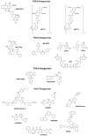
Receptor-selective P2X receptor antagonists. IP5I (diinosine pentaphosphate), RO3, and RO4 likely bind allosteric recognition sites on their respective receptors. Competitive receptor antagonism has been demonstrated for TNP-ATP, A-317491, A-438079, and A-804598. Several new receptor-selective antagonists for P2X2 (PSB-10211, NF770, NF778) and for P2X4 (PSB-12062) have been recently reported. AACBA is also know as (adamantan-1-ylmethyl)-5-[(3R-amino-pyrrolidin-1-yl)methyl]-2-chloro-benzamide (GSK314181A). See text for further details.
Fig. 4.
Fig. 4.
P2X receptor structure. (A) Two dolphin-shaped subunits are depicted. ATP binding is associated with an upward movement of the dorsal fin (chain A, at back), a downward movement of the head region (chain B, at front), and a retraction of the left flipper (chain A, at front) in the directions shown by the arrows. The lower body (light blue) is flexed outward, carrying with it transmembrane domains (green). Reproduced (with permission) from Hattori and Gouaux (2012). (B) ATP binding pocket. Residues involved in direct interactions with ATP are shown from homology model of the rat P2X2 receptor, based on PDB ID 4DW1. Atoms identified are as follows: chain A (blue) N288 ND2, K308 NZ, R290 NH1; chain B (pink) K71 NZ, K69 NZ, T184 OG1, I226 CG2, L186 CD2, L211 CB, K188 NZ. (C) Lateral portals that form the ion entry pathway into extracellular vestibule. The lateral portal between two subunits (blue and pink) is outlined by a green oval. The third subunit (yellow) is visible through the open portal. Arrowhead marks level of section shown in (D). Rat P2X2 receptor. (D) Dilation of outer part of transmembrane domains. Positions of TM1 (filled gray circle) and TM2 (open gray circle) are indicated. Residues shown as space fill are I328 at outer end of TM2: dashed circle passes through Cα atoms. Rat P2X2 homology model.

Similar articles

Cited by

References

    1. Ahlquist RP. (1948) A study of the adrenotropic receptors. Am J Physiol 153:586–600 - PubMed
    1. Albers-Schönberg G, Arson BH, Chabala JC, Douglas AW, Eskola P, Fisher MH, Lusi A, Mrozik H, Smith JL, Tolman RL. (1981) Avermectins: structure determination. J Am Chem Soc 103:4216–4221
    1. Alexander SPH, Mathie A, Peters JA.(2011) Guide to receptors and channels (GRAC), 5th edition. Br J Pharmacol 164:S1–S324 - PMC - PubMed
    1. Allsopp RC, Evans RJ. (2011) The intracellular amino terminus plays a dominant role in desensitization of ATP-gated P2X receptor ion channels. J Biol Chem 286:44691–44701 - PMC - PubMed
    1. Arunlakshana O, Schild HO. (1959) Some quantitative uses of drug antagonists. Br Pharmacol Chemother 14:48–58 - PMC - PubMed

Publication types

MeSH terms

LinkOut - more resources