. 2013 Jan;72 Suppl 1(0 1):115-23.
doi: 10.1227/NEU.0b013e3182753093.

Role of cranial and spinal virtual and augmented reality simulation using immersive touch modules in neurosurgical training

Affiliations

Role of cranial and spinal virtual and augmented reality simulation using immersive touch modules in neurosurgical training

Ali Alaraj et al. Neurosurgery. 2013 Jan.

Erratum in

  • Neurosurgery. 2013 Nov;73(5):E913. Tobin, Mathew [corrected to Tobin, Matthew]

Abstract

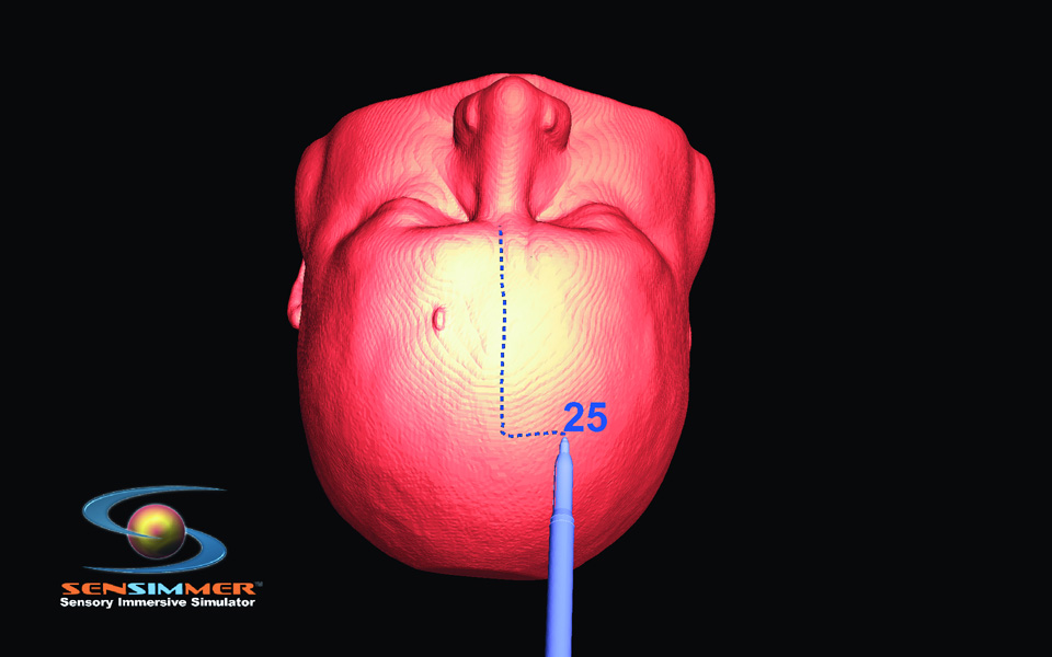
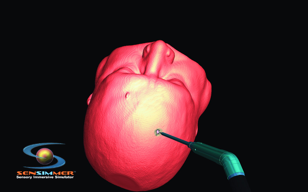
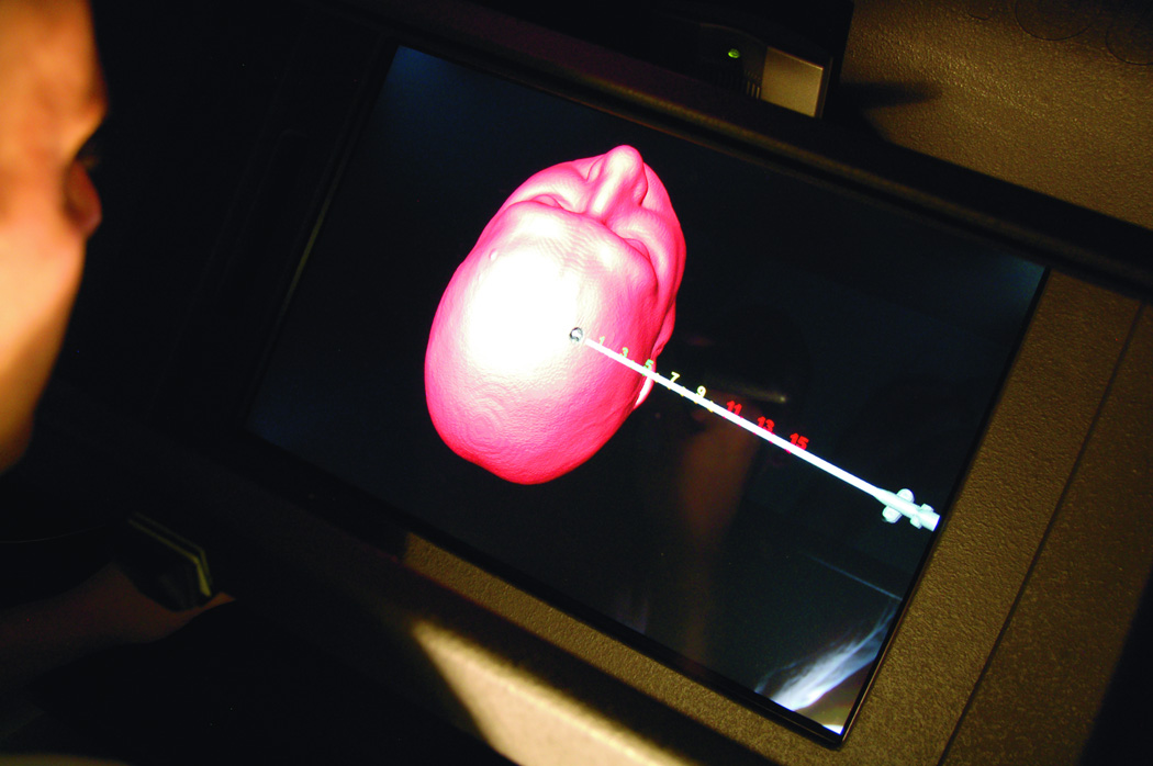
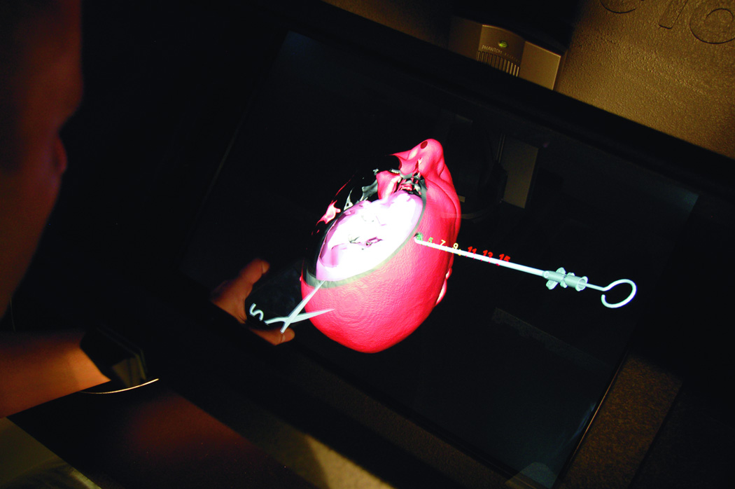
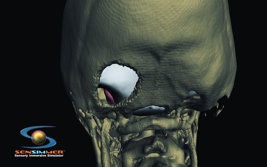
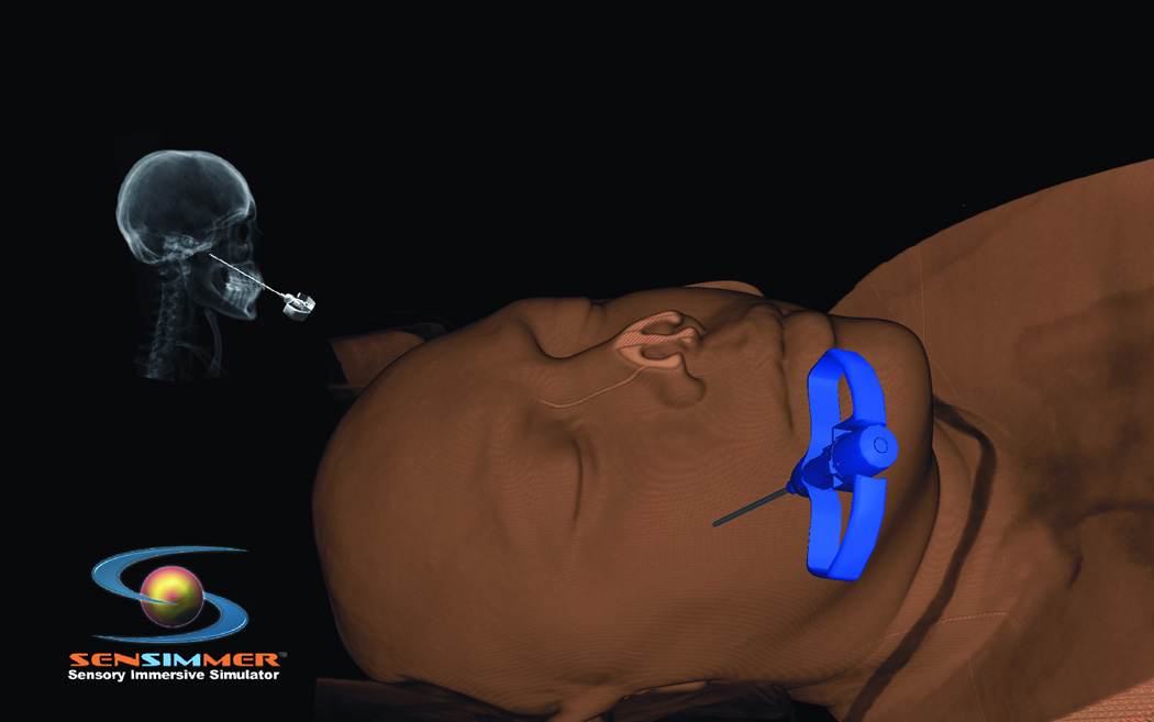
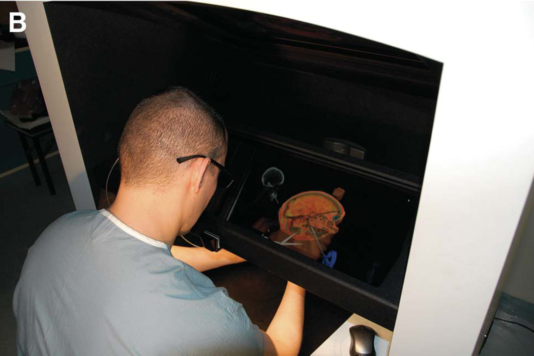
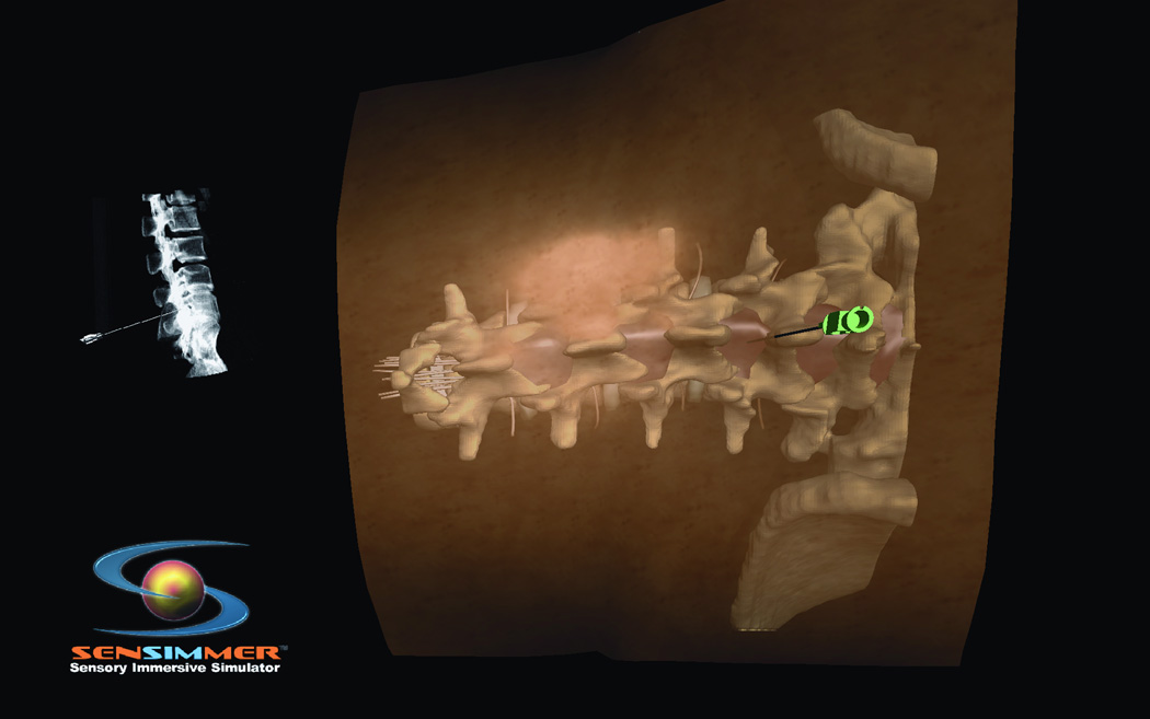
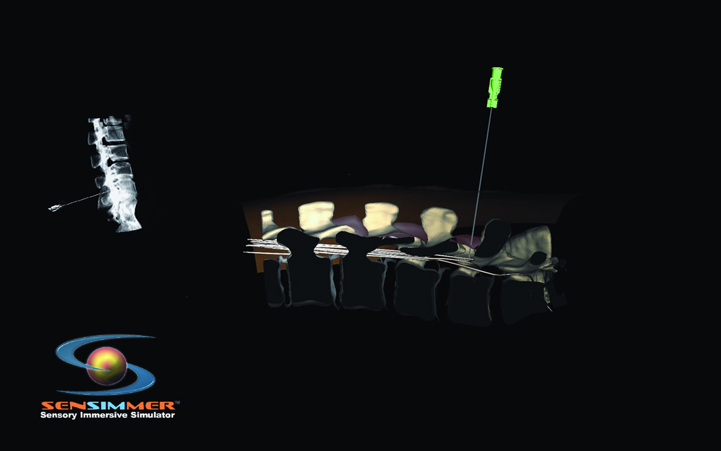
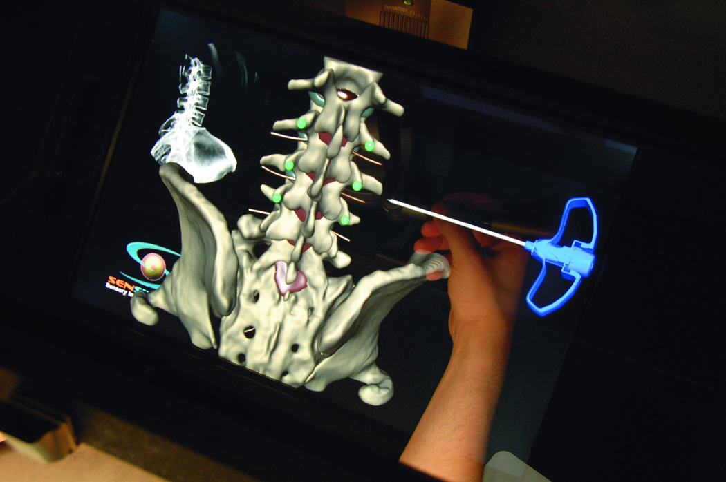
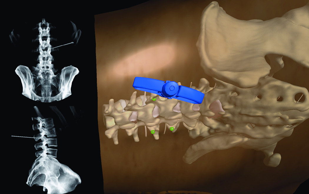
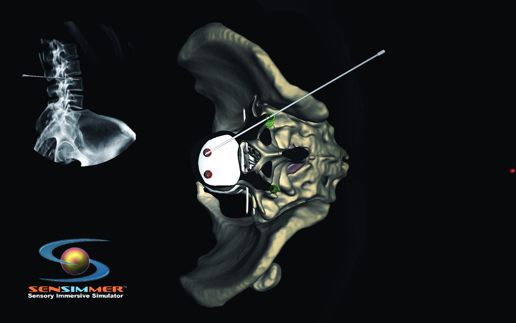
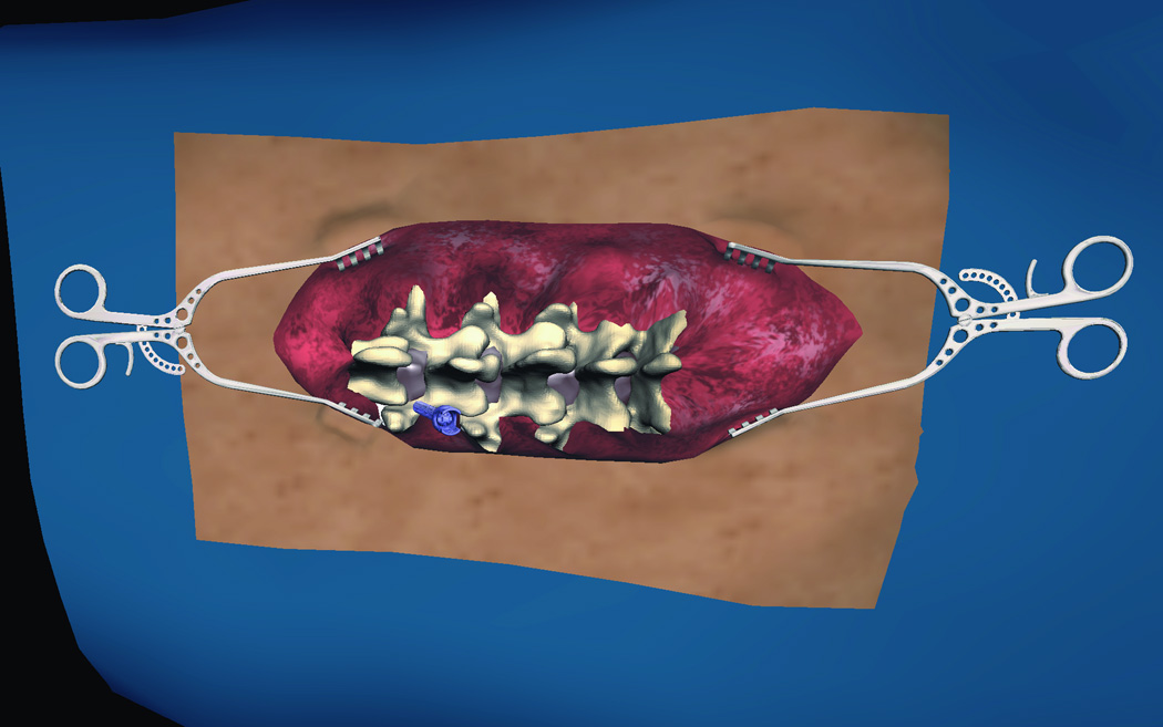
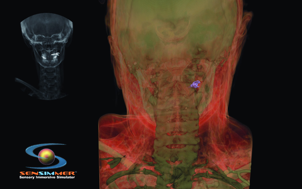
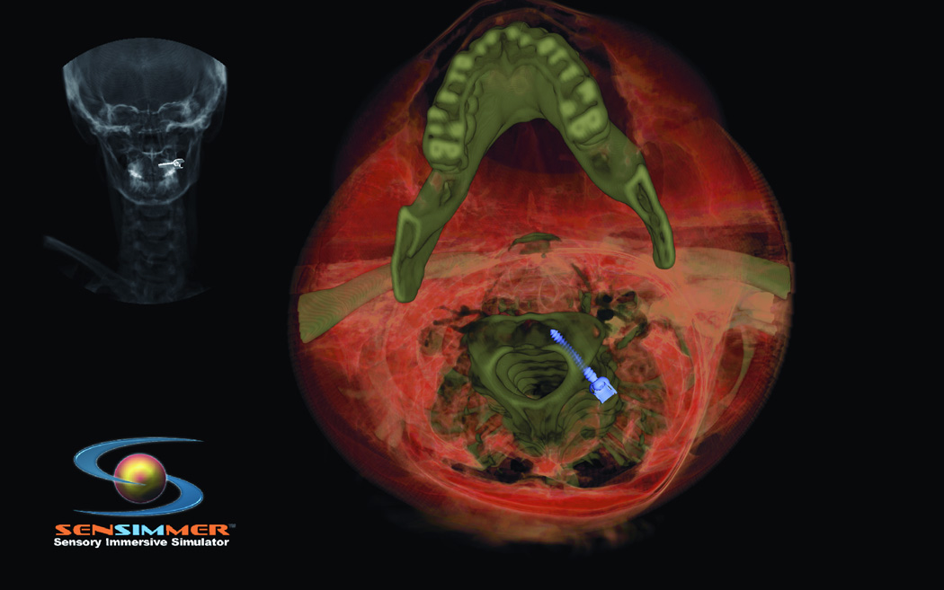
Recent studies have shown that mental script-based rehearsal and simulation-based training improve the transfer of surgical skills in various medical disciplines. Despite significant advances in technology and intraoperative techniques over the last several decades, surgical skills training on neurosurgical operations still carries significant risk of serious morbidity or mortality. Potentially avoidable technical errors are well recognized as contributing to poor surgical outcome. Surgical education is undergoing overwhelming change, as a result of the reduction of work hours and current trends focusing on patient safety and linking reimbursement with clinical outcomes. Thus, there is a need for adjunctive means for neurosurgical training, which is a recent advancement in simulation technology. ImmersiveTouch is an augmented reality system that integrates a haptic device and a high-resolution stereoscopic display. This simulation platform uses multiple sensory modalities, re-creating many of the environmental cues experienced during an actual procedure. Modules available include ventriculostomy, bone drilling, percutaneous trigeminal rhizotomy, and simulated spinal modules such as pedicle screw placement, vertebroplasty, and lumbar puncture. We present our experience with the development of such augmented reality neurosurgical modules and the feedback from neurosurgical residents.


Figures

Figure 1
Figure 1
Steps of ventriculostomy insertion, the trainee position the virtual head in space, the burr hole location can be decided based on anatomical landmarks or on measurements. (A) The skin is marked for midline identification (10cm from the glabella, and 2.5 cm from the midline). (B) High speed drill simulation with vibration/drill haptic feedback creating a burr hole for ventriculostomy insertion. (C) A trainee plans his ventriculostomy insertion. Centimeter markers are close to the virtual catheter. (D) Post catheter insertion, the operator lock the catheter in space, and virtually cut through the head to identify visually the trajectory and location of the tip of the catheter in relationship to the ventricular system.
Figure 1
Figure 1
Steps of ventriculostomy insertion, the trainee position the virtual head in space, the burr hole location can be decided based on anatomical landmarks or on measurements. (A) The skin is marked for midline identification (10cm from the glabella, and 2.5 cm from the midline). (B) High speed drill simulation with vibration/drill haptic feedback creating a burr hole for ventriculostomy insertion. (C) A trainee plans his ventriculostomy insertion. Centimeter markers are close to the virtual catheter. (D) Post catheter insertion, the operator lock the catheter in space, and virtually cut through the head to identify visually the trajectory and location of the tip of the catheter in relationship to the ventricular system.
Figure 2
Figure 2
(A) Axial and coronal (B) CT scan of the corresponding virtual head where the trainee will practice on. CT scan showing a normal size ventricular system.
Figure 2
Figure 2
(A) Axial and coronal (B) CT scan of the corresponding virtual head where the trainee will practice on. CT scan showing a normal size ventricular system.
Figure 3
Figure 3
Illustrative CT scan one of CT from the 15 different sincere library. (A) Axial and (B) sagital CT scan showing a compressed and shifted right frontal horn.
Figure 3
Figure 3
Illustrative CT scan one of CT from the 15 different sincere library. (A) Axial and (B) sagital CT scan showing a compressed and shifted right frontal horn.
Figure 4
Figure 4
Posterior view at virtual head model illustrating location of a suboccipital craniotectomy.
Figure 5
Figure 5
(A) Virtual head model for rhizotomy procedure, needed is aimed towards the foramen ovale. In the left upper corner of the screen a lateral fluoroscopy image of the head is seen showing the location of the needed as the needed is being inserted. (B) Training resident cut through the virtual head and identify the location of the tip of the needle to learn how to improve his trajectory.
Figure 5
Figure 5
(A) Virtual head model for rhizotomy procedure, needed is aimed towards the foramen ovale. In the left upper corner of the screen a lateral fluoroscopy image of the head is seen showing the location of the needed as the needed is being inserted. (B) Training resident cut through the virtual head and identify the location of the tip of the needle to learn how to improve his trajectory.
Figure 6
Figure 6
(A) Virtual model for a patient’s back with the lumbar spine incorporated in the haptics of the model. The needle is aimed through the inter-laminar space. The location of the tip is seen on a virtual fluoroscopy screen. (B) At the end of the needle insertion, the operator perform a virtual sagital cut through the model to verify the trajectory and location of the tip of the needle.
Figure 6
Figure 6
(A) Virtual model for a patient’s back with the lumbar spine incorporated in the haptics of the model. The needle is aimed through the inter-laminar space. The location of the tip is seen on a virtual fluoroscopy screen. (B) At the end of the needle insertion, the operator perform a virtual sagital cut through the model to verify the trajectory and location of the tip of the needle.
Figure 7
Figure 7
(A) Lumbar spine model for training trans-pedicular vertebroplasty procedure. The trocar needle is aimed at the left L3 pedicle. The pedicle target entry zone is highlighted as green. (B) Trocar introduced into the right L 3 pedicle, with corresponding AP and lateral X-ray imaging identifying the trajectory of the trocar. (C) Virtual Axial cut of the vertebral body made to check for the location of the trocar at the end of the procedure, green circles indicate the ideal site of entry through the pedicle, and red circles indicate the ideal target for the tip of the trocar, deviation from those points affect the scoring of the procedure. (D) Virtual model for open surgical approach to teach pedicluar spinal instrumentation. The skin and muscles are retracted to the side to facilitate the open trajectory to the pedicle.
Figure 7
Figure 7
(A) Lumbar spine model for training trans-pedicular vertebroplasty procedure. The trocar needle is aimed at the left L3 pedicle. The pedicle target entry zone is highlighted as green. (B) Trocar introduced into the right L 3 pedicle, with corresponding AP and lateral X-ray imaging identifying the trajectory of the trocar. (C) Virtual Axial cut of the vertebral body made to check for the location of the trocar at the end of the procedure, green circles indicate the ideal site of entry through the pedicle, and red circles indicate the ideal target for the tip of the trocar, deviation from those points affect the scoring of the procedure. (D) Virtual model for open surgical approach to teach pedicluar spinal instrumentation. The skin and muscles are retracted to the side to facilitate the open trajectory to the pedicle.
Figure 8
Figure 8
(A) Posterior cervical instrumentation training module. A right C2 transpedicular screw is inserted into right C2. Real time fluoroscopy image is seen in the right upper corner of the screen, demonstrating the trajectory of the C2 screw. (B) The trainee perform a virtual cut of the spine model (here showing axial cut) to check the trajectory as well as the distal tip of the screw.
Figure 8
Figure 8
(A) Posterior cervical instrumentation training module. A right C2 transpedicular screw is inserted into right C2. Real time fluoroscopy image is seen in the right upper corner of the screen, demonstrating the trajectory of the C2 screw. (B) The trainee perform a virtual cut of the spine model (here showing axial cut) to check the trajectory as well as the distal tip of the screw.

References

    1. Aggarwal R, Black SA, Hance JR, Darzi A, Cheshire NJ. Virtual reality simulation training can improve inexperienced surgeons' endovascular skills. Eur J Vasc Endovasc Surg. 2006 Jun;31(6):588–593. - PubMed
    1. Fried MP, Sadoughi B, Gibber MJ, et al. From virtual reality to the operating room: the endoscopic sinus surgery simulator experiment. Otolaryngology--head and neck surgery : official journal of American Academy of Otolaryngology-Head and Neck Surgery. 2010 Feb;142(2):202–207. - PubMed
    1. Grantcharov TP. Is virtual reality simulation an effective training method in surgery? Nature clinical practice. Gastroenterology & hepatology. 2008 May;5(5):232–233. - PubMed
    1. Palter VN, Graafland M, Schijven MP, Grantcharov TP. Designing a proficiency-based, content validated virtual reality curriculum for laparoscopic colorectal surgery: a Delphi approach. Surgery. 2012 Mar;151(3):391–397. - PubMed
    1. Arabi Y, Memish ZA, Balkhy HH, et al. Ventriculostomy-associated infections: incidence and risk factors. American journal of infection control. 2005 Apr;33(3):137–143. - PubMed

Publication types

MeSH terms