. 2013 Apr;37(4):715-21.
doi: 10.1007/s00264-012-1743-y. Epub 2012 Dec 20.

Achilles tendon: functional anatomy and novel emerging models of imaging classification

Affiliations

Achilles tendon: functional anatomy and novel emerging models of imaging classification

Angelo Del Buono et al. Int Orthop. 2013 Apr.

Abstract

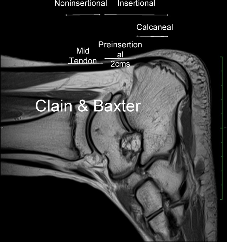
Purpose: Ideally, a classification should have some prognostic value, and should therefore include precise information upon extent and location of the Achilles tendon disorders. We propose a new imaging and anatomical system to classify Achilles tendon disorders at imaging using US and MRI.

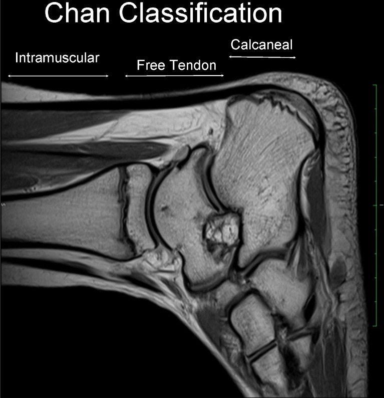
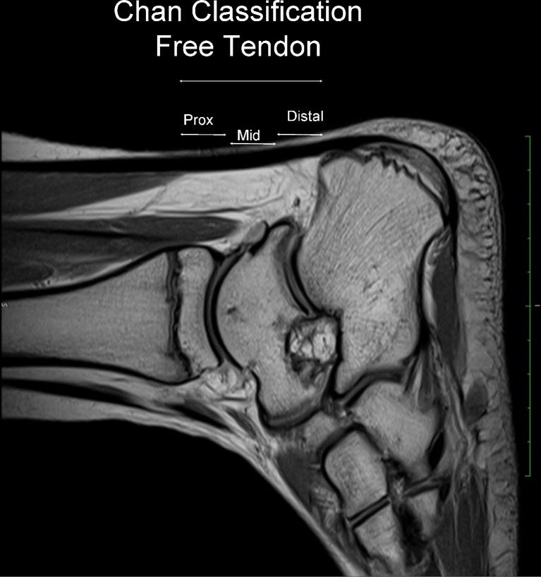
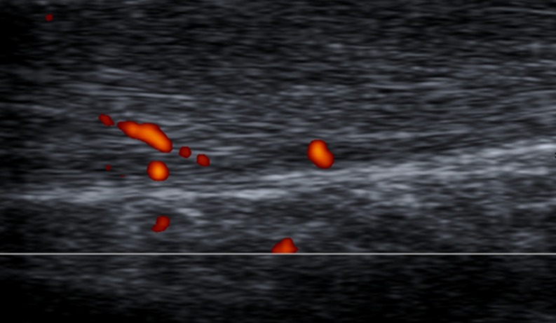
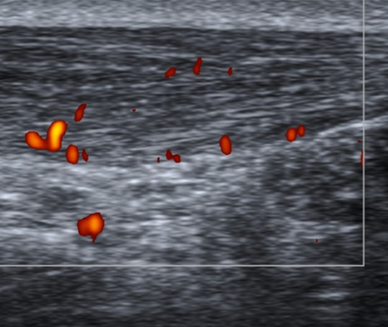
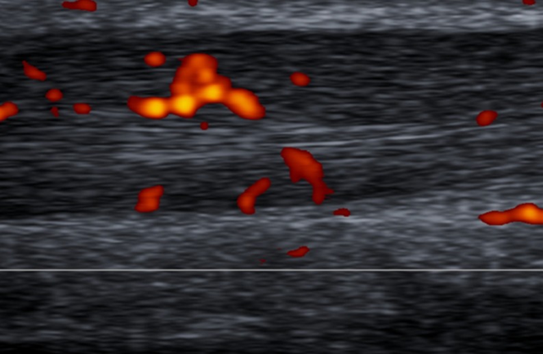
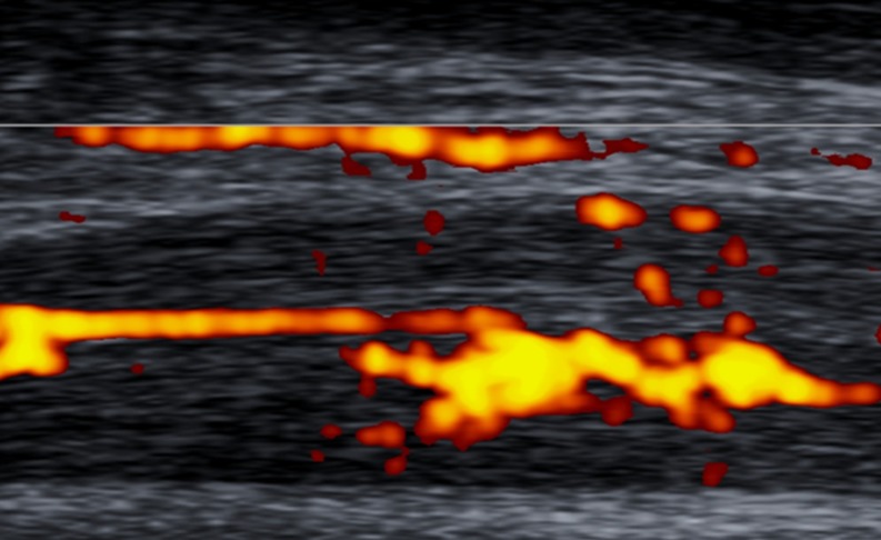
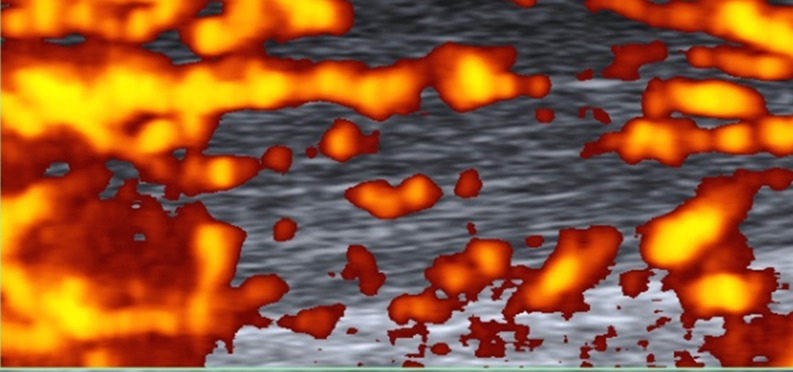
Approach: We consider the non-insertional region as the tendon mid-portion, and distinguish the insertional component into a pre-insertion site, located about two centimetres above the calcaneum, and a calcaneal insertion, where the tendon is attached to the bone. On sagittal scans, we introduced a new classification which considers two main portions: "musculotendinous" and "insertional". In the context of the muscolotendinous portion, it is possible to find muscle fibres proximally, and the free tendon distally. This latter is made up of proximal, middle and distal portions. We also propose a 5 grade Doppler classification system to quantify blood flow, in which Grades I and II are respectively characterised by the presence of one and two vessels within the tendon; in Grades III, IV and V, the neovascularisation respectively involves less than 50 %, from 50 to 90 %, and more than 90 % of the tendon tissue. These proposed systems will require validation and possible modification to be applied to different tendons.


Figures

Fig. 1
Fig. 1
Sagittal T2 MRI scan in a 29-year-old track runner with 2-year history of Achilles tendinopathy. Calin & Baxter classification according which the Achilles tendon contains two main portions: non-insertional (proximal) and insertional (distal)
Fig. 2
Fig. 2
Sagittal T2 MRI scan. We present the new classification by Chan et al. that considers three main portions: “intramuscular”, “insertional” and “calcaneal” insertion
Fig. 3
Fig. 3
Sagittal T2 MRI scan. In the context of the free tendon, Chan et al. distinguished proximal, middle, and distal components, according to the involvement of which management changes
Fig. 4
Fig. 4
Sagittal US scan of the Achilles tendon in a 33-year-old male who was an asymptomatic soccer player. New Doppler classification By Chan et al.; Grade I vascularisation: only a single vessel may be appreciated within the tendon
Fig. 5
Fig. 5
Sagittal US scan of the tendon in a 27-year-old male rugby player with six months history of recurrent tendinopathy, located at the proximal and middle portions of the free tendon. On Doppler assessment, evidence of two vessels into the tendon (Grade II vascularisation)
Fig. 6
Fig. 6
Sagittal US Doppler assessment in a 31-year-old male who was an elite jumper with 15-month history of pain and stiffness to the free portion of the left Achilles tendon. The presence of vessels in more than 50 % of the thickness of the Achilles tendon is indicative of Grade III vascularisation
Fig. 7
Fig. 7
Sagittal US Doppler imaging in a 47-year-old female who was an amatuer runner complaining of symptoms to the insertion and free portion of the Achilles tendon. Grade IV vascularisation: evidence of vascularisation within 50–90 % of the tendon thickness
Fig. 8
Fig. 8
Sagittal US Doppler assessment in a 34-year-old male basketball player who was symptomatic for more than 18 months. Imaging shows tendon thickening and vascularisation involving more than 90 % of the tendon itself (Grade V)

References

    1. Ahmed IM, Lagopoulos M, McConnell P, Soames RW, Sefton GK. Blood supply of the Achilles tendon. J Orthop Res. 1998;16:591–596. doi: 10.1002/jor.1100160511. - DOI - PubMed
    1. Apaydin N, Bozkurt M, Loukas M, Vefali H, Tubbs RS, Esmer AF. Relationships of the sural nerve with the calcaneal tendon: an anatomical study with surgical and clinical implications. Surg Radiol Anat. 2009;31(10):775–780. doi: 10.1007/s00276-009-0520-0. - DOI - PubMed
    1. Astrom M, Gentz CF, Nilsson P, Rausing A, Sjoberg S, Westlin N. Imaging in chronic achilles tendinopathy: a comparison of ultrasonography, magnetic resonance imaging and surgical findings in 27 histologically verified cases. Skeletal Radiol. 1996;25:615–620. doi: 10.1007/s002560050146. - DOI - PubMed
    1. Astrom M, Rausing A. Chronic Achilles tendinopathy. A survey of surgical and histopathologic findings. Clin Orthop Relat Res. 1995;316:151–164. - PubMed
    1. Benjamin M, McGonagle D. The anatomical basis for disease localisation in seronegative spondyloarthropathy at entheses and related sites. J Anat. 2001;199:503–526. doi: 10.1046/j.1469-7580.2001.19950503.x. - DOI - PMC - PubMed

LinkOut - more resources