. 2013 Mar;23(3):381-94.
doi: 10.1093/glycob/cws166. Epub 2012 Dec 18.

KSGal6ST generates galactose-6-O-sulfate in high endothelial venules but does not contribute to L-selectin-dependent lymphocyte homing

Affiliations

KSGal6ST generates galactose-6-O-sulfate in high endothelial venules but does not contribute to L-selectin-dependent lymphocyte homing

Michael L Patnode et al. Glycobiology. 2013 Mar.

Abstract

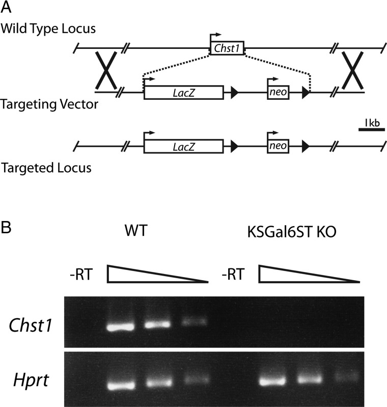
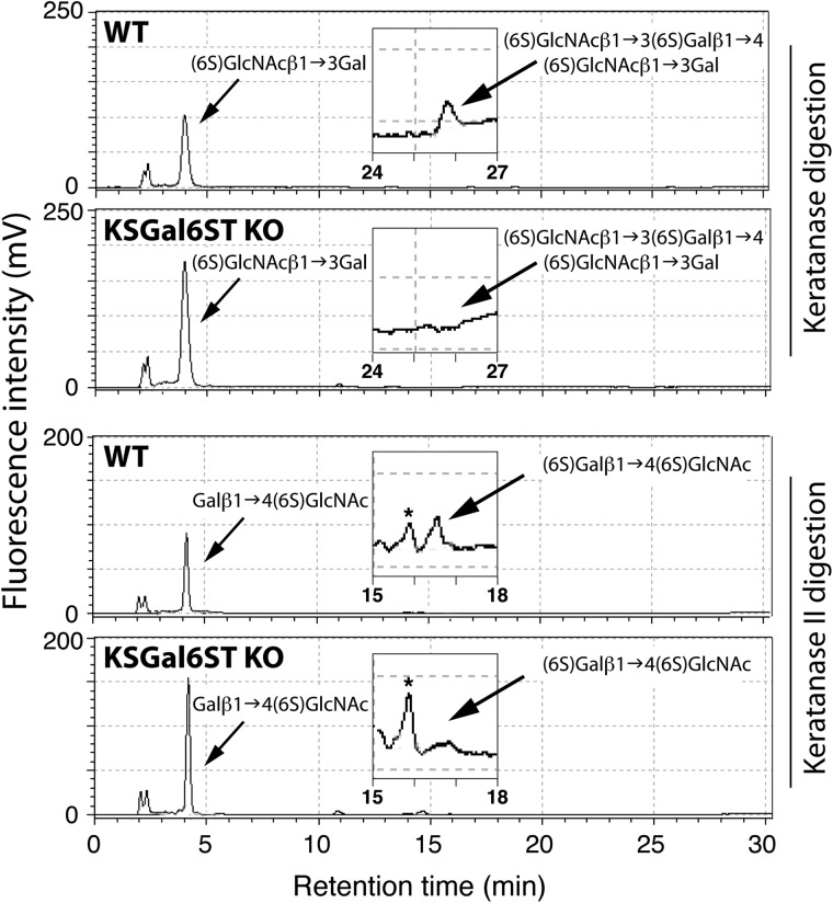
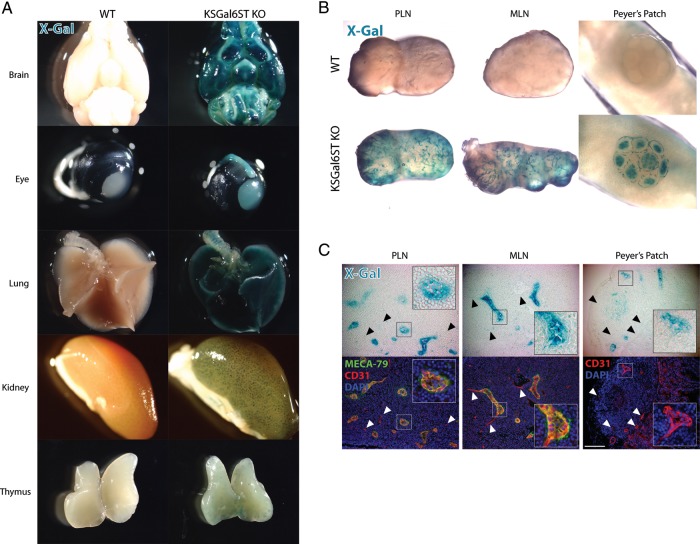
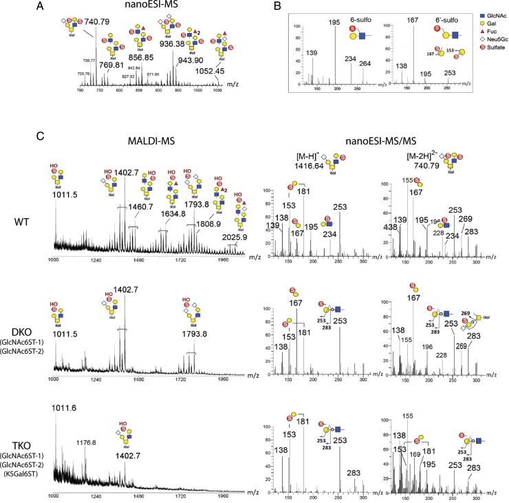
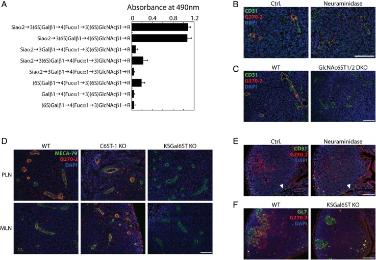
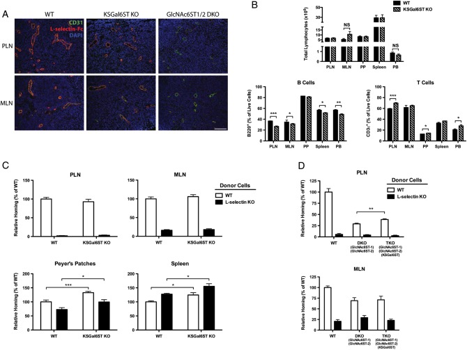
The addition of sulfate to glycan structures can regulate their ability to serve as ligands for glycan-binding proteins. Although sulfate groups present on the monosaccharides glucosamine, uronate, N-acetylglucosamine and N-acetylgalactosamine are recognized by defined receptors that mediate important functions, the functional significance of galactose-6-O-sulfate (Gal6S) is not known. However, in vitro studies using synthetic glycans and sulfotransferase overexpression implicate Gal6S as a binding determinant for the lymphocyte homing receptor, L-selectin. Only two sulfotransferases have been shown to generate Gal6S, namely keratan sulfate galactose 6-O-sulfotransferase (KSGal6ST) and chondroitin 6-O-sulfotransferase-1 (C6ST-1). In the present study, we use mice deficient in KSGal6ST and C6ST-1 to test whether Gal6S contributes to ligand recognition by L-selectin in vivo. First, we establish that KSGal6ST is selectively expressed in high endothelial venules (HEVs) in lymph nodes and Peyer's patches. We also determine by mass spectrometry that KSGal6ST generates Gal6S on several classes of O-glycans in peripheral lymph nodes. Furthermore, KSGal6ST, but not C6ST-1, is required for the generation of the Gal6S-containing glycan, 6,6'-disulfo-3'sLN (Siaα2→3[6S]Galβ1→4[6S]GlcNAc) or a closely related structure in lymph node HEVs. Nevertheless, L-selectin-dependent short-term homing of lymphocytes is normal in KSGal6ST-deficient mice, indicating that the Gal6S-containing structures we detected do not contribute to L-selectin ligand recognition in this setting. These results refine our understanding of the biological ligands for L-selectin and introduce a mouse model for investigating the functions of Gal6S in other contexts.


Figures

Fig. 1.
Fig. 1.
Generation of KSGal6ST-deficient mice. (A) Schematic of the Chst1 locus and the BAC targeting vector created by Regeneron, Inc., which replaces the entire protein-coding region of Chst1 with the LacZ and neomycin phosphotransferase (neo) genes. Boxes represent protein-coding regions of exons. Arrows represent transcriptional start sites. Arrowheads represent loxP sites. Xs denote regions of homologous recombination. The scale bar represents 1 kb (kilobase). (B) PCR products generated with primers specific for Chst1 (206 bp product) and Hprt (252 bp product) using 5-fold serial dilutions of cDNA from WT or KSGal6ST KO mouse cortex as a template. No products were observed when RT was omitted from the cDNA synthesis reaction. RT, reverse transcriptase; WT, wild type.
Fig. 2.
Fig. 2.
KS analysis in KSGal6ST-deficient mice. Reversed-phase ion-pair chromatography analysis of ocular KS from eyes of WT and KSGal6ST KO mice. Standard substances were eluted at the peak positions indicated by arrows. Elution profiles around the peak positions of (6S)GlcNAcβ1→3(6S)Galβ1→4(6S)GlcNAcβ1→3Gal and (6S)Galβ1→4(6S)GlcNAc are magnified in insets. Peaks indicated by asterisks were not identified. WT, wild type.
Fig. 3.
Fig. 3.
KSGal6ST expression in HEVs (A), (B) Tissues from WT or KSGal6ST KO mice analyzed for β-galactosidase activity using X-Gal (blue). (C) Bright field images (top) of cryostat-cut sections of lymphoid organs from KSGal6ST KO mice stained with X-Gal (blue). Fluorescent images (bottom) of the same sections stained with MECA-79 (green), anti-CD31 (red) and DAPI (blue). Insets show HEVs magnified 3×. Arrowheads highlight examples of vessels with weak X-Gal staining. The scale bar represents 100 μm and applies to adjacent images. Isotype-matched control antibodies were used to establish background fluorescence. WT, wild type; PLN, peripheral lymph nodes; MLN, mesenteric lymph nodes.
Fig. 4.
Fig. 4.
MS analyses of permethylated sulfated O-glycans released from lymph nodes of KSGal6ST-deficient mice. (A) NanoLC-nanoESI-MS spectrum for WT di-sulfated O-glycan fractions (summed over a period of elution time for the major signals). Both sulfates were preserved under nanoESI-MS to give doubly charged [M–2H]2− molecular ions. (B) Spectra for a panel of sulfated LacNAc standards analyzed to establish the diagnostic ions produced under nanoESI-HCD MS/MS fragmentation. The singly charged low mass ions at m/z 195 and 234 were reproducibly detected for all internal GlcNAc6S-containing glycans. In contrast, the 3,5A ion at m/z 167 was detected for Gal6S-containing glycans, which could be distinguished from terminal Gal3S by the ions at m/z 153 and 181 (not shown). (C) Spectra of the di-sulfated O-glycan fractions from WT, GlcNAc6ST-1/-2 DKO and GlcNAc6ST-1/-2/KSGal6ST TKO lymph nodes analyzed in negative ion mode by MALDI-MS (left). The di-sulfated O-glycans were prone to losing a sulfate by in-source fragmentation to produce [M–H] molecular ions carrying one sulfate and one free OH group. Low mass regions of the nanoESI-HCD MS/MS spectra (right) of mono- and di-sulfated mono-sialylated core 2 structures from WT, DKO and TKO lymph nodes, annotated with diagnostic ions for sulfated structures (see B). Additional ions at m/z 253 (E ion) and 283 (B ion) were indicative of terminal sulfated Gal whilst m/z 269 (D ion) was derived from an internal sulfated Gal, as illustrated. The full spectra of each (data not shown) carried other neutral loss ions and nonreducing terminal fragment ions, which supported the assigned overall structures but did not allow differentiation of the positions of sulfates. Assignment of the major peaks for all spectra is as annotated using the standard cartoon symbols. WT, wild type; DKO, double KO; TKO, triple KO.
Fig. 5.
Fig. 5.
Expression of sialylated 6,6′-disulfated glycans in HEVs and germinal centers of lymphoid organs. (A) Reactivity of G270-2 as determined by ELISA using 5 ng immobilized neoglycolipids per well. (B) Cryostat-cut serial sections of PLN from WT mice treated with neuraminidase or buffer alone (ctrl.), then stained with anti-CD31 (green), G270-2 (red) and DAPI (blue). (C) Cryostat-cut sections of PLN from WT and GlcNAc6ST-1/-2 DKO mice stained as in (B). (D) Cryostat-cut sections of PLN and MLN from WT, C6ST-1 KO and KSGal6ST KO mice stained with MECA-79 (green), G270-2 (red) and DAPI (blue). (E) Cryostat-cut serial sections of Peyer's patches from WT mice treated with neuraminidase or buffer alone (ctrl.), and then stained as in (B). Arrowheads highlight examples of CD31+ vessels. (F) Cryostat-cut sections of Peyer's patches from WT and KSGal6ST KO mice stained with GL7 (green), G270-2 (red) and DAPI (blue). All scale bars represent 100 μm and apply to adjacent images. Isotype-matched control antibodies were used to establish background fluorescence. PLN, peripheral lymph nodes; MLN, mesenteric lymph nodes; WT, wild type; DKO, double KO.
Fig. 6.
Fig. 6.
L-selectin ligand expression, lymphocyte numbers and lymphocyte homing in KSGal6ST-deficient mice. (A) Cryostat-cut sections of PLN and MLN from WT, KSGal6ST KO and GlcNAc6ST-1/-2 DKO mice stained with anti-CD31 (green), L-selectin-Fc (red) and DAPI (blue). Background fluorescence was established by staining separate sections with L-selectin-Fc in the presence of 10 mM EDTA. The scale bar represents 100 μm and applies to adjacent images. (B) Total numbers (top) and percentages (bottom) of CD3ε+ T cells and B220+ B cells in peripheral blood and lymphoid organs from WT (n = 4) and KSGal6ST KO (n = 3) mice. For MLN, n = 5 for WT, and n = 6 for KSGal6ST KO (P = 0.14). (C) Short-term lymphocyte homing to PLN, MLN, Peyer's patches and spleens of WT (n = 11) and KSGal6ST KO (n = 15) mice. Pooled results from three independent experiments are shown. (D) Short-term homing of splenocytes to PLN and MLN of WT (n = 6), GlcNAc6ST-1/-2 DKO (n = 7) and GlcNAc6ST-1/-2/KSGal6ST TKO (n = 8) mice. Pooled results from two independent experiments are shown. Differences between mouse genotypes are not significant unless otherwise noted. Homing of L-selectin KO splenocytes was significantly reduced relative to WT splenocytes in all cases, except in homing to the spleen, where it was significantly enhanced (P values between 0.01 and 0.0001). Homing of WT splenocytes to PLN (P < 0.0001) and MLN (P < 0.005) was significantly reduced in DKO recipients relative to WT recipients. PLN, peripheral lymph nodes; MLN, mesenteric lymph nodes; PP, Peyer's patches; SPLN, spleen; PB, peripheral blood; WT, wild type; DKO, double KO; TKO, triple KO; NS, not significant; *P < 0.05; **P < 0.005; ***P < 0.0005.

References

    1. Akama TO, Nishida K, Nakayama J, Watanabe H, Ozaki K, Nakamura T, Dota A, Kawasaki S, Inoue Y, Maeda N, et al. Macular corneal dystrophy type I and type II are caused by distinct mutations in a new sulphotransferase gene. Nat Genet. 2000;26:237–241. doi:10.1038/79987. - DOI - PubMed
    1. Arata-Kawai H, Singer MS, Bistrup A, Zante A, Wang YQ, Ito Y, Bao X, Hemmerich S, Fukuda M, Rosen SD. Functional contributions of N- and O-glycans to L-selectin ligands in murine and human lymphoid organs. Am J Pathol. 2011;178:423–433. doi:10.1016/j.ajpath.2010.11.009. - DOI - PMC - PubMed
    1. Arbones ML, Ord DC, Ley K, Ratech H, Maynard-Curry C, Otten G, Capon DJ, Tedder TF. Lymphocyte homing and leukocyte rolling and migration are impaired in L-selectin-deficient mice. Immunity. 1994;1:247–260. doi:10.1016/1074-7613(94)90076-0. - DOI - PubMed
    1. Baenziger JU. Glycoprotein hormone GalNAc-4-sulphotransferase. Biochem Soc Trans. 2003;31:326–330. doi:10.1042/BST0310326. - DOI - PubMed
    1. Bertozzi CR, Fukuda S, Rosen SD. Sulfated disaccharide inhibitors of L-selectin: Deriving structural leads from a physiological selectin ligand. Biochemistry. 1995;34:14271–14278. doi:10.1021/bi00044a001. - DOI - PubMed

Publication types