Review
. 2012 Dec:Chapter 8:Unit8.8.
doi: 10.1002/0471142700.nc0808s51.

Binding to the DNA minor groove by heterocyclic dications: from AT-specific monomers to GC recognition with dimers

Affiliations
Review

Binding to the DNA minor groove by heterocyclic dications: from AT-specific monomers to GC recognition with dimers

Rupesh Nanjunda et al. Curr Protoc Nucleic Acid Chem. 2012 Dec.

Abstract

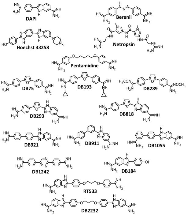
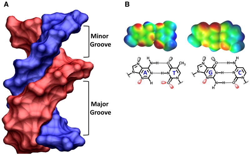
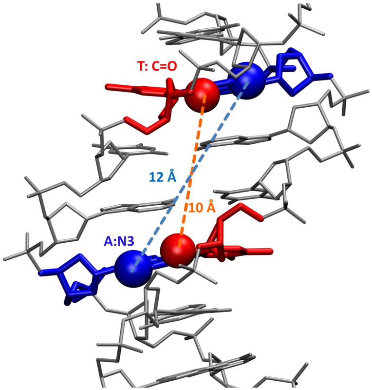
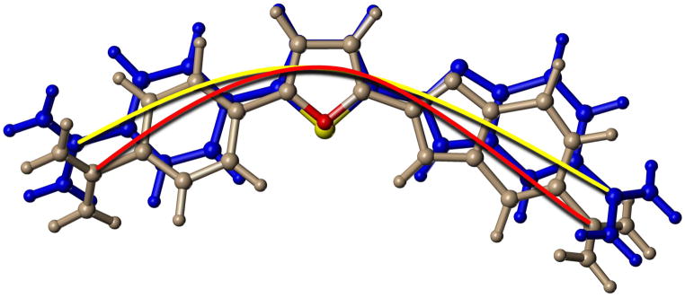
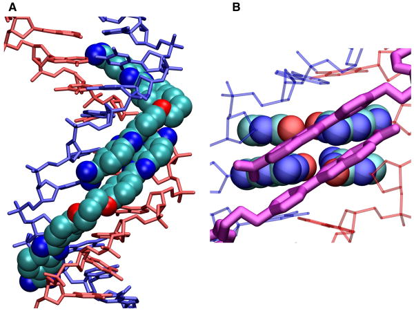
Compounds that bind in the DNA minor groove have provided critical information on DNA molecular recognition, have found extensive uses in biotechnology, and are providing clinically useful drugs against diseases as diverse as cancer and sleeping sickness. This review focuses on the development of clinically useful heterocyclic diamidine minor groove binders. These compounds have shown us that the classical model for minor groove binding in AT DNA sequences must be expanded in several ways: compounds with nonstandard shapes can bind strongly to the groove, water can be directly incorporated into the minor groove complex in an interfacial interaction, and the compounds can form cooperative stacked dimers to recognize GC and mixed AT/GC base pair sequences.


Figures

Figure 1
Figure 1
Compound structures for the heterocyclic compounds discussed in this paper as well as some well-known reference compounds: DAPI, netropsin, Hoechst 33258, berenil and pentamidine. The heterocyclic diamidines are listed from top (DB75) to bottom as classical binders, nonclassical binders, and GC recognizing compounds.
Figure 2
Figure 2
(A) A B-form DNA model with the chains colored blue and pink is tilted so that the width of both the minor and major grooves can be seen and compared. (B) Space-filling models of AT and GC base pairs (top) is shown with electrostatic potential coloring: from blue for positive to red for negative partial charges. The same base pairs are shown as line models (bottom) with the base pairs labeled and lone pair electrons shown on H-bond accepting groups.
Figure 3
Figure 3
The minor groove of a B-form DNA model with the central sequence 5′-AATT-3′ shown with the DNA as a gray stick model. The connecting distances between the 5′-As and 3′-Ts on opposite chains are shown.
Figure 4
Figure 4
(A) A space filling crystal model of a cyclopropyl substituted diamidine derivative (DB193 in Fig. 1) bound to an AATT sequences is shown with the DNA strands colored blue and red and the compound shown as a tube model. (B) The complex is shown tilted so that the compound (space filling model) can be seen bound in the DNA (tube model) minor groove.
Figure 5
Figure 5
An overlay tube model of optimized structures for DB293 (grey) and DB818 (blue) is shown with the furan O in red and the thiophene S in yellow. A red arc connects the amidine carbons of the furan, DB293, while a yellow arc connects the equivalent atoms in the thiophene, DB818. The slight difference in bond angles in the furan and thiophene, due to the size difference of O and S, creates a significant curvature difference between the compounds that gives DB818 a better binding constant for the DNA minor groove. The two arcs clearly show the differences in radius of curvatures for the compounds.
Figure 6
Figure 6
In a Biacore T100 biosensor surface plasmon resonance (SPR) instrument, the DNA hairpin shown in the figure was immobilized on a Biacore SA streptavidin coated sensor chip by biotin capture. After cleaning the instrument and chip with standard protocols (Nanjunda et al., 2011), buffer flow was started until a stable baseline was obtained. To monitor the compound-DNA association reaction, DB921 (A) and DB911 (B) was then injected over the DNA surface (the bracket region in both panels) at concentrations from 1- 100 nM and binding was monitored by the change in SPR signal (RU) observed in real time. After sample injection, buffer flow was again started and the dissociation reaction was observed. Fitting these curves with a global 1:1 kinetic fit model (black lines through the curves in both panels) provides the association and dissociation rate constants and the equilibrium constants for binding of both compounds (Nanjunda et al., 2011). The binding in the Figure is only shown to 10 nM to illustrate the much stronger binding of DB921. The flow rate in the experiment was 100 μL/min at 25 °C and 0.01 M MES buffer at pH 6.5 with 0.2 M NaCl.
Figure 7
Figure 7
The figure is from a crystal structure by Neidle and coworkers (2B0K. pdb). (A) The DNA duplex is shown in space filling with one strand in blue and one in red. DB921 is shown as large tubes in the AATT minor groove sequence. The phenyl-amidine that rises off the floor of the groove points towards the reader in this view. The interfacial water that connects this end of the DB921 complex to the floor of the groove can be seen near the floor of the groove with important DNA contact atoms in space filling representation, T-C=O in red and AN3 in blue. H-bonds are shown as dashed lines. (B) The contacts that help with the strong affinity of DB921 are highlighted. Starting at the phenyl-amidine end (bottom of the diagram) there is the interfacial water-AN3 H-bond (this is the first A of AATT) and the water is closely H-bonded to other water molecules in the minor groove. The phenyl proton that is meta to the amidine points into the groove and makes a close contact with the interfacial water. The central phenyl has a close –CH ••AN3 contact that certainly provides a stabilizing interaction. The benzimidazole –NH points into the center of the groove and forms a bifurcated H-bond with the two middle T C2=O of AATT. The benzimidazole amidine –NH that points into the groove forms an H-bond with the last T C2=O of AATT. These contacts as well as the van der Waals contacts with the floor and walls of the groove and the amidine positive charges, result in a very high binding for DB921 binding to the AATT sequence. These features could not be seen without the structural model.
Figure 8
Figure 8
Two-dimensional COSY spectra of the -ATGA- sequence and with DB293 complex showing the T-CH3 and T-H6 correlations at different ratios of DB293. The DNA sequence contains six thymine residues and all the six methyl-aromatic proton scalar couplings are observed in the absence of DB293 (0:1, top). At the intermediate ratio (1:1, middle), a doubling in the number of signals is observed suggesting the presence of two distinct species. One set of these peaks correspond to the free DNA (indicated with solid lines between 0:1 and 1:1). The other set of peaks correspond to the 2:1 complex based on the spectra at 2:1 ratio (bottom, indicated with broken lines between 1:1 and 2:1). The absence of any free DNA cross peaks at 2:1 highly suggests the complete saturation of free DNA at 2 molar equivalents of DB293. The presence of two distinct sets of signals at the intermediate ratio of 1:1 for the free DNA and the 2:1 species clearly shows the formation of a strong and highly cooperative stacked complex by DB293 with the –ATGA– sequence as also observed in SPR studies.
Figure 9
Figure 9
A docked model for the stacked complex of DB1242 with the 5′-GCTCG-3′ sequence is shown. (A) The DNA helix is shown in a space filling model with one chain in blue and one in red. The two DB1242 molecules are shown as tube models with CPK color schemes. Hydrogens are not shown for clarity. (B) Several of the major interactions that help in the stabilization of the complex are highlighted. The amidine of the top molecule (bottom of the figure) makes strong contact with the outer nitrogen of the pyrimidine of the bottom molecule. The inner nitrogen of the same pyrimidine makes strong contacts with the G-NH2 in the minor groove with the 5′G of GCTCG (red colored DNA strand). Similar interactions are observed with the amidine of the bottom molecule to the pyrimidine nitrogens of the top molecule and to the G-NH2 of the first G of the 5′-CGAGC-3′ sequence of the complementary strand (blue colored DNA strand). The other amidine of the top molecule (top of the figure) makes a strong interaction with the carbonyl group in the minor groove of the first C in -CGAGC- (blue strand). All the DNA-ligand and the inter-ligand interactions help in the formation of a very strong and highly cooperative dimer in the wider minor groove of this DNA sequence.
Figure 10
Figure 10
A model for the stacked complex of DB2232 docked into the AATTGCAATT sequence is shown. (A) The DNA helix is shown as a tube model with one chain in blue and one in red. DB2232 is shown in space fill with carbons in light blue, nitrogens in dark blue and oxygens in red. Hydrogens are not shown for clarity. (B) The two central GC base pairs are shown in space filling representation with the same atom colors as in (A). The two stacked amidine-benzimidazole-phenyl modules are shown as purple tube models. The other DNA base pairs and backbone atoms are shown as thin red or blue tubes. The close interactions of the compounds with the GC base pairs and widened minor groove to accommodate the stacked modules are easily seen in this view. In the AATT sequences only single modules fit snugly into the narrow A-tract type groove structure. This model clearly provides the rationale for the 2:1 model of DB2232 at AATTGCAATT.

Similar articles

Cited by

References

    1. Athri P, Wilson WD. Molecular dynamics of water-mediated interactions of a linear benzimidazole-biphenyl diamidine with the DNA minor groove. J Am Chem Soc. 2009;131:7618–7625. - PMC - PubMed
    1. Bailly C, Tardy C, Wang L, Armitage B, Hopkins K, Kumar A, Schuster GB, Boykin DW, Wilson WD. Recognition of ATGA sequences by the unfused aromatic dication DB293 forming stacked dimers in the DNA minor groove. Biochemistry. 2001;40:9770–9779. - PubMed
    1. Bailly C, Waring MJ. Transferring the purine 2-amino group from guanines to adenines in DNA changes the sequence-specific binding of antibiotics. Nucleic Acids Res. 1995;23:885–892. - PMC - PubMed
    1. Bajic M, Kumar A, Boykin DW. Synthesis of 2,5-bis-(4-cyanophenyl)-furan. Heterocycl Commun. 1996;2:135–140.
    1. Balasubramanian S, Hurley LH, Neidle S. Targeting G-quadruplexes in gene promoters: a novel anticancer strategy. Nat Rev Drug Discov. 2011;10:261–275. - PMC - PubMed

Publication types

LinkOut - more resources