. 2013 Feb 1;88(2):51.
doi: 10.1095/biolreprod.112.103135. Print 2013 Feb.

A Wt1-Dmrt1 transgene restores DMRT1 to sertoli cells of Dmrt1(-/-) testes: a novel model of DMRT1-deficient germ cells

Affiliations

A Wt1-Dmrt1 transgene restores DMRT1 to sertoli cells of Dmrt1(-/-) testes: a novel model of DMRT1-deficient germ cells

Valentine A Agbor et al. Biol Reprod. .

Abstract

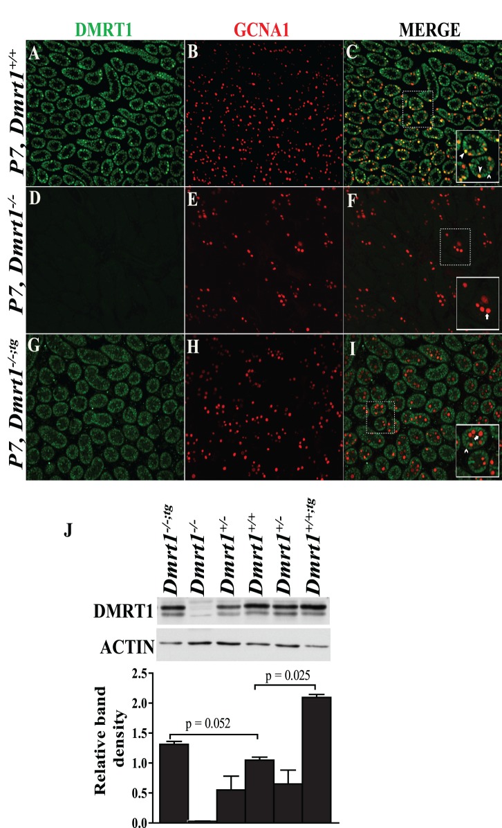
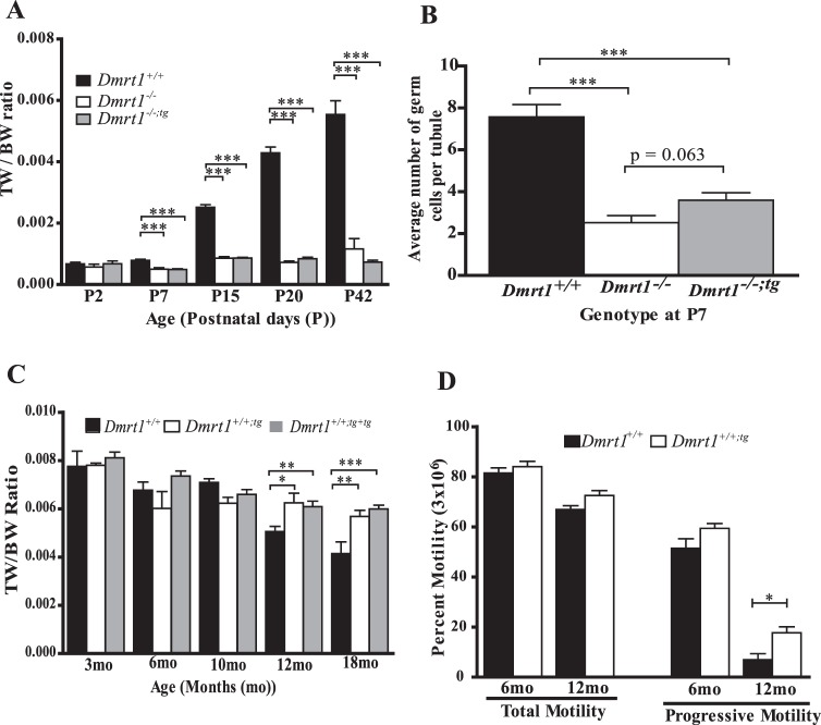
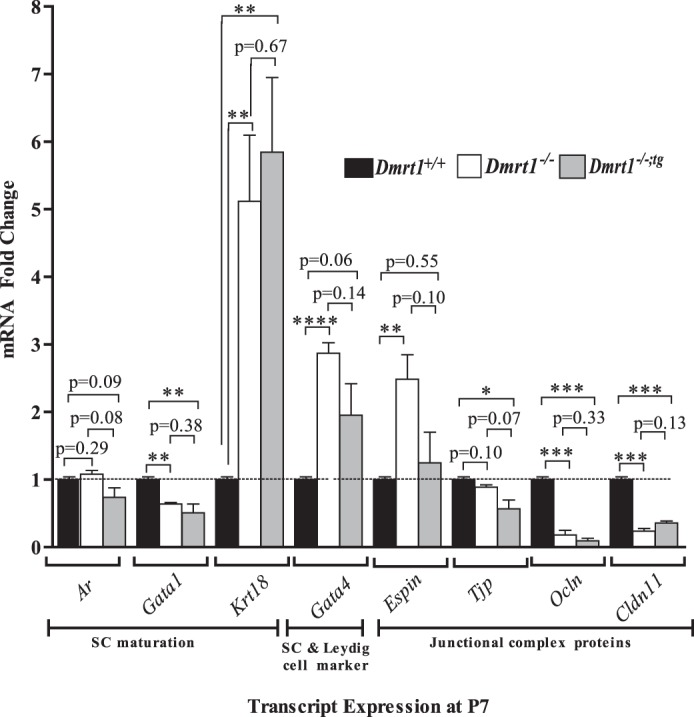
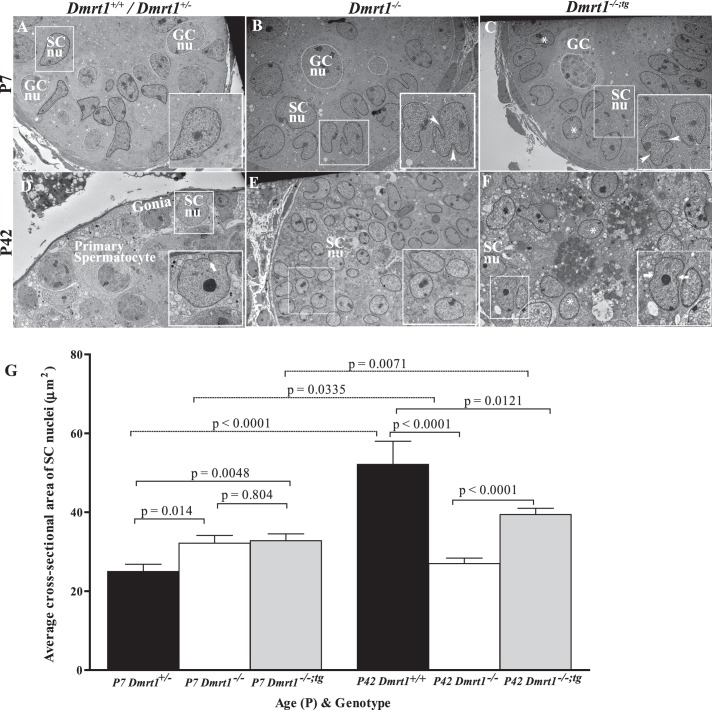
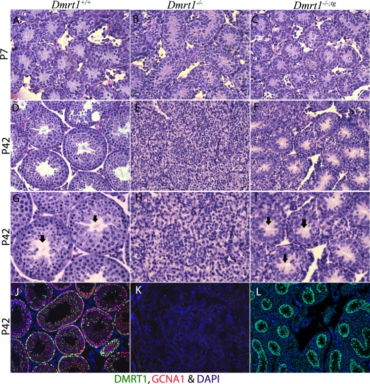
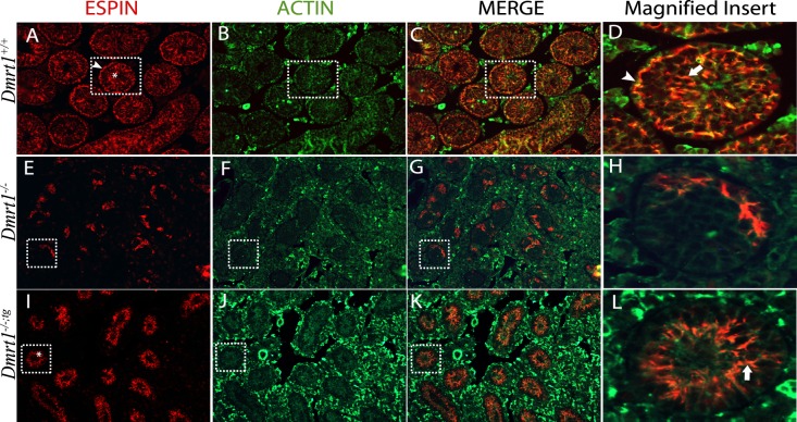
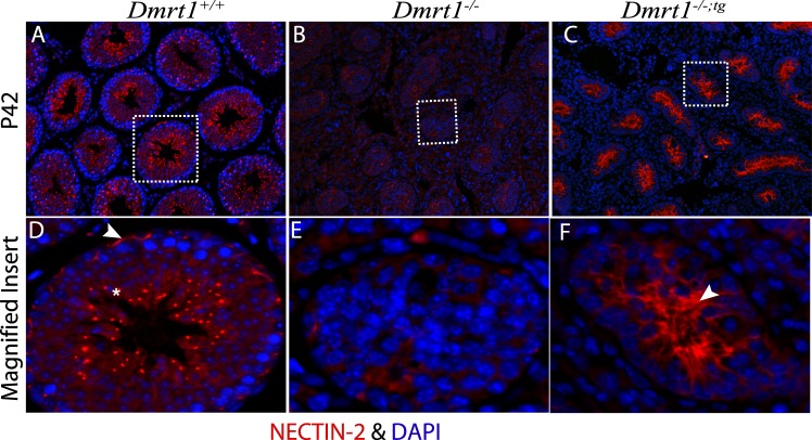
DMRT1 is an evolutionarily conserved transcriptional factor expressed only in the postnatal testis, where it is produced in Sertoli cells and germ cells. While deletion of Dmrt1 in mice demonstrated it is required for postnatal testis development and fertility, much is still unknown about its temporal- and cell-specific functions. This study characterized a novel mouse model of DMRT1-deficient germ cells that was generated by breeding Dmrt1-null (Dmrt1(-/-)) mice with Wt1-Dmrt1 transgenic (Dmrt1(+/-;tg)) mice, which express a rat Dmrt1 cDNA in gonadal supporting cells by directing it from the Wilms tumor 1 locus in a yeast artificial chromosome transgene. Like Dmrt1(-/-) mice, male Dmrt1(-/-) transgenic mice (Dmrt1(-/-;tg)) were infertile, while female mice were fertile. Immunohistochemistry and Western blot analysis showed transgenic DMRT1 expressed in supporting cells of the newborn gonads of both sex and in Sertoli cells of the testis afterbirth. Sertoli cells were evaluated by electron microscopy, revealing that maturation of Dmrt1(-/-;tg) Sertoli cells was incomplete. Morphological analysis of testes from 42-day-old mice showed that, compared to Dmrt1(-/-) mice, Dmrt1(-/-;tg) mice have improved seminiferous tubule structure, with lumens present in many. Immunohistochemistry of the polarity markers ESPIN and NECTIN-2 showed that DMRT1 in Sertoli cells is required for NECTIN-2 expression and influences organization of ectoplasmic specializations. Further functional analyses of the transgene on a Dmrt1(-/-) background showed that it did not rescue the decrease in Dmrt1(-/-) testis size, but when expressed on a wild-type background, exogenous DMRT1 prevented the normal age-related decline in testis size and enhanced sperm progressive motility. The studies suggest that DMRT1 in Sertoli cells regulates tubule morphology, spermatogenesis, and sperm function via its effects on Sertoli cell maturation and polarity. Furthermore, expression and function of transgenic DMRT1 in Sertoli cells establishes a novel mouse model of DMRT1-deficient germ cells generated by breeding Dmrt1-null mice with Wt1-Dmrt1 transgenic mice (rescue; Dmrt1(-/-;tg)).


Figures

FIG. 1
FIG. 1
Generation and tissue-specific expression of the Wt1-Dmrt1 transgene. A) Generation of the Wt1-Dmrt1 transgene. The rat Dmrt1 cDNA was inserted in frame with the translational start ATG of mouse Wilms tumor gene 1 (Wt1) within the yeast artificial chromosome YAC620mWt1. Top: YAC620mWt1; exons = boxed with numbers, URA/Acentric, and TRP/Centric = the pYAC4 acentric vector arms with uracil and tryptophan selectable markers, respectively. Middle: targeting vector, pBKS-Wt1-Dmrt1-HPRT1-LTS2. Includes the rat Dmrt1 cDNA, exons 8 and 9, intron 8 and polyadenylation site of HPRT1, lysine 2 selectable marker (LYS2), 825 bp of Wt1 sequence 5′ of translational start site (Wt15′), and 776 bp of Wt1 sequence 3′ of translational start site (Wt13′). Bottom: Wt1-Dmrt1 transgene after homologous recombination in yeast (AB1380). B and C) RT-PCR of RNA prepared from different organs from wild-type (wt superscript) and transgenic (tg superscript) mice. B) Transgene expression in the kidneys (K) and ovaries (O) and testis (T) of newborn littermates. M and F subscripts indicate male and female, respectively. C) Transgene expression in P15 male mice. Tissues are denoted as testis (T), brain (B), heart (H), spleen (Sp), stomach (St), lung (Lu), kidney (K), and liver (Li). The plasmid pBKS-Wt1 5′-Dmrt1-HPRT1-LYS2-Wt1 3′ (P) was included as positive control and water (H2O) as negative control. cDNA was synthesized in the presence (+) or absence (−) of reverse transcriptase. A ∼450-bp band denotes cDNA amplification, while the ∼1105-bp band bottom is amplified genomic DNA.
FIG. 2
FIG. 2
DMRT1 expression in P7 testes. Coimmunofluorescence of DMRT1 (green) and the GC marker GCNA1 (red) in P7 testes from wild-type (AC), Dmrt1−/− (DF), and Dmrt1−/−;tg (GI) mice. Individual panels for DMRT1 (A, D, and G) and GCNA1 (B, E, and H) staining and their merged images (C, F, and I) are shown. The solid boxed areas in C, F, and I represent enlarged merged images of areas denoted by the dashed boxes. Within the boxed areas, solid arrowheads denote DMRT1-positive germ cells (yellow), open arrowheads denote DMRT1-positive SCs (green), and arrows denote DMRT1-negative germ cells (red). Note that DMRT1-positive germ cells were observed only in wild-type mice (C). Final magnification for all micrographs is ×200. Relative expression levels of DMRT1 in P30 testes from Dmrt1+/+, Dmrt1−/−, Dmrt1−/−;tg, and Dmrt1+/+;tg mice (J). DMRT1 and ACTIN levels were analyzed by Western blot analysis (top). The bands were quantified by densitometry, and DMRT1 band intensities, relative to that of ACTIN, are represented graphically (bottom). Graphed is the mean DMRT1/ACTIN ratio of three mice/genotype. Error bars represent the SEM.
FIG. 3
FIG. 3
The Wt1-Dmrt1 transgene helps maintain testis weights and sperm motility in aging mice. A) TW/BW ratios of Dmrt1+/+ (black bar), Dmrt1−/− (white bar), and Dmrt1−/−;tg (gray bar) mice at Postnatal Day 2 (P2), 7 (P7), 15 (P15), 20 (P20), and 42 (P42). Graphed is the mean TW/BW + SEM of 3–20 animals. B) Average GC number per seminiferous tubule in Dmrt1+/+ (black bar), Dmrt1−/− (white bar), and Dmrt1−/−;tg (gray bar) P7 testes. Graphed is the mean number of GCs + SEM from at least 30 tubules/testes in a minimum of three mice per genotype. C) Effect of exogenous Wt1-Dmrt1 transgene on testis weights of wild-type mice. TW/BW ratios for Dmrt1+/+ mice (black bar) and Dmrt1+/+ mice with one copy (Dmrt1+/+;tg; white bar) or two copies of the Wt1-Dmrt1 transgene (Dmrt1+/+;tg+tg; gray bar) at 3, 6, 10, 12, and 18 mo of age. D) Total and progressive motility of epididymal sperm from Dmrt1+/+ (black bar) and Dmrt1+/+;tg (white bar) mice at 6 and 12 mo of age. Graphed is the mean + SEM of 3–20 animals per genotype. *P < 0.05, **P < 0.001, and ***P < 0.0001 (two-tailed Student t-test).
FIG. 4
FIG. 4
Expression of genes in response to Dmrt1 loss. Espin, Ocln, Cldn11, Tjp, Gata1, Ar, and Krt18 mRNA levels were quantified by quantitative RT-PCR using RNA isolated from testes of Dmrt1+/+ (black bar), Dmrt1−/− (white bar), and Dmrt1−/−;tg (gray bar) mice at P7. *P < 0.05, **P < 0.005, and ***P < 0.0005 (two-tailed Student t-test).
FIG. 5
FIG. 5
Electron microscopic evaluation of SC nuclei. Testes from Dmrt1+/+ or Dmrt1+/− (A and D), Dmrt1 −/− (B and E), and Dmrt1−/−;tg (C and F) mice were evaluated at P7 (AC) and P42 (DF). Some GC (GCnu) and SC nuclei (SCnu) are traced by white and black lines, respectively. AC) Electron micrographs of seminiferous tubules from P7 Dmrt1+/−, Dmrt1−/−, and Dmrt1−/−;tg testes. A) Dmrt1+/− tubule showing normal ultrastructure of SC nucleus (inset). Magnification ×3040. B) Dmrt1−/− tubule showing abnormal SC nuclei. Boxed SC nuclei denoted the irregular shaped and multilobulated nuclei containing deep invaginations (arrowheads). Magnification ×3040. C) Dmrt1−/−;tg tubule showing a mixed population of normal- and abnormal-looking SC nuclei. The normal (asterisk) and abnormal (arrowhead) nuclei shared characteristics with Dmrt1+/+ and Dmrt1−/− mice, respectively. Magnification ×2280. DF) Electron micrographs of seminiferous tubules from P42 Dmrt1+/+, Dmrt1−/−, and Dmrt1−/−;tg testes. D) Dmrt1+/+ tubule showing normal ultrastructure of SC and GCs. Note that SC nuclei are larger with considerable perinuclear cytoplasm and have an irregular ovoid shape with invaginations (arrow) and significant nucleolus. Magnification ×2280. E) Dmrt1−/− tubule showing SCs with many regular round nuclei (asterisk) and scanty amount of cytoplasm (inset). Note that there are no GCs present. Magnification ×1900. F) Dmrt1−/−;tg tubule showing many regular shaped, round SC nuclei (asterisk) with a few irregular, indented (arrows) nuclei, and prominent nucleolus. Magnification ×3040. G) The average cross-sectional area of SC nuclei in P7 and P42 testes of control (black bars), Dmrt1−/− (white bars), and Dmrt1−/−;tg (gray bars) testes. Graphed is the mean cross-sectional area of ≥60 nuclei per genotype per time point. Error bars represent the SEM. Statistical significance was determined using a two-tailed Student t-test with P-values denoted between relevant pairings.
FIG. 6
FIG. 6
Morphological analysis of P7 and P42 mouse testes. Periodic acid Schiff staining of P7 (AC) and P42 (DI) testis section from wild-type (A, D, and G), Dmrt1−/− (B, E, and H), and Dmrt1−/−;tg (C, F, and I) mice. Note that the addition of the transgene (i.e., in Dmrt1−/−;tg mice) partially restored seminiferous tubule morphology. Magnification ×400 (AF) and ×600 (GI). Arrows (G and I) denote the presence of defined lumens, which were absent from the Dmrt1−/− testis (H). Immunohistochemical analysis of wild-type (J), Dmrt1−/− (K), and Dmrt1−/−;tg (L) at P42 (magnification ×200). Testis sections were evaluated using fluorescently labeled antibodies against DMRT1 (green) and GCNA1 (red) and DAPI for nuclei (blue).
FIG. 7
FIG. 7
Immunolocalization of ESPIN and ACTIN in P42 mouse testes. Coimmunofluorescence of ESPIN (red) and ACTIN (green) in testes sections from wild-type (AD), Dmrt1−/− (EH), and Dmrt1−/−;tg (IL) mice. Shown are individual panels for ESPIN (A, E, and I) and ACTIN (B, F, and J) staining, their merged (C, G, and K) images, and enlarged merged images of areas denoted by boxes (D, H, and L). Arrowhead in A and asterisk in A and I show basal and apical ES, respectively. Arrowhead in D and arrow in D and L show colocalization of ESPIN and ACTIN at the basal and apical ES, respectively. Final magnification of all panels except for magnified inserts was ×200.
FIG. 8
FIG. 8
Immunolocalization of NECTIN-2 in P42 mouse testes. Immunofluorescence of NECTIN-2 (red) and DAPI stain (blue) in testes sections from wild-type (A), Dmrt1−/− (B), and Dmrt1−/−;tg (C) mice. DF show enlarged images of the areas denoted by boxes in AC. Arrowhead and asterisk in D show basal and apical ES, respectively. Arrowhead in F shows ES in the adluminal compartment of the seminiferous tubule. Final magnification of all panels except for magnified inserts was ×200.

Comment in

References

    1. Ferguson-Smith M. The evolution of sex chromosomes and sex determination in vertebrates and the key role of DMRT1. Sex Dev 2007; 1: 2 11 - PubMed
    1. Kim S, Bardwell VJ, Zarkower D. Cell type-autonomous and non-autonomous requirements for Dmrt1 in postnatal testis differentiation. Dev Biol 2007; 307: 314 327 - PMC - PubMed
    1. Krentz AD, Murphy MW, Kim S, Cook MS, Capel B, Zhu R, Matin A, Sarver AL, Parker KL, Griswold MD, Looijenga LH, Bardwell VJ. et al. The DM domain protein DMRT1 is a dose-sensitive regulator of fetal germ cell proliferation and pluripotency. Proc Natl Acad Sci U S A 2009; 106: 22323 22328 - PMC - PubMed
    1. Marchand O, Govoroun M, D'Cotta H, McMeel O, Lareyre J, Bernot A, Laudet V, Guiguen Y. DMRT1 expression during gonadal differentiation and spermatogenesis in the rainbow trout, Oncorhynchus mykiss. Biochim Biophys Acta 2000; 1493: 180 187 - PubMed
    1. Matson CK, Murphy MW, Griswold MD, Yoshida S, Bardwell VJ, Zarkower D. The mammalian doublesex homolog DMRT1 is a transcriptional gatekeeper that controls the mitosis versus meiosis decision in male germ cells. Dev Cell 2010; 19: 612 624 - PMC - PubMed

Publication types

MeSH terms

LinkOut - more resources