. 2013 Feb 1;190(3):970-6.
doi: 10.4049/jimmunol.1202805. Epub 2012 Dec 19.

Recirculating memory T cells are a unique subset of CD4+ T cells with a distinct phenotype and migratory pattern

Affiliations

Recirculating memory T cells are a unique subset of CD4+ T cells with a distinct phenotype and migratory pattern

Shannon K Bromley et al. J Immunol. .

Abstract

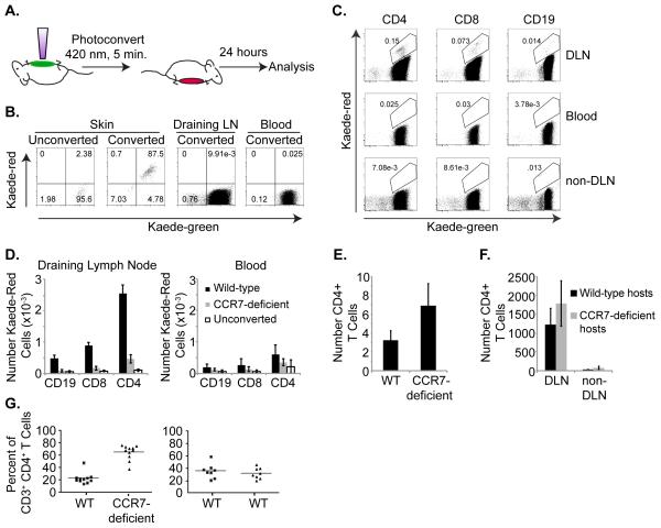
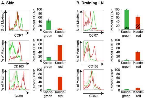
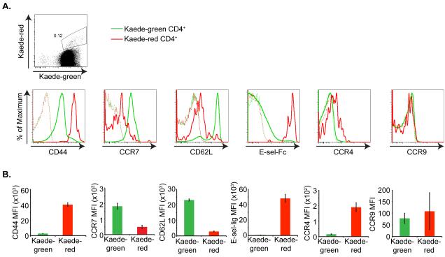
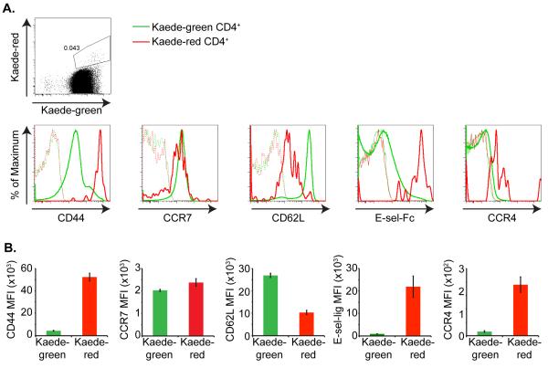
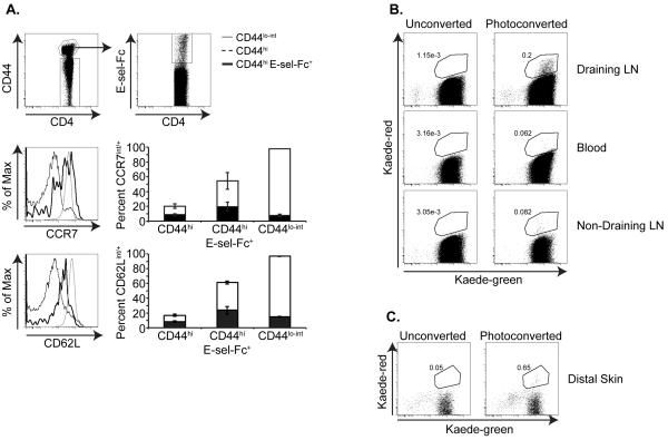
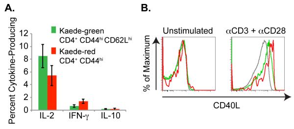
Several populations of memory T cells have been described that differ in their migration and function. In this study, we have identified a unique subset of memory T cells, which we have named recirculating memory T cells (T(RCM)). By exposing Kaede transgenic mouse skin to violet light, we tracked the fate of cutaneous T cells. One population of memory CD4(+) T cells remained in the skin. A second population migrated from the skin into draining lymph nodes (LNs) in a CCR7-dependent manner. These migrating CD4(+) T cells expressed a novel cell surface phenotype (CCR7(int/+)CD62L(int)CD69(-)CD103(+/-) E-selectin ligands(+)) that is distinct from memory T cell subsets described to date. Unlike memory T cell subsets that remain resident within tissues long-term, or that migrate either exclusively between lymphoid tissues or into peripheral nonlymphoid sites, CD4(+) T(RCM) migrate from the skin into draining LNs. From the draining LNs, CD4(+) T(RCM) reenter into the circulation, distal LNs, and sites of non-specific cutaneous inflammation. In addition, CD4(+) T(RCM) upregulated CD40L and secreted IL-2 following polyclonal stimulation. Our results identify a novel subset of recirculating memory CD4(+) T cells equipped to deliver help to both distal lymphoid and cutaneous tissues.


Figures

Figure 1
Figure 1. Lymphocyte migration from skin to draining LNs in the steady-state is CCR7-dependent
(a) Schematic of Kaede transgenic mouse photoconversion. Shaved abdominal skin of Kaede transgenic mice is exposed to 420 nm light for 5 minutes. After 24 h, leukocytes are recovered from tissues and analyzed by flow cytometry for CD3, CD4, CD8, CD19 and Kaede-green or Kaede-red expression. (b) Flow cytometric analysis of Kaede-green and Kaede-red expression by CD3+ CD4+ T cells recovered from unconverted skin or photoconverted skin, blood and draining axillary LN immediately after exposure to violet light. (c) Flow cytometric analysis of Kaede-green and Kaede-red expression by draining axillary LN CD3+ CD4+ T cells, CD3+ CD8+ T cells and CD3CD19+ B cells 24 h after photoconversion of Kaede transgenic mouse abdominal skin. Data are representative of 3 experiments with 6 mice total. (d) Numbers of Kaede-red CD3+ CD4+ T cells, CD3+ CD8+ T cells and CD3 CD19+ B cells within draining axillary LNs (left) or blood (right) 24 h after photoconversion of WT-(black bars) or CCR7-deficient-(gray bars) Kaede transgenic abdominal skin. Open bars are lymphocytes that fall within the Kaede-red gate from draining axillary LNs or blood of control unconverted Kaede transgenic mice. Data are from 3 experiments with 6 mice total for each genotype. A total of 3 control unconverted Kaede transgenic mice were analyzed. (e) The shaved abdominal skin of WT or CCR7-deficient mice was isolated, frozen in OCT, and 10 μm sections were stained with anti-CD4 antibodies. Slides were imaged and numbers of CD4+ T cells in 200× fields were counted. Data are the average of 4-6 mice, with at least 3 fields counted per mouse. (f) Five million WT CD4+ Thy1.1+ T cells were injected s.c. into the right footpads of either WT congenic Thy1.2+ or CCR7-deficient Thy1.2+ hosts. After 16 h, right draining popliteal LNs (DLN) and left contralateral popliteal LNs (non-DLN) were isolated, recovered cells were counted, stained with anti-CD3, anti-CD4 and anti-Thy1.1 antibodies, and analyzed by flow cytometry. Data are from 3 experiments with 9 mice total per genotype. (g) Bone marrow chimeras were generated by reconstituting irradiated WT CD45.1 mice with a 1:1 mix of either WT CD45.1 CD45.2 and CCR7-deficient CD45.2 bone marrow cells or with WT CD45.1 CD45.2 and WT CD45.2 bone marrow cells. After eight weeks, leukocytes were recovered from skin and the percent of WT or CCR7-deficient CD3+ CD4+ T cells of total cutaneous CD3+ CD4+ T cells was calculated. Data are compiled from three experiments with WT: CCR7-deficient chimeras (left graph) and WT:WT chimeras (right graph) and include 11 or 8 chimeric mice total, respectively.
Figure 2
Figure 2. Kaede-red CD4+ T cells residing in skin are CCR7 CD103+ CD69+
Flow cytometric analysis of CCR7, CD103 and CD69 expression by Kaede-green CD3+ CD4+ T cells (green solid line) and Kaede-red CD3+ CD4+ T cells (red solid line) remaining within the abdominal skin (a) or migrated from skin to draining LN (b) 24 h after abdominal skin photoconversion. Dashed lines are control “fluorescence minus one” stains of Kaede-green- and Kaede-red CD3+ CD4+ T cells. Graphs depict the average percent of Kaede-green (green bars) and Kaede-red (red bars) CD3+ CD4+ T cells in the skin or draining LN that express CCR7, CD103 or CD69. For CCR7 expression in the draining LN, striped bars depict the percent of CD3+ CD4+ T cells that express intermediate CCR7 and solid bars show the percent of CD3+ CD4+ T cells that express high CCR7. Data are representative of 3 experiments with 9 mice total.
Figure 3
Figure 3. Kaede-red CD4+ T cells express lymph node- and skin-homing receptors
(a) Top panel illustrates Kaede-green and Kaede-red expression by CD3+ CD4+ T cells within the draining LN 24 h after photoconversion of Kaede transgenic mouse abdominal skin. Bottom panels depict flow cytometric analysis of CD44, CCR7, CD62L, E-selectin ligands, CCR4 and CCR9 expression by Kaede-green CD3+ CD4+ (green solid line) and Kaede-red CD3+ CD4+ T cells (red solid line) within the draining LNs 24 h after photoconversion of Kaede transgenic mouse abdominal skin. Dashed lines are control “fluorescence minus one” stains of Kaede-green- and Kaede-red CD3+ CD4+ T cells. (b) MFI for receptor expression on Kaede-green CD3+ CD4+ T cells and Kaede-red CD3+ CD4+ T cells are displayed for one experiment with three mice per receptor analyzed and is representative of three experiments with at least eight mice total per receptor.
Figure 4
Figure 4. Kaede-red CD4+ T cells up-regulate CCR7 expression over time
Shaved abdominal skin of Kaede transgenic mice was exposed to 420 nm light for 5 minutes on 3 consecutive days. (a) Top panel illustrates Kaede-green and Kaede-red expression by CD3+ CD4+ T cells within the draining LNs 8 d after the final photoconversion of Kaede transgenic mouse abdominal skin. Bottom panel depicts flow cytometric analysis of CD44, CCR7, CD62L, E-selectin ligands, and CCR4 expression on these Kaede-green CD3+ CD4+ (green solid line) and Kaede-red CD3+ CD4+ T cells (red solid line). Dashed lines are control “fluorescence minus one” stains of Kaede-green- and Kaede-red CD3+ CD4+ T cells. b) MFI for receptor expression on Kaede-green CD3+ CD4+ T cells and Kaede-red CD3+ CD4+ T cells are displayed for one experiment with three mice per receptor analyzed and is representative of three experiments with at least eight mice per receptor.
Figure 5
Figure 5. Kaede-red CD4+ T cells reenter the circulation from draining LNs
(a) CCR7 and CD62L expression on CD4+ CD44hi E-sel-Fc+ T cells within the blood of resting C57Bl/6 mice. Leukocytes recovered from the blood were gated on CD3+ CD4+ CD44hi memory T cells and on CD3+ CD4+ CD44lo-int naïve T cells (top left panel). The CD44hi T cell population was then gated on E-sel-Fc+ cells (top right panel). Middle and bottom histograms depict CCR7 and CD62L expression on CD4+ CD44lo-int naïve T cells, CD4+ CD44hi bulk memory T cells, and on CD4+ CD44hi E-sel-Fc+ memory T cells. Each plot is from 6 mice pooled and is representative of 3 independent experiments. Graphs depict the average percent of each CD3+ CD4+ T cell population in the blood that express CCR7 or CD62L. Black bars depict the percent of CD3+ CD4+ T cells that express intermediate CCR7 or CD62L and white bars show the percent of CD3+ CD4+ T cells that express high CCR7 or CD62L. (b) Shaved abdominal skin of Kaede transgenic mice was exposed to 420 nm light for 5 minutes on 3 consecutive days. Flow cytometric analysis of Kaede-green and Kaede-red expression on CD3+ CD4+ T cells recovered from the blood, non-draining cervical LNs and draining axillary LNs 48 h after the final photoconversion. Data are representative of 3 experiments with 8 mice/group total. (c) The right ear of Kaede transgenic mice was injected with 10 μL of a 1:1 PBS:CFA emulsion, and then the shaved abdominal skin was photoconverted as in (a). Flow cytometry plots depict Kaede-green and Kaede-red expression by CD3+ CD4+ T cells recovered from the CFA-injected distal ear skin 48 h after the final photoconversion. Numbers in plots are average percent Kaede-red T cells of CD3+ CD4+ T cells recovered from the ear, and are calculated from 3 experiments with 8-14 mice total/group. Data shown are of 6 mice pooled/group.
Figure 6
Figure 6. Recirculating Kaede-red CD3+ CD4+ CD44hi T cells up-regulate CD40L and secrete IL-2
Shaved abdominal skin of Kaede transgenic mice was exposed to 420 nm light for 5 minutes. After 24 h, draining LNs were isolated and (a) recovered cells were stimulated for 4 h with plate-bound anti-CD3 and soluble anti-CD28. Cytokine secretion by Kaede-green CD3+ CD4+ CD44hi CD62Lhi memory T cells and Kaede-red CD3+ CD4+ CD44hi memory T cells was analyzed by flow cytometry. Data are pooled from 3 experiments with 9 mice total for each cytokine. (b) Or, CD4+ T cells were purified from draining LNs and stimulated for 2 h with PMA and ionomycin, or left untreated. Then, surface CD40L expression was analyzed on Kaede-green CD44lo naïve T cells (gray lines), Kaede-green CD44hi memory T cells (green lines) or Kaede-red CD44hi memory T cells (red lines). Data are representative of 2 experiments with 6 mice total.

References

    1. Sallusto F, Lenig D, Forster R, Lipp M, Lanzavecchia A. Two subsets of memory T lymphocytes with distinct homing potentials and effector functions. Nature. 1999;401:708–712. - PubMed
    1. Clark RA, Chong B, Mirchandani N, Brinster NK, Yamanaka K, Dowgiert RK, Kupper TS. The vast majority of CLA+ T cells are resident in normal skin. J Immunol. 2006;176:4431–4439. - PubMed
    1. Gebhardt T, Wakim LM, Eidsmo L, Reading PC, Heath WR, Carbone FR. Memory T cells in nonlymphoid tissue that provide enhanced local immunity during infection with herpes simplex virus. Nat Immunol. 2009;10:524–530. - PubMed
    1. Klonowski KD, Williams KJ, Marzo AL, Blair DA, Lingenheld EG, Lefrancois L. Dynamics of blood-borne CD8 memory T cell migration in vivo. Immunity. 2004;20:551–562. - PubMed
    1. Jiang X, Clark RA, Liu L, Wagers AJ, Fuhlbrigge RC, Kupper TS. Skin infection generates non-migratory memory CD8+ TRM cells providing global skin immunity. Nature. 2012;483:227–231. - PMC - PubMed

Publication types

MeSH terms