

Discrete mineralisation of the acetabular labrum: a novel marker of femoroacetabular impingement?
- PMID: 23255539
- PMCID: PMC3615393
- DOI: 10.1259/bjr.20120182
Discrete mineralisation of the acetabular labrum: a novel marker of femoroacetabular impingement?
Abstract
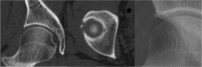
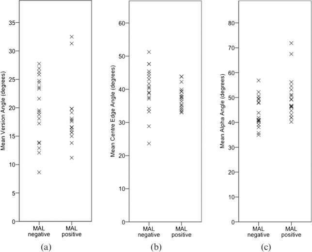
Femoroacetabular impingement (FAI) is increasingly thought to play a role in the development of hip osteoarthritis, but is difficult to define clinically and on imaging. This study investigates mineralisations of the acetabular labrum (MALs), which are small, discrete foci of dense radio-opacity within the region of the acetabular labrum. The study aims to characterise MALs and test the hypothesis that MALs are associated with FAI. CT images and radiographs of 106 hips in 66 individuals without known FAI were reviewed for the presence of MALs. The anatomical locations of the MALs in the acetabular labrum were measured. Three current radiographic markers of FAI were recorded in hips with MALs and in age- and gender-matched hips without MALs: centre-edge angle and acetabular version angle as measures of pincer impingement, and alpha angle as a measure of cam impingement. MALs were identified in 18% of hips (n=19). Hips with MAL had a larger mean alpha angle (p=0.013) than those without. MALs were found to be located anterosuperiorly and posterosuperiorly within the labrum, consistent with coup and contrecoup impingement lesion locations reported for FAI. No significant association was found between MAL and centre-edge angle or version angle. Our data demonstrate that MALs are associated with increased alpha angle and thus may be linked to cam-type FAI. MALs have not previously been associated with FAI. This correlation may give further insight into the disease process underlying hip osteoarthritis and might represent a future radiographic marker of cam-type FAI.
Figures


References
-
- Ganz R, Parvizi J, Beck M, Leunig M, Nötzli H, Siebenrock KA. Femoroacetabular impingement: a cause for osteoarthritis of the hip. Clin Orthop Relat Res 2003:112–20. - PubMed
-
- Tannast M, Siebenrock KA, Anderson SE. Femoroacetabular impingement: radiographic diagnosis—what the radiologist should know. AJR Am J Roentgenol 2007;188:1540–52. - PubMed
-
- Beall DP, Sweet CF, Martin HD, Lastine CL, Grayson DE, Ly JQ, et al. Imaging findings of femoroacetabular impingement syndrome. Skeletal Radiol 2005;34:691–701. - PubMed
-
- Lohan DG, Seeger LL, Motamedi K, Hame S, Sayre J. Cam-type femoral-acetabular impingement: is the alpha angle the best MR arthrography has to offer? Skeletal Radiol 2009;38:855–62. - PubMed
-
- Domayer SE, Ziebarth K, Chan J, Bixby S, Mamisch TC, Kim YJ. Femoroacetabular cam-type impingement: diagnostic sensitivity and specificity of radiographic views compared to radial MRI. Eur J Radiol 2011;80:805–10. - PubMed
MeSH terms
Substances
LinkOut - more resources
Full Text Sources
Other Literature Sources
Medical
Research Materials
