. 2013 Feb 1;288(5):3322-33.
doi: 10.1074/jbc.M112.420497. Epub 2012 Dec 19.

Function of focal adhesion kinase scaffolding to mediate endophilin A2 phosphorylation promotes epithelial-mesenchymal transition and mammary cancer stem cell activities in vivo

Affiliations

Function of focal adhesion kinase scaffolding to mediate endophilin A2 phosphorylation promotes epithelial-mesenchymal transition and mammary cancer stem cell activities in vivo

Huaping Fan et al. J Biol Chem. .

Abstract

Tyrosine kinases have been shown to play critical roles in cancer development and progression, and their inhibitors hold the potential as effective targeted therapies for breast and other cancers. However, some of these kinases like focal adhesion kinase (FAK) also possess scaffolding functions in intracellular signaling, but such kinase-independent functions of FAK or other kinases have not been examined in cancer directly in vivo. Here, we report that disruption of the function of FAK scaffolding through its Pro-878/881 motif suppressed mammary tumor growth and metastasis in a well characterized murine model of human breast cancer. P878A/P881A mutation in the endogenous FAK gene decreased the expression of markers for epithelial-mesenchymal transition (EMT) and mammary cancer stem cell (MaCSC) activities in tumors derived from mutant mice. This mutation disrupted the function of FAK scaffolding to mediate endophilin A2 phosphorylation at Tyr-315 by Src, leading to the decreased surface expression of MT1-MMP, as observed previously in transformed fibroblasts in vitro. Inhibition of the downstream components of this FAK scaffolding function by Y315F endophilin A2 mutant or MT1-MMP knockdown reduced markers for EMT and MaCSC activities. Conversely, bypass of the scaffolding function using the phosphorylation mimic mutant Y315E endophilin A2 or endophilin A2 knockdown rescued the decreased markers for EMT and MaCSCs as well as surface expression of MT1-MMP in tumor cells harboring the P878A/P881A mutation. Together, these results identify a novel role of FAK scaffolding function in breast cancer, which could serve as a new target in combination with kinase inhibition for more effective treatment strategies.


Figures

FIGURE 1.
FIGURE 1.
PA/PA-MT mice had comparable tumor appearance time but suppressed mammary tumor growth and lung metastasis. A, Kaplan-Meier analysis of mammary tumor development in the +/PA-MT (n = 30) and PA/PA-MT (n = 28) mice. B, mean cumulative mammary tumor volume per mouse at different times after primary tumor appearance for +/PA-MT (n = 30) and PA/PA-MT (n = 28). PA/PA-MT versus +/PA-MT, p < 0.05 by the two-way ANOVA. C and D, lung sections (four to six sections per mouse) were prepared 8 weeks after the primary mammary tumor onset and stained with H&E (C), and the micrometastatic nodules (arrows) were quantitated under a microscope for percentages of mice of the indicated genotype with <1, 1–5, or >5 metastases per lung section (D). *, p < 0.05, when the percentage of each group in PA/PA-MT mice is compared with that in +/PA-MT mice. Scale bars, 1 mm. E, 1 × 10⁵ freshly isolated primary tumor cells from +/PA-MT or PA/PA-MT mice were injected into the tail vein of 8-week-old nude mice (n = 3 for each group). The micrometastatic nodules in their lungs were quantitated 4 or 6 weeks after injection. **, p < 0.01, when the number of nodules after injection of tumor cells from PA/PA-MT mice is compared with that from +/PA-MT mice.
FIGURE 2.
FIGURE 2.
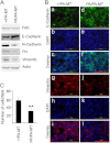
Tumor cell growth, markers for EMT, and migration were suppressed in PA/PA-MT mice. A, lysates of four tumors from two different +/PA-MT or PA/PA-MT mice were prepared and analyzed by immunoblotting using antibodies against various proteins, as indicated. B, cultured primary tumor cells from +/PA-MT or PA/PA-MT mice were analyzed by immunofluorescent labeling using antibody against E-cadherin (panels a–f) or vimentin (panels g–l). Scale bars, 100 μm. C, cell mobility was measured by Boyden chamber assay for the freshly isolated primary tumor cells from +/PA-MT or PA/PA-MT mice attracted by fibronectin. Mean ± S.E. of the number of migrated cells per field from three independent experiments are shown. **, p < 0.01, when the migrated cell number from PA/PA-MT mice are compared with that from +/PA-MT mice.
FIGURE 3.
FIGURE 3.
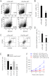
Decreased pool of MaCSCs and their tumorigenicity in PA/PA-MT mice. A and B, Aldefluor assay of freshly isolated tumor cells from +/PA-MT or PA/PA-MT mice. The percentage of ALDH+ cells was determined under the same gating criteria. Results are representative of two separate experiments. *, p < 0.05, when the percentage of ALDH+ cells from PA/PA-MT mice are compared with that from +/PA-MT mice. C and D, freshly isolated tumor cells from +/PA-MT or PA/PA-MT mice were depleted of CD45-positive and CD31-positive cells and labeled with CD24, CD29, and CD61 antibodies. The MaCSC population in each strain of mice was gated as LinCD24+CD29hiCD61hi (indicated as R4 quad) under the same gating criteria. Results are representative of two separate experiments. **, p < 0.01, when the percentage of LinCD24+CD29hiCD61hi cells from PA/PA-MT mice are compared with that from +/PA-MT mice. E, sorted ALDH+ or LinCD24+CD29hiCD61hi tumor cells from +/PA-MT or PA/PA-MT mice were analyzed for primary formation of tumor spheres. Results are generated from eight separated incubations for each sample and are representative of two independent experiments. **, p < 0.01, when the number of spheres by the ALDH+ or LinCD24+CD29hiCD61hi cells from PA/PA-MT mice are compared with that from +/PA-MT mice. F, mean cumulative mammary tumor volume per mouse at different times after different numbers of LinCD24+CD29hiCD61hi cells from +/PA-MT or PA/PA-MT mice injected into mammary fat pad of 8-week-old nude mice (n = 3 for each group).
FIGURE 4.
FIGURE 4.
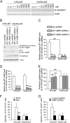
FAK interaction with endophilin A2 controls the surface expression of MT1-MMP in mammary tumor cells. A–C, primary tumor cell lysates from +/PA-MT or PA/PA-MT mice were immunoprecipitated (IP) with anti-FAK (A) or anti-endophilin (Endo) A2 (B and C) followed by Western blotting with antibodies as indicated. Aliquots of lysates were also analyzed by Western blotting directly as indicated. IB, immunoblot. D, freshly isolated primary tumor cells from +/PA-MT or PA/PA-MT mice were infected with recombinant lentiviruses encoding endophilin A2 shRNA or a control shRNA. Three days after infection, surface expression of MT1-MMP as labeled by biotinylation followed by streptavidin precipitation was determined. Aliquots of the lysates were also analyzed directly by Western blotting with antibodies as indicated. E–H, intensity of the endophilin A2 (E), surface MT1-MMP (F), total MT1-MMP (G), and E-cadherin (H) bands was quantified from two independent experiments by densitometry. The mean ± S.D. of relative intensity (normalized to cells from +/PA-MT mice treated with control (Con) shRNA) are shown. **, p < 0.01 compared with cells treated with control shRNA. **, p < 0.01 (linked by lines in F and H) when cells from +/PA-MT and PA/PA-MT mice were compared (both treated with control shRNA).
FIGURE 5.
FIGURE 5.
Regulation of EMT and tumor sphere formation by MT1-MMP in mammary tumor cells. A, freshly isolated primary tumor cells from +/PA-MT or PA/PA-MT mice were treated with different concentrations of Marimastat as indicated for 8 h. After treatment, total cell lysates were analyzed directly by Western blotting with antibody anti-E-cadherin or anti-actin as a loading control. B, freshly isolated primary tumor cells from +/PA-MT or PA/PA-MT mice were infected with recombinant lentiviruses encoding MT1-MMP shRNA or a control shRNA. Three days after infection, surface expression of MT1-MMP as labeled by biotinylation followed by streptavidin precipitation was determined. Aliquots of the lysates were also analyzed directly by Western blotting with antibodies as indicated. IB, immunoblot. C–E, intensity of the surface MT1-MMP (C), total MT1-MMP (D), and E-cadherin (E) bands was quantified from two independent experiments by densitometry. The mean ± S.D. of relative intensity (normalized to cells from +/PA-MT mice treated with control (Con) shRNA) are shown. **, p < 0.01 compared with cells treated with control shRNA. **, p < 0.01 (linked by lines in C and E) when cells from +/PA-MT and PA/PA-MT mice are compared (both treated with control shRNA). F, sorted ALDH+ tumor cells from +/PA-MT or PA/PA-MT mice were analyzed for primary formation of tumor spheres in the medium contained 100 nm Marimastat. Results are generated from six separated incubations for each sample and are representative of two independent experiments. **, p < 0.01, when the number of spheres by the ALDH+ cells from +/PA-MT mice treated with 100 nm Marimastat are compared with that without treatment. G, sorted ALDH+ tumor cells from +/PA-MT or PA/PA-MT mice were infected with recombinant lentiviruses encoding MT1-MMP shRNA or a control shRNA and then were analyzed for primary formation of tumor spheres. *, p < 0.05, when the number of spheres by the ALDH+ cells from +/PA-MT mice infected with MT1-MMP shRNA virus are compared with that infected with control shRNA virus.
FIGURE 6.
FIGURE 6.
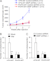
Regulation of MaCSC and metastatic activities by MT1-MMP. A, mean cumulative mammary tumor volume per mouse at different times after 5000 ALDH+ cells from +/PA-MT or PA/PA-MT mice infected with recombinant lentiviruses encoding MT1-MMP shRNA or a control shRNA were injected into mammary fat pads of 8-week-old nude mice (n = 3 for each group). B, 5000 ALDH+ tumor cells from +/PA-MT or PA/PA-MT mice were injected into the tail vein of 8-week-old nude mice (n = 3 for each group). Marimastat were administered orally at a dose of 10 mg/kg daily to the recipient mice right after injection. The micrometastatic nodules in their lungs were quantitated 4 weeks after injection. **, p < 0.01, when the number of nodules after injection of tumor cells from +/PA-MT mice into Marimastat-treated recipient mice compared with that into control recipient mice. C, 5000 ALDH+ tumor cells from +/PA-MT or PA/PA-MT mice were infected with recombinant lentiviruses encoding MT1-MMP shRNA or a control shRNA. Then these cells were injected into the tail vein of 8-week-old nude mice (n = 3 for each group). The micrometastatic nodules in their lungs were quantitated 4 weeks after injection. *, p < 0.05, when the number of nodules after injection of tumor cells from +/PA-MT mice infected with MT1-MMP shRNA virus are compared with that infected with control shRNA virus.
FIGURE 7.
FIGURE 7.
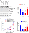
Regulation of EMT and MaCSC activities by endophilin A2 phosphorylation at Tyr-315 in mammary tumor cells. A, freshly isolated primary tumor cells from +/PA-MT or PA/PA-MT mice were infected with recombinant adenoviruses encoding endophilin A2 mutant Y315F or Y315E or GFP as a control. Three days after infection, surface expression of MT1-MMP was determined. Aliquots of the lysates were also analyzed directly by Western blotting with antibodies as indicated. B–D, freshly isolated primary tumor cells from +/PA-MT or PA/PA-MT mice were sorted for ALDH+ cells and then infected with recombinant adenoviruses encoding endophilin A2 mutants Y315F or Y315E or GFP as a control. B, infected ALDH+ cells were subjected to analysis for tumor sphere formation. **, p < 0.01, when compared with the cells from +/PA-MT mice infected with GFP virus. C, mean cumulative mammary tumor volume per mouse at different times after 5000 infected ALDH+ cells were injected into mammary fat pad of nude mice. +/PA-MT ALDH+ cells expressing Y315F versus +/PA-MT ALDH+ cells expressing GFP, p < 0.01 was done by the two-way ANOVA; PA/PA-MT ALDH+ cells expressing Y315E versus PA/PA-MT ALDH+ cells expressing GFP, p < 0.01 was done by the two-way ANOVA. D, number of nodules per lung section 4 weeks after injection of 5000 ALDH+ tumor cells from +/PA-MT or PA/PA-MT mice infected with different adenoviruses as indicated into tail vein of 8-week-old nude mice, **, p < 0.01, when compared with +/PA-MT ALDH+ cells expressing GFP.

Similar articles

Cited by

References

    1. Sjöblom T., Jones S., Wood L. D., Parsons D. W., Lin J., Barber T. D., Mandelker D., Leary R. J., Ptak J., Silliman N., Szabo S., Buckhaults P., Farrell C., Meeh P., Markowitz S. D., Willis J., Dawson D., Willson J. K., Gazdar A. F., Hartigan J., Wu L., Liu C., Parmigiani G., Park B. H., Bachman K. E., Papadopoulos N., Vogelstein B., Kinzler K. W., Velculescu V. E. (2006) The consensus coding sequences of human breast and colorectal cancers. Science 314, 268–274 - PubMed
    1. Vogelstein B., Kinzler K. W. (2004) Cancer genes and the pathways they control. Nat. Med. 10, 789–799 - PubMed
    1. Luo J., Solimini N. L., Elledge S. J. (2009) Principles of cancer therapy: oncogene and non-oncogene addiction. Cell 136, 823–837 - PMC - PubMed
    1. Parsons J. T. (2003) Focal adhesion kinase. The first ten years. J. Cell Sci. 116, 1409–1416 - PubMed
    1. Schlaepfer D. D., Mitra S. K. (2004) Multiple connections link FAK to cell motility and invasion. Curr. Opin. Genet. Dev. 14, 92–101 - PubMed

Publication types

MeSH terms