. 2013 Feb 1;288(5):3655-67.
doi: 10.1074/jbc.M112.428219. Epub 2012 Dec 19.

The sulfonylurea receptor 1 (Sur1)-transient receptor potential melastatin 4 (Trpm4) channel

Affiliations

The sulfonylurea receptor 1 (Sur1)-transient receptor potential melastatin 4 (Trpm4) channel

Seung Kyoon Woo et al. J Biol Chem. .

Abstract

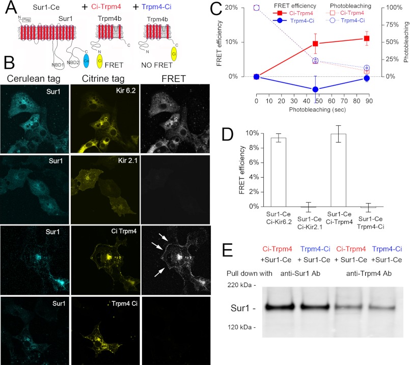
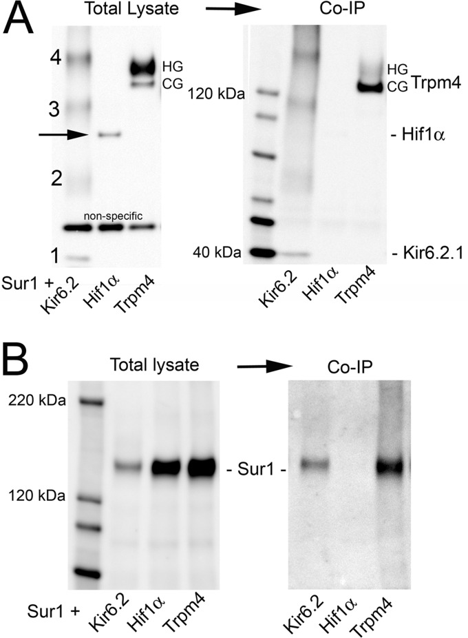
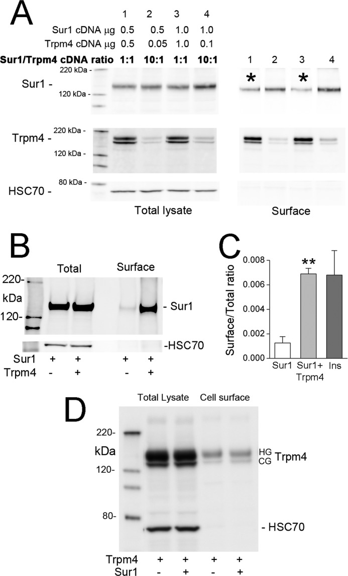
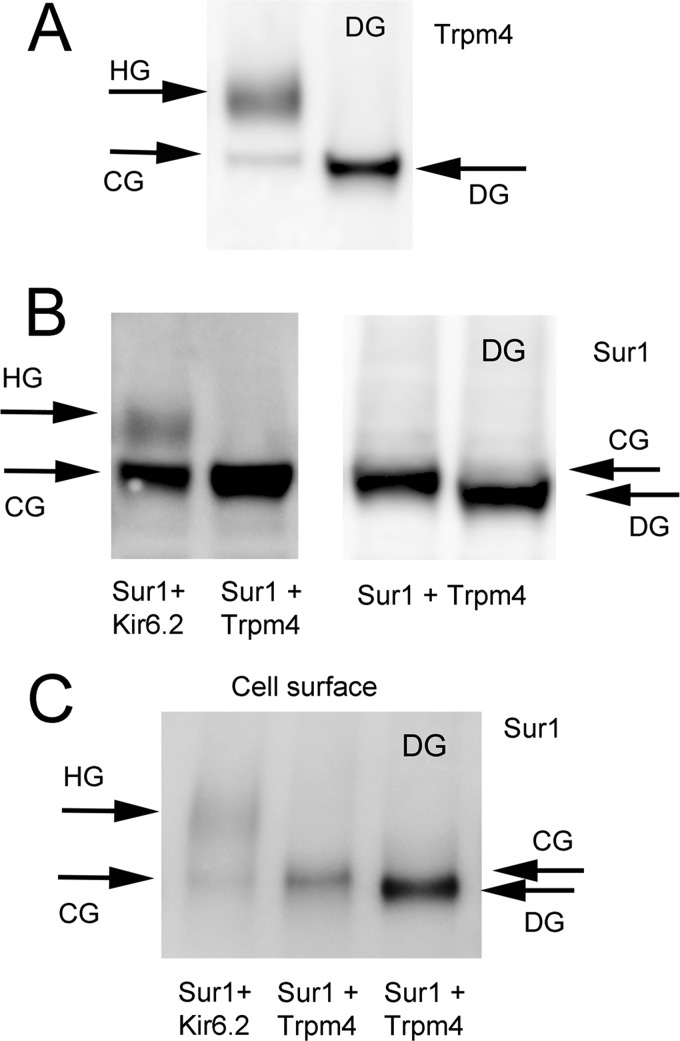
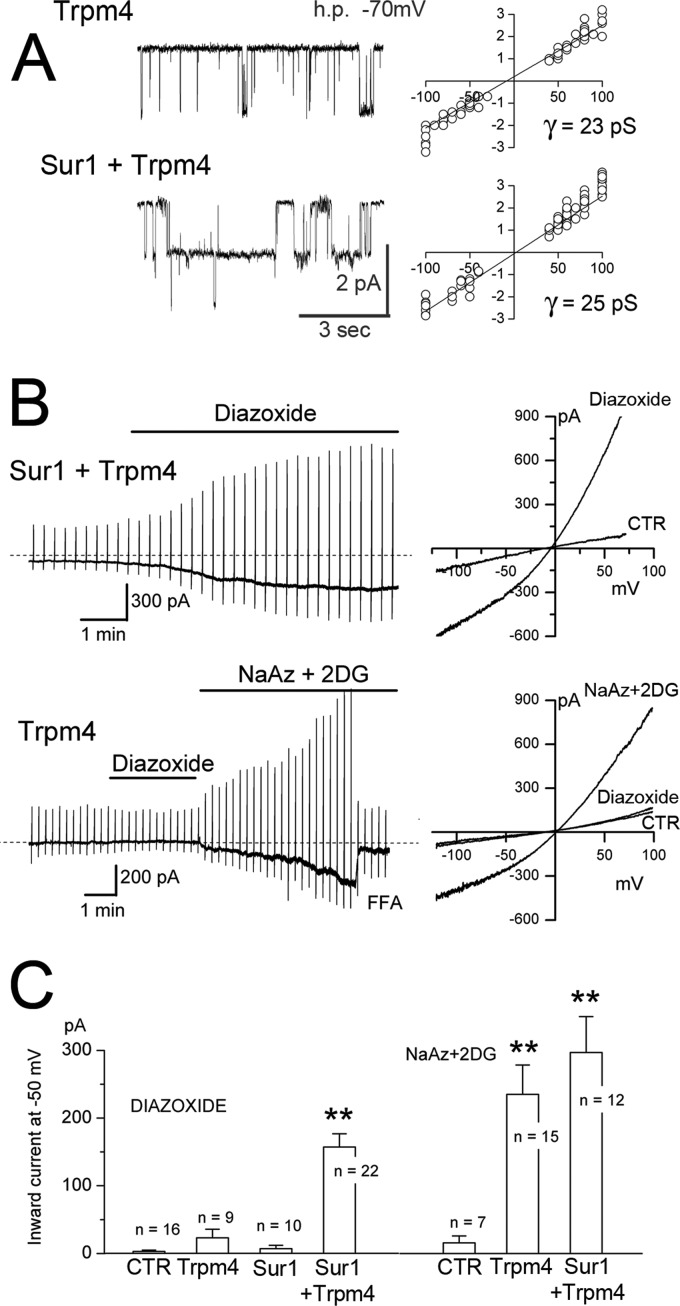
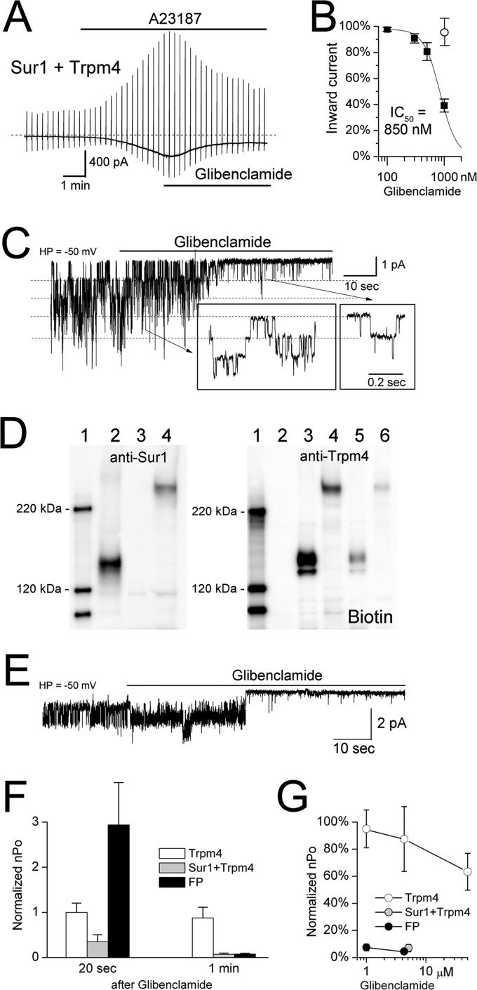
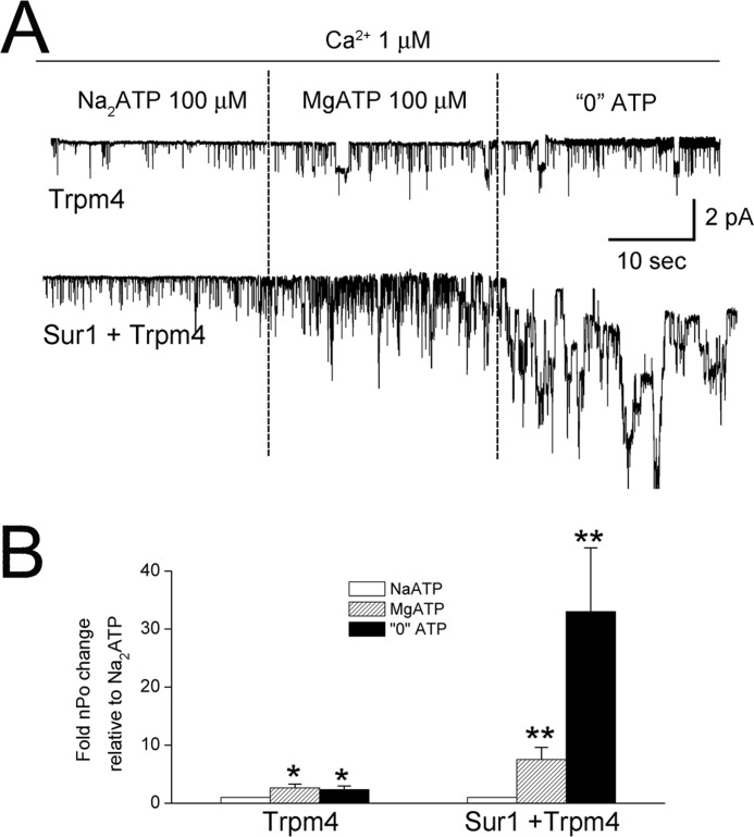
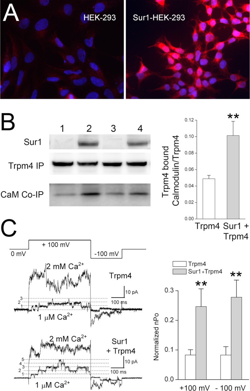
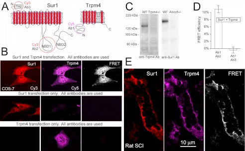
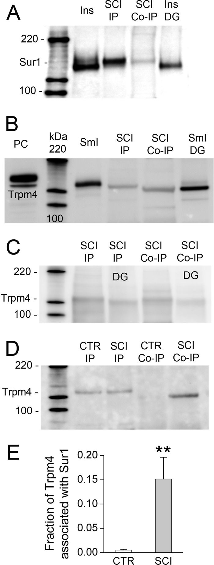
The sulfonylurea receptor 1 (Sur1)-NC(Ca-ATP) channel plays a central role in necrotic cell death in central nervous system (CNS) injury, including ischemic stroke, and traumatic brain and spinal cord injury. Here, we show that Sur1-NC(Ca-ATP) channels are formed by co-assembly of Sur1 and transient receptor potential melastatin 4 (Trpm4). Co-expression of Sur1 and Trpm4 yielded Sur1-Trpm4 heteromers, as shown in experiments with Förster resonance energy transfer (FRET) and co-immunoprecipitation. Co-expression of Sur1 and Trpm4 also yielded functional Sur1-Trpm4 channels with biophysical properties of Trpm4 and pharmacological properties of Sur1. Co-assembly with Sur1 doubled the affinity of Trpm4 for calmodulin and doubled its sensitivity to intracellular calcium. Experiments with FRET and co-immunoprecipitation showed de novo appearance of Sur1-Trpm4 heteromers after spinal cord injury in rats. Our findings depart from the long-held view of an exclusive association between Sur1 and K(ATP) channels and reveal an unexpected molecular partnership with far-ranging implications for CNS injury.


Figures

FIGURE 1.
FIGURE 1.
Evidence from FRET that Sur1 and Trpm4 co-associate. A, schematic diagram of Sur1 and Trpm4 proteins, showing the positions of Ci and Ce fluorescent tags. B, fluorescence images (left and middle columns) of each fluorophore and FRET signal (right column) for COS-7 cells expressing FLAG-Sur1-Ce and Ci-Myc-Kir6.2 (positive control), FLAG-Sur1-Ce and Ci-Myc-Kir2.1 (negative control), FLAG-Sur1-Ce and Ci-Myc-Trpm4, and FLAG-Sur1-Ce and Myc-Trmp4-Ci; FRET within the cell membrane is indicated (arrows); results shown are representative of 3–7 replicates. C and D, average FRET efficiency as a function of time (C) and at steady state (D) for the four conditions in B; n = 32–45; Sur1:Trpm4 plasmid ratio, 16:1. Error bars indicate mean ± S.E. E, immunoblot of COS-7 cells expressing FLAG-Sur1-Ce and Ci-Myc-Trpm4, or FLAG-Sur1-Ce and Myc-Trmp4-Ci, as indicated. Immunoisolation was performed using anti-Sur1-b antibody (anti-Sur1-b Ab) or anti-Trpm4-b antibody (anti-Trpm4-b Ab), as indicated, and proteins were detected with anti-Sur1-a antibody. Sur1:Trpm4 plasmid ratio, 13:1.
FIGURE 2.
FIGURE 2.
Evidence from co-immunoprecipitation (Co-IP) that Sur1 and Trpm4 co-associate. A and B, immunoblots of COS-7 cells expressing FLAG-Sur1 and either Myc-Kir6.2 (positive control) or Myc-Hif1α (negative control) or Myc-Trpm4. Left and right, immunoblots of total lysate (left) and after Co-IP (right), with immunoisolation performed using anti-Sur1-a antibody and protein detected with anti-Myc antibody (A) or with immunoisolation performed using anti-Myc antibody and protein detected with anti-Sur1-a antibody (B). HG and CG, highly glycosylated and core-glycosylated Trpm4, respectively. Results shown are representative of 7 replicates for Sur1-Trpm4 and Sur1-Kir6.2 and 3 replicates for Hif1α. Sur1:Trpm4 plasmid ratio, 1:1.
FIGURE 3.
FIGURE 3.
Surface expression of Sur1 requires Trpm4, but is reduced by overexpression of Trpm4. A, immunoblots of COS-7 cells co-expressing His6-Sur1 and Trpm4-EGFP using different amounts and different ratios of plasmids, as indicated. Left four lanes, immunoblots for Sur1 and Trpm4 of total lysates; Right four lanes, immunoblots for surface expressed protein. Surface expression of Sur1 was assessed using biotinylation of surface proteins, with HSC70 used as a negative control for surface proteins. Note that overexpression of Trpm4 yields a less favorable ratio of Sur1 to Trpm4 at the surface (asterisks). Results shown are representative of 3 replicates; immunoblot was performed using anti-Sur1-a antibody and anti-Trpm4-a antibody. B, immunoblot of COS-7 cells expressing His6-Sur1 with and without co-expression of Myc-Trpm4, as indicated. Left two lanes, total expression of Sur1; right two lanes, surface expression of Sur1; surface expression was assessed as in A. Note that Trpm4 expression is required for appreciable surface expression of Sur1; immunoblot was performed using anti-Sur1-a antibody. Sur1:Trpm4 plasmid ratio, 8:1. C, densitometric analysis of the experiment in B showing the surface-to-total expression ratio (mean ± S.E.) for Sur1 without and with co-expression of Trpm4; n = 3. For comparison, the surface-to-total expression ratio for Sur1 in insulinoma cells (Ins) is also shown. n = 2; **, p < 0.01. D, immunoblots of COS-7 cells expressing Myc-Trpm4 with and without co-expression of His6-Sur1, as indicated. Left two lanes, total expression of Trpm4; right two lanes, surface expression of Trpm4; surface expression was assessed as in A. Note that Sur1 expression has no effect on surface expression of Trpm4. Results shown are representative of 4 replicates; immunoblot was performed using anti-Trpm4-a antibody. Sur1:Trpm4 plasmid ratio, 2:1.
FIGURE 4.
FIGURE 4.
Co-associated Sur1 and Trpm4 are predominantly core-glycosylated. A, immunoblot for Trpm4 of total lysate from COS-7 cells expressing Myc-Trpm4, before (left lane) and after (right lane) deglycosylation with peptide:N-glycosidase F (DG). Note the occurrence of both HG (147 kDa) and CG (135 kDa) forms of Trpm4. Immunoblot was performed using anti-Trpm4-a antibody; results shown are representative of 7 replicates. B, immunoblot for Sur1 of total lysate from COS-7 cells co-expressing FLAG-Sur1 and Myc-Kir6.2, or FLAG-Sur1 and Myc-Trpm4, as indicated, before (three left lanes) and after (far right lane) deglycosylation with peptide:N-glycosidase F (DG). Note the occurrence of both highly glycosylated (HG; 165 kDa) and core-glycosylated (CG; 150 kDa) forms of Sur1 when co-expressed with Kir6.2, but the occurrence of only the CG form of Sur1 when co-expressed with Trpm4. Immunoblot was performed using anti-Sur1-a antibody; results shown are representative of 3–7 replicates. Sur1:Trpm4 plasmid ratio, 1:1. C, immunoblot of surface Sur1 from COS-7 cells co-expressing His6-Sur1 and Myc-Kir6.2, or His6-Sur1 and Myc-Trpm4, as indicated. The third lane is of total lysate after deglycosylation with peptide:N-glycosidase F (DG). Note that with Kir6.2, both glycosylation forms are present at the surface, whereas with Trpm4, the CG form of Sur1 is dominant at the surface. Surface expression of Sur1 was assessed using biotinylation of surface proteins; immunoblot was performed using anti-Sur1-a antibody. Sur1:Trpm4 plasmid ratio, 1.5:1.
FIGURE 5.
FIGURE 5.
Co-expression with Sur1 endows Trpm4 with sensitivity to the Sur1 activator, diazoxide. A, single channel recordings (left) and single channel conductances (right) from inside-out patches from COS-7 cells expressing Myc-Trpm4 alone or co-expressing FLAG-Sur1 and Myc-Trpm4. Conditions were as follows: 11–15 patches per condition; Ca2+, 1 μm; ATP, 0; charge carrier, Cs+. h.p., holding potential; pS, picosiemens. B, representative whole-cell membrane currents in COS-7 cells co-expressing FLAG-Sur1 and Myc-Trpm4, or expressing Myc-Trpm4 alone, as indicated, before and after exposure to diazoxide and, for Trpm4 alone, after ATP depletion. Left and right, currents shown at low (left) and high (right) temporal resolution during ramp pulses (−100 to +100 mV in 500 ms, repeated every 15 s; holding potential, −50 mV). CTR, control cells without transfection. C, magnitude (mean ± S.E.) of the inward current at −50 mV activated by diazoxide (left) or by ATP depletion (right) during experiments performed as in B, in control cells without transfection (CTR), cells expressing Myc-Trpm4 alone, FLAG-Sur1 alone or co-expressing FLAG-Sur1 and Myc-Trpm4; n denotes number of cells tested. The currents were activated by diazoxide (100 μm) or by an ATP-depleting mixture (sodium azide (NaAz), 1 mm plus 2-deoxyglucose (2DG), 10 mm). Charge carrier, Cs+; **, p < 0.01; for all experiments. Sur1:Trpm4 plasmid ratio, 2.3:1.
FIGURE 6.
FIGURE 6.
Co-expression with Sur1 endows Trpm4 with sensitivity to the Sur1 inhibitor, glibenclamide. A, representative whole-cell current from a COS-7 cell co-expressing FLAG-Sur1-Ce and Ci-Myc-Trpm4; the current was activated by A23187 (10 μm) and was inhibited by subsequent application of glibenclamide (1 μm). B, concentration-response data (mean ± S.E., filled squares) for glibenclamide inhibition in COS-7 cells co-expressing FLAG-Sur1-Ce and Ci-Myc-Trpm4 were obtained from whole-cell recordings and were fitted with a standard logistic equation with IC50 = 850 nm; 5–7 cells at each concentration. The empty circle shows the inhibitory effect of 1 μm glibenclamide in cells that expressed Ci-Myc-Trpm4 alone. Whole-cell currents were activated by A23187 (10 μm). Charge carrier, Cs+; HP, holding potential. Sur1:Trpm4 plasmid ratio, 12:1. C, single channel activity in a responsive inside-out patch from a COS-7 cell co-expressing FLAG-Sur1-Ce and Ci-Myc-Trpm4, before and after application of glibenclamide (5 μm), shown at low and high (box insets) temporal resolution. Conditions were as follows: Ca2+, 1 μm; ATP, 0; charge carrier, Cs+. D, immunoblots of total lysate from COS-7 cells expressing His6-Sur1 alone (lane 2), Myc-Trpm4 alone (lane 3), or the His6-Sur1-Trpm4 fusion protein (lane 4), labeled with anti-Sur1 antibody-a (left panel) or anti-Trpm4-a antibody (right panel); also shown is surface expression (Biotin) of Trpm4 alone (lane 5) and of the Sur1-Trpm4 fusion protein (lane 6). Surface expression was assessed using biotinylation of surface proteins; results shown are representative of 4 replicates. E, single channel activity in an inside-out patch from a COS-7 cell expressing the His6-Sur1-Trpm4 fusion protein, before and after application of glibenclamide (5 μm). Conditions were as follows: Ca2+, 1 μm; ATP, 0; charge carrier, Cs+. F, the effect of glibenclamide (1 or 5 μm) on open channel probability (nPo) at 20 s and at 1 min after application of glibenclamide to COS-7 cells expressing Ci-Myc-Trpm4 alone (empty bars), co-expressing FLAG-Sur1-Ce and Ci-Myc-Trpm4 (gray bars), or expressing the His6-Sur1-Trpm4 fusion protein (FP, black bars); 3–5 patches for each condition. G, concentration-response data (mean ± S.E.) for steady-state glibenclamide inhibition in COS-7 cells expressing Ci-Myc-Trpm4 alone (empty circles), co-expressing FLAG-Sur1-Ce and Ci-Myc-Trpm4 (gray circles), or expressing the His6-Sur1-Trpm4 fusion protein (black circles). The same patches as in F plus additional patches for Ci-Myc-Trpm4 alone were used.
FIGURE 7.
FIGURE 7.
Co-expression with Sur1 changes the sensitivity of Trpm4 to Mg2+. A, single channel recordings of inside-out patches from COS-7 cells expressing Ci-Myc-Trpm4 alone or co-expressing FLAG-Sur1-Ce and Ci-Myc-Trpm4, as indicated. Patches were exposed first to Na2-ATP (100 μm), then to Mg2+-ATP (100 μm), and then to 0 ATP. All solutions contained Ca2+ (1 μm). B, bar graph showing the mean (±S.E.) open channel probability (nPo) relative to that in Na2-ATP for the three conditions in A, as indicated. Conditions were as follows: charge carrier, Cs+; 10 patches per condition. Sur1:Trpm4 plasmid ratio, 12:1; *, p < 0.05; **, p < 0.01.
FIGURE 8.
FIGURE 8.
Co-expression with Sur1 increases the sensitivity of Trpm4 to Ca2+. A, immunolabeling for Sur1 in His6-Sur1-overexpressing HEK-293 (Sur1-HEK-293) cells, showing prominent expression in all cells (right panel) as compared with nontransfected HEK-293 cells (left panel). The nuclei were labeled with 4′-6-diamidino-2-phenylindole (DAPI; blue). B, immunoblots for Sur1, Trpm4, and CaM from HEK-293 cells or Sur1-HEK-293 cells transiently transfected to express Myc-Trpm4. Note that the abundance of calmodulin associated with Myc-Trpm4 (CaM Co-IP) is greater in cells co-expressing Sur1 and Trpm4 (lanes 2 and 4; replicate experiments) as compared with cells expressing Trpm4 alone (lanes 1 and 3). Sur1 was immunoblotted using total lysate, and was detected using anti-Sur1-a antibody. Myc-Trpm4 was immunoisolated using anti-Myc antibody, and immunoisolated proteins were detected using anti-Trpm4-a antibody and anti-CaM antibody. Bar graph: densitometric analysis showing the mean (±S.E.) abundance of calmodulin co-immunoprecipitated with Trpm4, normalized to the total amount of Trpm4, in the absence and presence of Sur1 co-expression; n = 6; **, p < 0.01. C, single channel recordings of inside-out patches from HEK-293 cells or Sur1-HEK-293 cells transiently transfected to express Ci-Myc-Trpm4; patches were pulled in a bath solution containing 1 μm Ca2+ to record base-line channel activity, after which the bath solution was changed to one containing 2 mm Ca2+ to maximally activate all channels. The patches were studied using the voltage clamp protocol depicted. Bar graph, mean (±S.E.) open channel probability of Trpm4, observed at +100 mV and at −100 mV, in 1 μm Ca2+ relative to that in 2 mm Ca2+, in cells expressing Trpm4 alone or co-expressing Sur1 and Trpm4. Charge carrier, Cs+; **, p < 0.01.
FIGURE 9.
FIGURE 9.
FRET analysis of Sur1-Trpm4 heteromers in spinal cord tissue after injury. A, schematic diagrams of Sur1 and Trpm4 proteins, showing the positions of epitopes for various antibodies (Ab) that were studied. B, COS-7 cells co-expressing FLAG-Sur1 and Myc-Trpm4, or expressing either one alone, as indicated, were incubated with both anti-Sur1-a and anti-Trpm4-e antibodies followed by both anti-rabbit Cy3-conjugated and anti-goat Cy5-conjugated secondary antibodies. Left and middle columns, fluorescence images of the two chromophores; right column, FRET images of the two chromophores. Results shown are representative of 6 replicates. Note that primary antibodies against Sur1 and Trpm4 do not cross-react. C, spinal cord tissue lysates were obtained from wild-type (WT), Trpm4−/−, and Abcc8−/− mice, as indicated. Anti-Sur1-b antibody and anti-Trpm4-b antibody were used to immunoisolate Sur1 and Trpm4, respectively, from the various tissue lysates; immunoisolated proteins were detected using anti-Sur1-a antibody (anti-Sur1 Ab) and anti-Trpm4-a antibody (anti-Trpm4 Ab). Note that primary antibodies against Sur1 and Trpm4 do not cross-react. D, average FRET efficiencies at steady state for the experiment in B using anti-Trpm4-e antibody (Ab1), detected with anti-goat Cy5-conjugated antibody versus intracellularly directed anti-Sur1-a antibody (Ab2) or extracellularly directed anti-FLAG antibody (Ab3), detected with Cy3-conjugated antibody, in COS-7 cells transfected to express FLAG-Sur1 and Myc-Trpm4; n = 58–64. E, immunolabeling and FRET images of a microvessel 24 h after spinal cord injury. Immunolabeling as in B; results shown are representative of 4 replicates.
FIGURE 10.
FIGURE 10.
Co-immunoprecipitation of Sur1-Trpm4 heteromers from spinal cord tissue after injury. A, immunoblot using anti-Sur1-a antibody of protein immunoisolated from insulinoma cells (Ins, positive control) and from spinal cord 6 h after injury using anti-Sur1-b antibody (SCI IP) or anti-Trpm4-b antibody (SCI Co-IP), and protein immunoisolated from insulinoma cells after treatment with peptide:N-glycosidase F (Ins DG). B, immunoblot using anti-Trpm4-a antibody of total lysate from COS-7 cells expressing Myc-Trpm4 (PC, positive control), of protein immunoisolated from small intestine (SmI, positive control) and spinal cord 6 h after injury using anti-Trpm4-b antibody (SCI IP) or anti-Sur1-b antibody (SCI Co-IP), and protein immunoisolated from small intestine after treatment with peptide:N-glycosidase F (SmI DG). C, immunoblot using anti-Trpm4-d antibody of protein immunoisolated from spinal cord 6 h after injury using anti-Trpm4-b antibody (SCI IP) or anti-Sur1-b antibody (SCI Co-IP), before and after treatment with peptide:N-glycosidase F (DG). Similar results were obtained using anti-Trpm4-b and antiTrpm4-c antibodies for detection. D, immunoblot using anti-Trpm4-a antibody of protein immunoisolated from uninjured spinal cord (CTR) or spinal cord 6 h after injury (SCI) using anti-Trpm4-b antibody (IP) or anti-Sur1-b antibody (Co-IP). Note that co-associated Sur1-Trpm4 is found only after spinal cord injury. E, densitometric analysis showing the mean (±S.E.) abundance of Trpm4 associated with Sur1 in the uninjured spinal cord versus 6 h after spinal cord injury (SCI) (n = 3; **, p < 0.01).

References

    1. Launay P., Fleig A., Perraud A. L., Scharenberg A. M., Penner R., Kinet J. P. (2002) TRPM4 is a Ca2+-activated nonselective cation channel mediating cell membrane depolarization. Cell 109, 397–407 - PubMed
    1. Nilius B., Prenen J., Tang J., Wang C., Owsianik G., Janssens A., Voets T., Zhu M. X. (2005) Regulation of the Ca2+ sensitivity of the nonselective cation channel TRPM4. J. Biol. Chem. 280, 6423–6433 - PubMed
    1. Song M. Y., Yuan J. X. (2010) Introduction to TRP channels: structure, function, and regulation. Adv. Exp. Med. Biol. 661, 99–108 - PubMed
    1. Vennekens R., Nilius B. (2007) Insights into TRPM4 function, regulation and physiological role. Handb. Exp. Pharmacol. 269–285 - PubMed
    1. Guinamard R., Sallé L., Simard C. (2011) The non-selective monovalent cationic channels TRPM4 and TRPM5. Adv. Exp. Med. Biol. 704, 147–171 - PubMed

Publication types

MeSH terms