. 2013 Apr;94(Pt 4):884-895.
doi: 10.1099/vir.0.046219-0. Epub 2012 Dec 19.

In vitro and in vivo replication of influenza A H1N1 WSN33 viruses with different M1 proteins

Affiliations

In vitro and in vivo replication of influenza A H1N1 WSN33 viruses with different M1 proteins

Zhiguang Ran et al. J Gen Virol. 2013 Apr.

Abstract

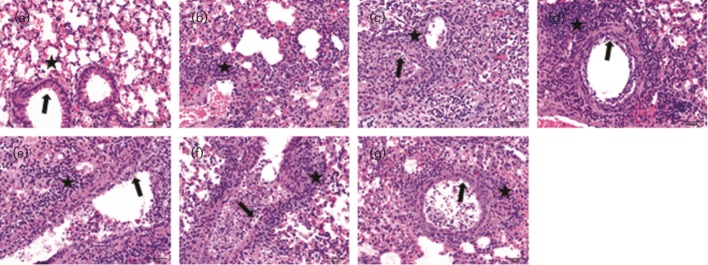
The M1 protein is a major structural protein that has multiple functions in various steps within the life cycle of the influenza A virus (IAV). However, little is currently known about the role of M1 in IAV replication in vivo and the associated pathogenesis. In this study, six isogenic H1N1 WSN33 viruses, constructed to express unique M1 proteins derived from various strains, subtypes or WSN33 itself, were tested to determine in vitro and in vivo functional exchangeability of M1 proteins in the replication and pathogenesis of the WSN33 virus. Despite five chimeric M1 viruses replicating to levels similar to those of the parental WSN33 virus in cell cultures, all M1 chimeras exhibited improved replication and enhanced virulence in mice when compared with the WSN33 virus. Interestingly, M1 proteins derived from swine viruses caused more severe clinical diseases than those from human or quail. These data indicate that the M1 protein is an important determinant of viral replication and pathogenic properties in mice, although the functions of M1 observed in vivo are not adequately reflected in simple infections of cultured cells. Chimeric M1 viruses that are variable in their clinical manifestations described here will aid future understanding of the role of M1 in IAV pathogenesis.


Figures

Fig. 1.
Fig. 1.
Construction of chimeric M segments. (a) Genomic structure of M segment of influenza A/WSN/33 virus (adapted and modified from Palese & Shaw, 2007). Numbering of the nucleotide positions (and in b) is based on the M1 coding sequence of WSN33 virus. Nucleotide positions 1 and 759 correspond, respectively, to the first and last nucleotides coding for M1 ORF. Nucleotides at positions 26 and 715 are splicing donor and acceptor sites, respectively, to generate M2 mRNA (intron is indicated by the V-shaped line). Nucleotide position 982 is the last coding nucleotide for M2 protein. (b) The overlapped coding sequences of M1 and M2 and the splicing site of M2. Nucleotides 715–759 are used to encode the amino acids for both M1 and M2, through a RNA splicing mechanism (Palese & Shaw, 2007). The most C-terminal 13 aa of M1 encoded by nucleotides 716–759 (bold) and the most N-terminal 9 aa of M2 encoded by nucleotides 1–26 and 715 (underlined) are identical among the six different M segments used in this study. The nucleotide at position 715 and upstream of the WSN33 M segment were replaced with its counterpart from each of these five viruses: A/swine/Iowa/15/30 (H1N1), A/Swine/Missouri/4296424/06 (H2N3), A/Swine/Texas/4199-2/98 (H3N2), A/California/04/09 (H1N1) and A/Quail/Hong Kong/G1/97 (H9N2) using the standard PCR and cloning technique, respectively, which does not alter the M2 amino acids of WSN33.
Fig. 2.
Fig. 2.
Replication kinetics of WT rWSN33, recombinant M-segment and chimeric M1 viruses in MDCK cells. The cultured MDCK cells were infected with either WT rWSN33 or the indicated recombinant M-segment (a) or chimeric M1 (b) viruses using an m.o.i. of 0.001. Virus replication following infection of MDCK cells was monitored every 12 h over a period of 3 days by measuring virus infectivity (p.f.u. ml−1) in the culture supernatant. The data shown represents the means±sd of two independent experiments.
Fig. 3.
Fig. 3.
Body weight changes (a) and survival rates (b) in mice infected with the WT rWSN and chimeric M1 viruses. Mice were infected intranasally with 5×10³ p.f.u. of the viruses indicated or mock infected with 50 µl MEM. Body weights were monitored daily up to 14 days p.i. Mice were euthanized after losing 25 % of their initial body weight.
Fig. 4.
Fig. 4.
Viral titres of homogenized lung tissues (a) and microscopic lung lesions (b) of mice infected with the WT rWSN and chimeric M1 viruses on 3 and 5 days p.i. The values are means±sem from each group (* P<0.05; ** P<0.01; *** P<0.001).
Fig. 5.
Fig. 5.
Haematoxylin and eosin staining of microscopic lung sections from mice infected with WT rWSN and M1 chimeric viruses on 5 days p.i. (a) Control: the bronchioles are lined by normal cuboidal epithelium (arrow) and the alveoli are clear (asterisk) in control mice. (b) WT rWSN33 virus: mild bronchiolar epithelial necrosis was seen and the alveolar interstitium is infiltrated by moderate numbers of lymphocytes and neutrophils (asterisk). Chimeric M1 viruses including (c) rWSN-CA09M1, (d) rWSN-IA30M1, (e) rWSN-MO06M1, (f) rWSN-TX98M1 and (g) rWSN-HK97M1: moderate bronchiolar epithelial degeneration and necrosis is visible (arrow) with sloughing of necrotic cells in the lumen. The peribronchiolar and interstitial areas are expanded by moderate numbers of lymphocytes and neutrophils (asterisk). Bars, 50 µm.
Fig. 6.
Fig. 6.
Analysis of M1 and NA proteins. (a, b) M1 protein expression levels in infected MDCK cells and virions harvested at 12 and 24 h p.i. M1 protein bands (resolved by SDS-PAGE and Western blot assay) were analysed by ImageJ software (http://rsbweb.nih.gov/ij/) to quantify the percentage of M1 protein of respective chimeric virus relative to that of WT rWSN virus. The expression level of WT rWSN M1 protein was arbitrarily set at 100 % and the M1 level for each chimeric virus was normalized by comparison with WT rWSN. Data shown are means±sd for three independent experiments. (c) Virion-associated NA activity. MDCK monolayers were infected with chimeric and WT rWSN viruses at a dose of 1 m.o.i. Supernatants were collected at 12 and 24 h p.i., and clarified with low-speed centrifugation. Clarified supernatants were reacted with substrate 2′-(4-methylumbelliferyl)-α-N-acetylneuraminic acid sodium salt hydrate (MUNANA), and relative fluorescence units (RFU) were measured. Data shown are means±sd for three independent experiments.
Fig. 7.
Fig. 7.
Alignment of the complete amino acid sequences of M1 proteins used in this study. Identical residues (*), highly conserved residues (:), and less conserved residues (.) are indicated. Note that amino acid variations at M1 positions 41, 115, 126, 137 and 204 only occur in WSN33 M1, not in the other five M1, and are marked by bold highlighting.

Similar articles

Cited by

References

    1. Akarsu H., Burmeister W. P., Petosa C., Petit I., Müller C. W., Ruigrok R. W., Baudin F. (2003). Crystal structure of the M1 protein-binding domain of the influenza A virus nuclear export protein (NEP/NS2). EMBO J 22, 4646–4655 10.1093/emboj/cdg449 - DOI - PMC - PubMed
    1. Ali A., Avalos R. T., Ponimaskin E., Nayak D. P. (2000). Influenza virus assembly: effect of influenza virus glycoproteins on the membrane association of M1 protein. J Virol 74, 8709–8719 10.1128/JVI.74.18.8709-8719.2000 - DOI - PMC - PubMed
    1. Avalos R. T., Yu Z., Nayak D. P. (1997). Association of influenza virus NP and M1 proteins with cellular cytoskeletal elements in influenza virus-infected cells. J Virol 71, 2947–2958 - PMC - PubMed
    1. Baudin F., Petit I., Weissenhorn W., Ruigrok R. W. (2001). In vitro dissection of the membrane and RNP binding activities of influenza virus M1 protein. Virology 281, 102–108 10.1006/viro.2000.0804 - DOI - PubMed
    1. Black B. L., Rhodes R. B., McKenzie M., Lyles D. S. (1993). The role of vesicular stomatitis virus matrix protein in inhibition of host-directed gene expression is genetically separable from its function in virus assembly. J Virol 67, 4814–4821 - PMC - PubMed

Publication types

MeSH terms

LinkOut - more resources