. 2013 Mar;87(5):2508-17.
doi: 10.1128/JVI.02319-12. Epub 2012 Dec 19.

Cellular entry of human papillomavirus type 16 involves activation of the phosphatidylinositol 3-kinase/Akt/mTOR pathway and inhibition of autophagy

Affiliations

Cellular entry of human papillomavirus type 16 involves activation of the phosphatidylinositol 3-kinase/Akt/mTOR pathway and inhibition of autophagy

Zurab Surviladze et al. J Virol. 2013 Mar.

Abstract

The mammalian target of rapamycin (mTOR) downstream of phosphatidylinositol 3-kinase (PI3K) in the growth factor receptor (GFR) pathway is a crucial metabolic sensor that integrates growth factor signals in cells. We recently showed that human papillomavirus (HPV) type 16 exposure activates signaling from GFRs in human keratinocytes. Thus, we predicted that the virus would induce the PI3K/mTOR pathway upon interaction with host cells. We detected activation of Akt and mTOR several minutes following exposure of human keratinocytes to HPV type 16 (HPV16) pseudovirions. Activated mTOR induced phosphorylation of the mTOR complex 1 substrates 4E-BP1 and S6K, which led to induction of the functional protein translational machinery. Blockade of epidermal GFR (EGFR) signaling revealed that each of these events is at least partially dependent upon EGFR activation. Importantly, activation of PI3K/Akt/mTOR signaling inhibited autophagy in the early stages of virus-host cell interaction. Biochemical and genetic approaches revealed critical roles for mTOR activation and autophagy suppression in HPV16 early infection events. In summary, the HPV-host cell interaction stimulates the PI3K/Akt/mTOR pathway and inhibits autophagy, and in combination these events benefit virus infection.


Figures

Fig 1
Fig 1
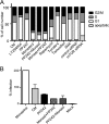
Analysis of the purity of HPV16 PsV preparations. Following generation of HPV16 PsVs via transfection in 293T cells, the particles were subjected to buoyant density centrifugation in CsCl or Optiprep (iodixanol) gradients. The visible bands were collected by side puncture and desalted into HSB buffer using Amicon Ultra-4 100,000 MWCO centrifugation filter units (Millipore). (A) PsV purity and L1 protein content were determined by SDS-10% PAGE and Coomassie brilliant blue staining against bovine serum albumin (BSA) standards. The asterisk (*) may represent an L1 cleavage product. MW, molecular weight (in thousands). (B) Virion morphology and quality of PsVs purified by CsCl gradient visualized by negative staining and transmission electron microscopy. Bar, 100 nm.
Fig 2
Fig 2
Activation of Akt and inhibition of PTEN by HPV16 PsVs. HaCaT cells were serum starved for 4 h. (A) Cells were treated with inhibitors (1 μM AG1478, 25 μM LY294002, or 1 μM wortmannin) or DMSO alone (Ø) for 1 h at 37°C. Cells were then exposed to 100 vge/cell HPV16 PsV for 15 min at 37°C in the presence of inhibitors. Cell lysates were fractionated by SDS-PAGE. Immunoblotting was performed using antibodies to p-Akt (Ser-473) and p-Akt (Thr-308). Serum-starved (4 h) HaCaT cells were incubated with 100 vge/cell HPV16 PsV, and the distribution of p-Akt (B and C) or PTEN (E) was assessed by confocal microscopy at 25 min postexposure. (D) Cells were mock treated or exposed to HPV16 for 25 min or 5 ng/ml EGF for 5 min. Postnuclear supernatants were analyzed by SDS-PAGE and immunoblotting for PTEN or p-PTEN (Ser-380). Numbers at the left of the gel represent kDa. Panel A includes lanes spliced together from the same exposure of the same films. Data are representative of 3 independent assays. (F) Akt and PTEN phosphorylation from panels B, C, and E was quantified with SlideBook; P values were determined by Student's t test.
Fig 3
Fig 3
HPV16 PsVs induce phosphorylation of mTOR and activation of mTORC1 substrates S6K and 4E-BP1. HaCaT cells starved in Tyrode's buffer for 4 h were incubated with HPV16 PsVs. (A) Cells were lysed at the indicated times, and postnuclear supernatants were analyzed by SDS-PAGE and immunoblotting for total mTOR and phosphorylation-mediated activation of mTOR on Ser-2448 and Ser-2481. (B) Cells were treated for 1 h with DMSO (Ø, lane 2) or inhibitors in DMSO (0.5, 1, and 5 μM rapamycin in lanes 3, 4, and 5, respectively; 50, 500, and 5,000 nM PP242 in lanes 6, 7, and 8, respectively) prior to exposure to virus for 30 min. Immunoblot analysis was performed for total mTOR and p-mTOR (Ser-2481). Panels include lanes spliced together from the same exposure of the same films. (C to F) Cells were exposed to HPV16 PsVs for the indicated times before lysis and SDS-PAGE and immunoblot analysis of mTORC1 substrate S6K phosphorylation on Thr-389 (C) and pS6K Ser-371 (D) and substrate 4E-BP1 phosphorylation on Thr-37/46 (E and F); actin (C and D) or total 4E-BP1 (E and F) was detected as loading control. (F) HaCaT cells were pretreated for 1 h with DMSO (Ø), EGFR inhibitors (1 μM AG1478; 0.5 μM PD168393), PI3K inhibitors (25 μM LY294002; 1 μM wortmannin), mTOR inhibitors (3 μM PP242; 1 μM and 5 μM rapamycin), Src inhibitor (5 μM PP2), and a paninhibitor of tyrosine kinases (100 μM genistein) prior to virus exposure for 30 min. (G and H) HPV16 PsV-exposed cell lysates were subjected to a 7-methyl-GTP cap-binding pulldown assay as described in Materials and Methods and analyzed by SDS-PAGE and immunoblotting. (G) Actin was detected as a loading control. (H) Starved cells were incubated with DMSO (Ø), 5 μM PP242, or 1 μM PD168393 for 1 h before addition of HPV16 PsVs and incubation for an additional 1 h in the presence of inhibitors. Immunoblot detection of eIF4G shows equal amounts of this protein in lysates (used for pulldown assay). Panels A, C, E, and F include lanes spliced together from the same exposure of the same films.
Fig 4
Fig 4
HPV16 interaction with keratinocytes suppresses autophagy. HaCaT cells grown in DMEM–10% FCS incubated with 100 vge/cell HPV16 PsVs and incubated at 37°C for 1 h. Controls included cells that were mock exposed to PsV, treated with 5 μM tamoxifen, or treated with 5 mM 3MA. (A) Cell lysates were analyzed by SDS-PAGE and immunoblotting for LC3, with actin detected as a loading control. Densities of LC3-I and LC3-II bands were analyzed with AlphaEaseFC software. (B) The graph represents data from six independent experiments quantifying the LC3-II/LC3-I ratio (two-tailed P value, 0.0145, indicating significance).
Fig 5
Fig 5
Chemical inhibitors have varied effects on HPV16 infectivity. HaCaT cells were pretreated for 1 h with DMSO (Ø) or inhibitors at the indicated concentrations. Cells were exposed to HPV16 PsVs or mock exposed (M), and infectivity was scored after 24 h of incubation at 37°C. (A) Infection in the presence of 5 μM PP2, 50 μM LY294002, 3 μM PP242, 100 μM rapamycin, 5 mM 3MA, or 10 μM tamoxifen. (B and C) Dose-response curves of infection to mTOR inhibitor PP242 (B) or Src kinase inhibitor PP2 (C). The 50% infective concentrations (IC50s) were calculated using GraphPad Prism software. (D) Immunoblot analysis of HPV16 L1 bound to HaCaT cells in the presence or absence of 5 μM PP2, 100 μM genistein, or 3 μM PP242. (E and F) Kinetics of HaCaT cell infection inhibition by PP242 and PP2. Cells were treated with DMSO (Ø), 4 μM PP242 (E), or 5 μM PP2 (F) for the indicated times pre- and postattachment of HPV16 PsVs (100 vge/cell), which was defined as 0 h; infection was analyzed 24 h following HPV exposure.
Fig 6
Fig 6
Autophagy plays a negative role in HPV infection. HaCaT cells were transfected with a control siRNA or siRNAs specific to mTOR, Beclin 1, or Atg7. Downregulation of these proteins was determined in transfected cells by immunoblot analyses using antibodies against mTOR (A) and Beclin 1 and Atg7 (F). Immunoblotting was used to determine the effect of mTOR depletion on p-4E-BP1 (C) and autophagy formation detected by analysis of LC3 (D). The LC3-II/LC3-I ratio (E) was quantified from three independent experiments by densitometry, and P values were determined by Student's t test. Actin (A, D, F) or 4E-BP1 (C) was detected as loading control. Infections were initiated 24 h posttransfection and proceeded for 24 h thereafter under normal conditions (B, G). Panel A includes lanes from the same exposure of the same films.
Fig 7
Fig 7
Cell cycle distribution fails to correlate with HPV16 infection inhibition. (A) HaCaT cells were transfected with mTOR siRNA or treated with drugs (50 μM LY294004, 1 μM PP242, 100 μM monastrol, 1 μM rapamycin, 10 μM tamoxifen, or 5 mM 3MA). Some cells were first incubated for 6 h with 1 μM PP242 and then with 100 μM monastrol (PP242+Monast); other cells were first incubated for 6 h with 100 μM monastrol and then with 1 μM PP242 (Monast+PP242). After 24 h of incubation at 37°C, cells were assayed with propidium iodide and examined using flow cytometry. The fractions of cells in G1 (1n), S (intermediate), and G2/M (2n) phases and apoptotic cells were expressed as percentages of the total cells counted. (B) HPV16 PsV infection levels (24 h postinfection) in the presence of complete media (CM) or inhibitors following pretreatment for 1 h with 100 μM monastrol, pretreatment with monastrol for 1 h plus 3 μM PP242 for the duration (Monast+PP242), or pretreatment with 3 μM PP242 for 1 h plus 100 μM monastrol for the duration (PP242+Monast).

Similar articles

Cited by

References

    1. Surviladze Z, Dziduszko A, Ozbun MA. 2012. Essential roles for soluble virion-associated heparan sulfonated proteoglycans and growth factors in human papillomavirus infections. PLoS Pathog. 8:e1002519 doi:10.1371/journal.ppat.1002519 - DOI - PMC - PubMed
    1. Marsh M, Helenius A. 2006. Virus entry: open Sesame. Cell 124:729–740 - PMC - PubMed
    1. Andjelkovic M, Alessi DR, Meier R, Fernandez A, Lamb NJ, Frech M, Cron P, Cohen P, Lucocq JM, Hemmings BA. 1997. Role of translocation in the activation and function of protein kinase B. J. Biol. Chem. 272:31515–31524 - PubMed
    1. Buchkovich NJ, Yu Y, Zampieri CA, Alwine JC. 2008. The TORrid affairs of viruses: effects of mammalian DNA viruses on the PI3K-Akt-mTOR signalling pathway. Nat. Rev. Microbiol. 6:266–275 - PMC - PubMed
    1. Cooray S. 2004. The pivotal role of phosphatidylinositol 3-kinase-Akt signal transduction in virus survival. J. Gen. Virol. 85:1065–1076 - PubMed

Publication types

MeSH terms