. 2013 Mar;87(5):2475-88.
doi: 10.1128/JVI.00776-12. Epub 2012 Dec 19.

Negevirus: a proposed new taxon of insect-specific viruses with wide geographic distribution

Affiliations

Negevirus: a proposed new taxon of insect-specific viruses with wide geographic distribution

Nikos Vasilakis et al. J Virol. 2013 Mar.

Abstract

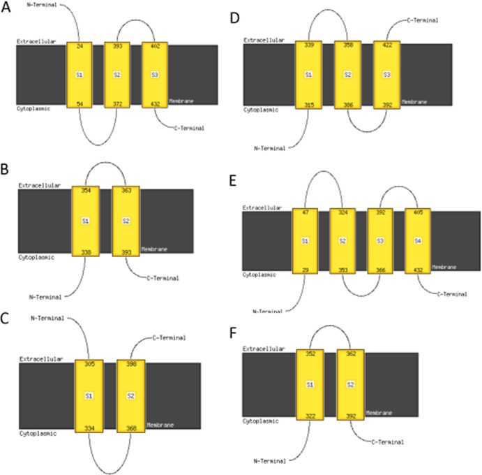
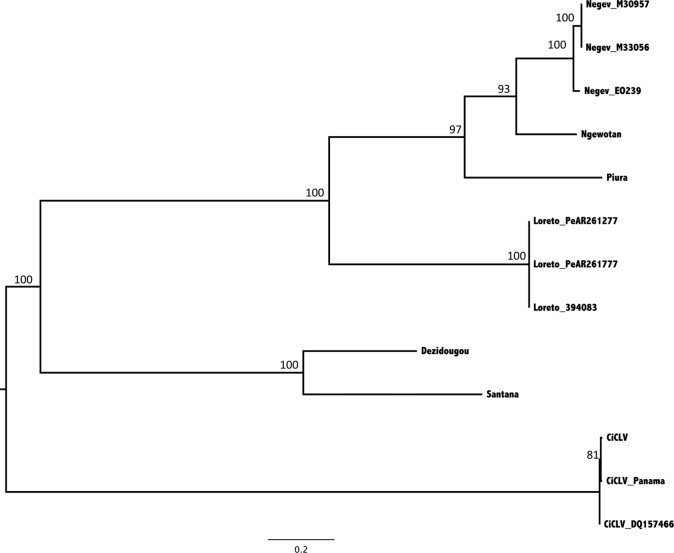
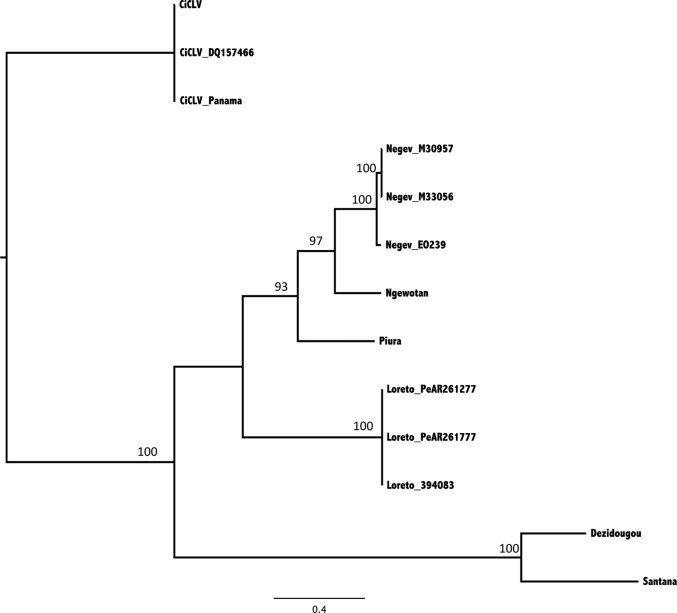
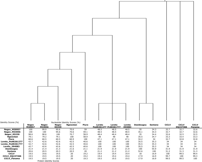
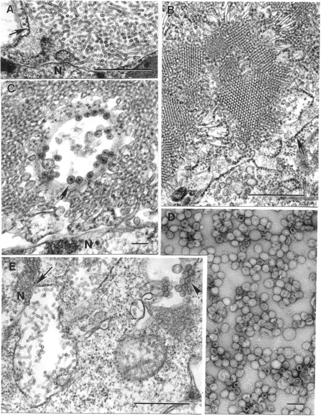
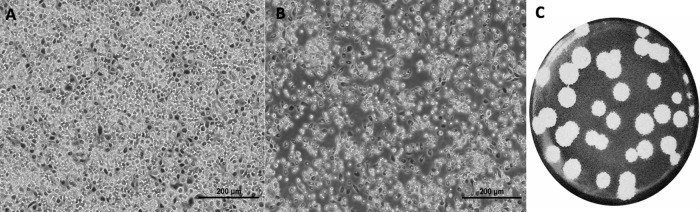
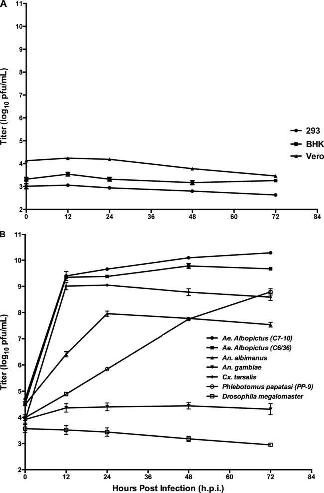
Six novel insect-specific viruses, isolated from mosquitoes and phlebotomine sand flies collected in Brazil, Peru, the United States, Ivory Coast, Israel, and Indonesia, are described. Their genomes consist of single-stranded, positive-sense RNAs with poly(A) tails. By electron microscopy, the virions appear as spherical particles with diameters of ∼45 to 55 nm. Based on their genome organization and phylogenetic relationship, the six viruses, designated Negev, Ngewotan, Piura, Loreto, Dezidougou, and Santana, appear to form a new taxon, tentatively designated Negevirus. Their closest but still distant relatives are citrus leposis virus C (CiLV-C) and viruses in the genus Cilevirus, which are mite-transmitted plant viruses. The negeviruses replicate rapidly and to high titer (up to 10(10) PFU/ml) in mosquito cells, producing extensive cytopathic effect and plaques, but they do not appear to replicate in mammalian cells or mice. A discussion follows on their possible biological significance and effect on mosquito vector competence for arboviruses.


Figures

Fig 1
Fig 1
Genome organization and position of the open reading frames (A) and the conserved protein domains (B) for Negev virus strain EO239. All 5 identified viruses showed similar genome organization and protein domains.
Fig 2
Fig 2
Predicted transmembrane domains and the orientation of ORF2 by MAMSAT-SVM for Negev virus (A), Piura virus (B), Loreto virus (C), Dezidougou virus (D), Santana virus (E), and Ngewotan virus (F).
Fig 3
Fig 3
Analysis of genomic RNA of NEGV and PIUV labeled with [3H]uridine in the presence of dactinomycin (ActD) for 12 h. Both viruses were purified via rate-zonal centrifugation. Viral RNA was analyzed by agarose gel electrophoresis. Lane 1, mock treatment; 2, NEGV; 3, PIUV.
Fig 4
Fig 4
Phylogenetic trees produced using maximum-likelihood methods of the 10 genomes determined in the study plus three genomes of CiCLV. The trees were rooted using the CiCLV viruses as an outgroup. The region of the genome corresponds to nt 626 to 2908 (Negev EO239), which corresponds to the helicase region of the genome. The model used was the TrN+G model with 1,000 bootstrap replications. Bootstrap replications are presented on the major branches.
Fig 5
Fig 5
Phylogenetic trees produced using maximum-likelihood methods of the 10 genomes determined in the study plus three genomes of CiCLV. The trees were midpoint rooted. The region of the genome corresponds to nt 4316 to 7309 (Negev virus EO239), which corresponds with the RNA-dependent RNA polymerase of the genome. The model used was the GTR+G model with 1,000 bootstrap replications. Bootstrap replications are presented on the major branches.
Fig 6
Fig 6
Cladistic tree showing the relationships between the viruses along with the nucleotide identity and the protein identity for ORF1 of the nine viruses and the RNA species one of CiCLV. Alignments were performed as proteins and then toggled back to nucleotide forms. The branch lengths of the tree do not reflect genetic distance but have the same topology as the trees shown in Fig. 3 and 4.
Fig 7
Fig 7
Transmission electron microscopy analysis of infected cells and purified suspensions. (A) Expanded perinuclear space (the arrow indicates its membrane) of an Ae. albopictus C6/36 cell infected with Negev (EO239) virus is filled with microtubules 20 nm in diameter and up to 160 nm long. Bar, 0.5 μm. (B) Portion of a tremendous perinuclear space-granular endoplasmic reticulum extension loaded with microtubules forming paracrystalline arrays in cross sections in a C6/36 cell infected with ArA 20086 virus. The arrow indicates a limiting membrane with ribosomes at the outer surface. Bar, 0.5 μm. (C) Cytopathic vacuole with spherules at its periphery (arrow) surrounded by microtubules in a perinuclear space of a C6/36 cell infected with ArA 20086 virus. Bar, 100 nm. (D) Negatively stained (2% uranyl acetate) suspension of purified suspension of P60 virus contains particles mostly ∼50 nm in diameter. Bar, 100 nm. (E) Expanded perinuclear space (arrow) filled with microtubules and a cytoplasmic vacuole with spherules (arrowhead) in cytoplasm of a C6/36 cell infected with JKT-9982 virus. N, host cell nucleus. Bar, 0.5 μm.
Fig 8
Fig 8
Cytopathic effects of Negev virus (EO239) infection in C7/10 cells. (A) Mock-infected C7/10 monolayers observed with bright-field microscopy at 12 hpi; (B) NEGV-infected C7/10 cells at an MOI of 10 at 12 hpi observed with bright-field microscopy; and (C) representative plaques of Negev virus-infected C7/10 cells 36 hpi. Cells were fixed with 10% formalin and stained with crystal violet dye.
Fig 9
Fig 9
Comparative replication curves of prototype Negev virus (EO239). (A) Virus outputs from 12 to 72 h following infection at an MOI of 10 by Negev virus EO239 in the vertebrate cell lines Vero (African green monkey kidney), BHK-21 (baby hamster kidney), and HEK293 (human embryonic kidney). (B) Virus outputs from 12 to 72 h following infection at an MOI of 10 by Negev virus in the insect cell lines Ae. albopictus (C6/36 and C7/10), An. albimanus, An. gambiae, Cx. tarsalis, P. papatasi, and D. melanogaster. The limit of detection of the assay is 1.0 log10 PFU/ml.

References

    1. Kuno G. 2004. A survey of the relationships among the viruses not considered arboviruses, vertebrates, and arthropods. Acta Virol. 48:135–143 - PubMed
    1. Hoshino K, Isawa H, Tsuda Y, Yano K, Sasaki T, Yuda M, Takasaki T, Kobayashi M, Sawabe K. 2007. Genetic characterization of a new insect flavivirus isolated from Culex pipiens mosquito in Japan. Virology 359:405–414 - PubMed
    1. Igarashi A, Harrap KA, Casals J, Stollar V. 1976. Morphological, biochemical, and serological studies on a viral agent (CFA) which replicates in and causes fusion of Aedes albopictus (Singh) cells. Virology 74:174–187 - PubMed
    1. Stollar V, Thomas VL. 1975. An agent in the Aedes aegypti cell line (Peleg) which causes fusion of Aedes albopictus cells. Virology 64:367–377 - PubMed
    1. Marklewitz M, Handrick S, Grasse W, Kurth A, Lukashev A, Drosten C, Ellerbrok H, Leendertz FH, Pauli G, Junglen S. 2011. Gouleako virus isolated from West African mosquitoes constitutes a proposed novel genus in the family Bunyaviridae. J. Virol. 85:9227–9234 - PMC - PubMed

Publication types

MeSH terms

LinkOut - more resources