. 2013 Mar;87(5):2721-34.
doi: 10.1128/JVI.02756-12. Epub 2012 Dec 19.

Tra2-mediated recognition of HIV-1 5' splice site D3 as a key factor in the processing of vpr mRNA

Affiliations

Tra2-mediated recognition of HIV-1 5' splice site D3 as a key factor in the processing of vpr mRNA

Steffen Erkelenz et al. J Virol. 2013 Mar.

Abstract

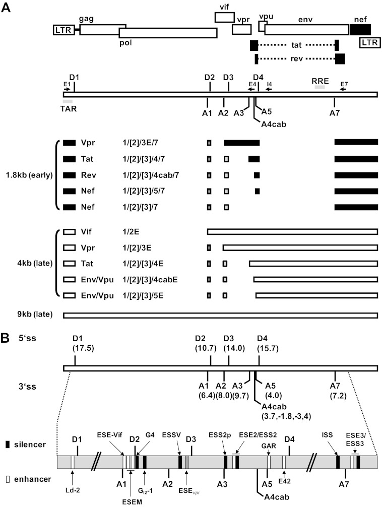
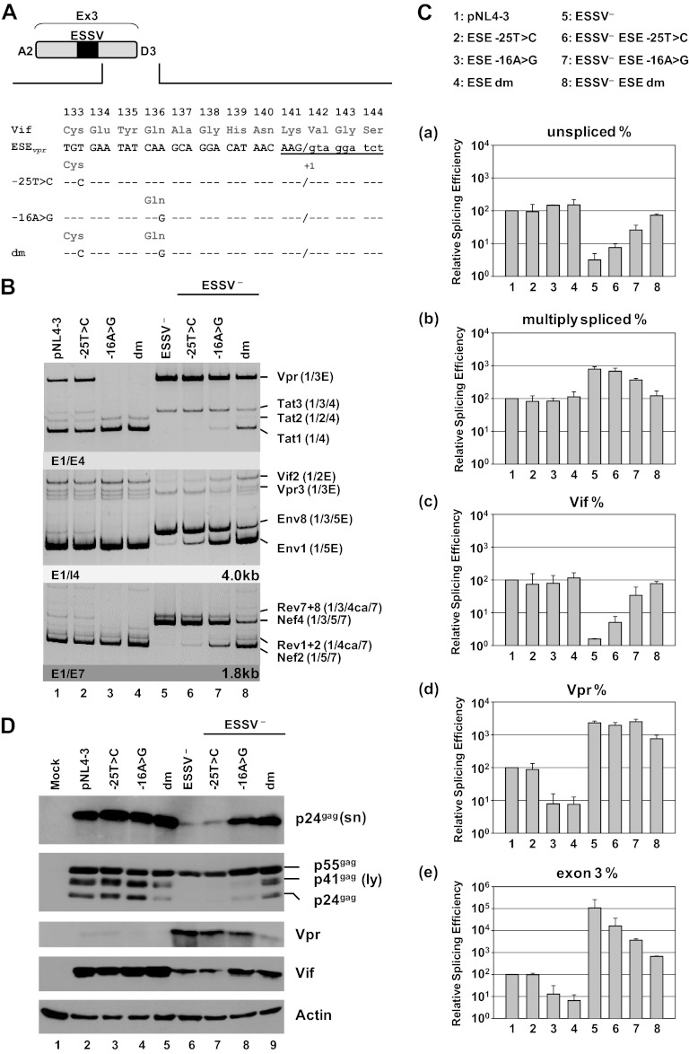
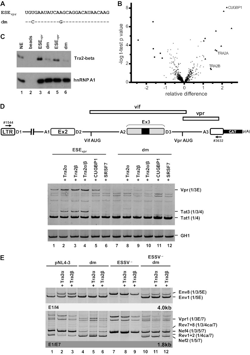
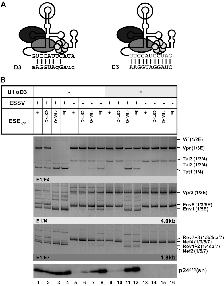
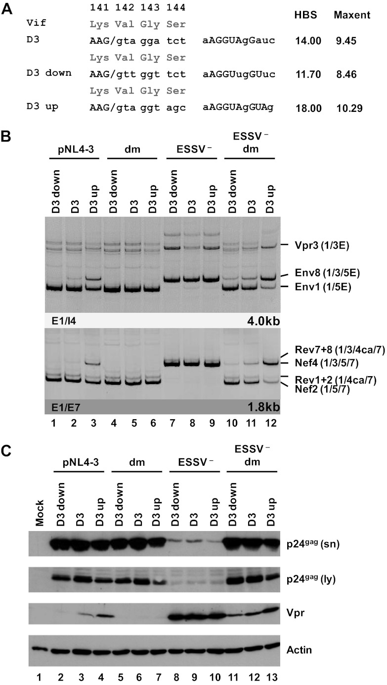
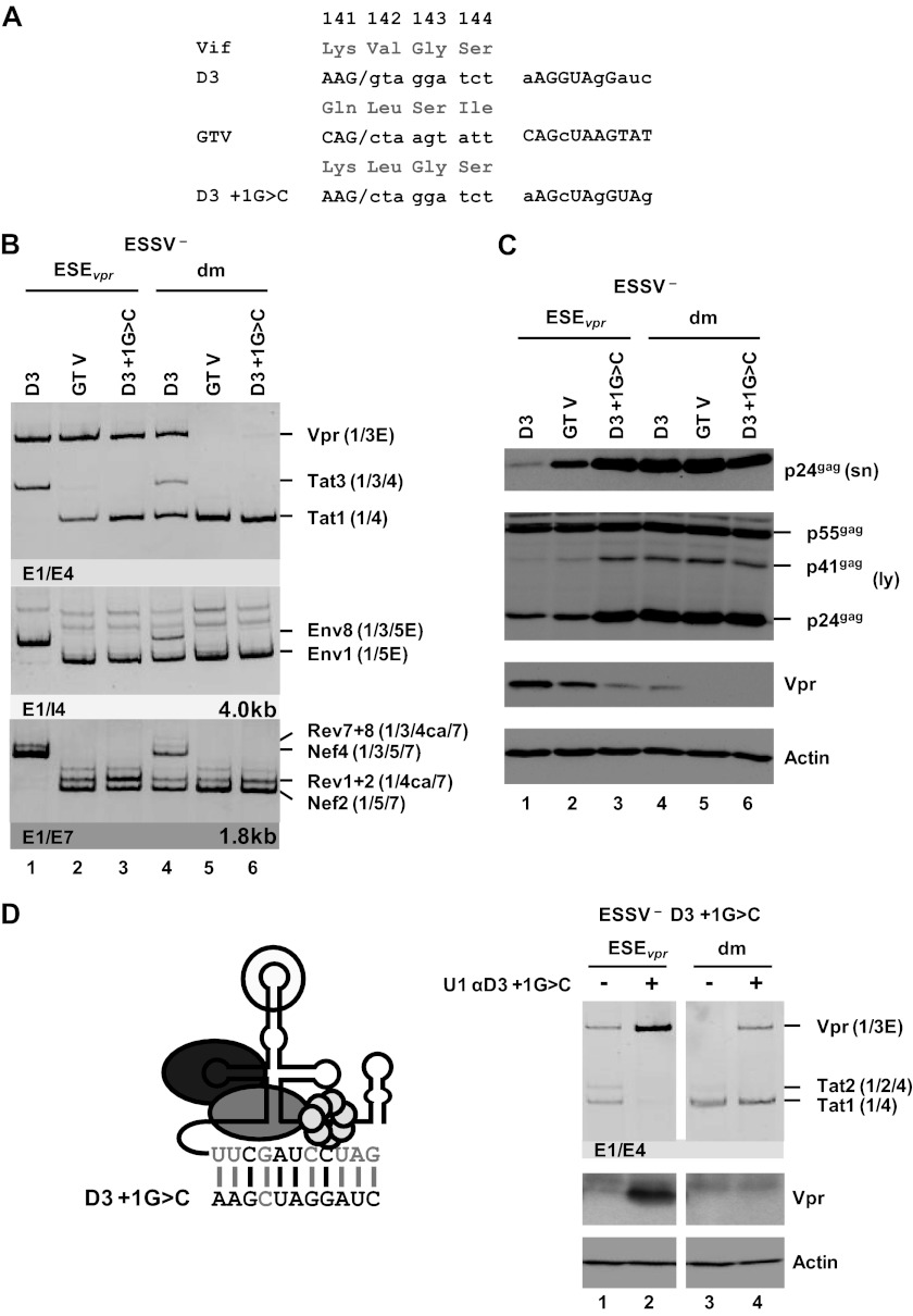
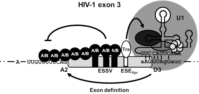
Small noncoding HIV-1 leader exon 3 is defined by its splice sites A2 and D3. While 3' splice site (3'ss) A2 needs to be activated for vpr mRNA formation, the location of the vpr start codon within downstream intron 3 requires silencing of splicing at 5'ss D3. Here we show that the inclusion of both HIV-1 exon 3 and vpr mRNA processing is promoted by an exonic splicing enhancer (ESE(vpr)) localized between exonic splicing silencer ESSV and 5'ss D3. The ESE(vpr) sequence was found to be bound by members of the Transformer 2 (Tra2) protein family. Coexpression of these proteins in provirus-transfected cells led to an increase in the levels of exon 3 inclusion, confirming that they act through ESE(vpr). Further analyses revealed that ESE(vpr) supports the binding of U1 snRNA at 5'ss D3, allowing bridging interactions across the upstream exon with 3'ss A2. In line with this, an increase or decrease in the complementarity of 5'ss D3 to the 5' end of U1 snRNA was accompanied by a higher or lower vpr expression level. Activation of 3'ss A2 through the proposed bridging interactions, however, was not dependent on the splicing competence of 5'ss D3 because rendering it splicing defective but still competent for efficient U1 snRNA binding maintained the enhancing function of D3. Therefore, we propose that splicing at 3'ss A2 occurs temporally between the binding of U1 snRNA and splicing at D3.


Figures

Fig 1
Fig 1
Alternative splicing of the HIV-1 pre-mRNA. (A) Schematic of the HIV-1 genome. The ORFs are indicated by open boxes. The LTRs are located at both ends of the provirus. All HIV-1 proteins are encoded in a single primary RNA. Alternative splicing allows all viral proteins to be efficiently translated within the host cell. The 5′ (SD) and 3′ (SA) splice sites are depicted. Alternatively spliced noncoding exons 2 and 3 within Rev-independent (1.8-kb size class) and Rev-dependent (4-kb size class) spliced mRNAs are shown as boxes (exon 2, dark gray; exon 3, light gray). The positions of the primers used in RT-PCRs for the analyses of viral mRNA splicing are indicated by arrows (E1 [fwd], exon1; E4 [rev], exon 4; I4 [rev], intron 4; E7 [rev], exon 7). (B) Intrinsic strength of the 5′ss (D1 to D4) and 3′ss (A1 to A7) distributed along the HIV-1 pre-mRNA. Each value in parentheses reflects the predicted intrinsic strength (5′ss, HBond score [www.uni-duesseldorf.de/rna]; 3′ss, MaxEnt score [http://genes.mit.edu/burgelab/maxent/Xmaxentscan_scoreseq_acc.html]). The nomenclature of the viral splice sites is from reference . Positions of known enhancer (white) and silencer (black) sequences within the HIV-1 pre-mRNA are shown. Exon 3 is flanked by 3′ss A2 and 5′ss D3. The positions of the Ld-2 (35), ESE-Vif (36), ESEM (14), G4 (36), GI2-1 (M. Widera, and H. Schaal, submitted for publication), ESSV (–13), ESS2p (37), ESE2 (38, 39), ESS2 (–42), GAR, guanosine-adenosine rich (GAR) ESE (16, 17, 27), E42 (27), ISS (15), ESE3 (43), and ESS3 (–; adapted from references and 46) sequences are shown.
Fig 2
Fig 2
ESEvpr is necessary for exon 3 inclusion and vpr mRNA processing. (A) The wild-type ESEvpr sequence and the amino acid sequence encoded by the overlapping vif ORF are shown below exon 3. Mutated ESEvpr nucleotide residues are denoted by their positions relative to the GT dinucleotide of viral 5′ss D3. The black box represents the upstream ESSV. Uppercase letters represent exonic positions, and lowercase letters represent intronic positions. (B) HEK 293T cells (2.5 × 10⁵) were transiently transfected with 1 μg of each of the proviral plasmids. At 30 h after transfection, total-RNA samples were collected and used for RT-PCR analyses with different sets of primer pairs. HIV-1 mRNA species are indicated to the right of the gels in accordance with the nomenclature published previously (5). (C) cDNA samples were prepared as described for panel B and used in real-time PCR assays to specifically quantitate the relative abundances of unspliced (a), multiply spliced (b), Vif (c), and Vpr (d) mRNA species and exon 3 inclusion ratios (e). For normalization, primers 3387 and 3388 were used to detect the total viral mRNA content of each sample. Data represent expression ratios relative to that of wild-type pNL4-3 (bar 1), which was set to 100%. Values and error bars show the average ± standard deviation of three independent transfection experiments. Bars correspond to lanes in panel B. (D) HEK 293T cells (2.5 × 10⁵) were transiently transfected with 1 μg of each of the proviral plasmids. At 48 h posttransfection, viral supernatants were collected, layered onto 20% sucrose solution, and centrifuged at 28,000 rpm for 90 min at 4°C to pellet the released virions. In addition, cells were harvested and resuspended in lysis buffer. Supernatants and cellular lysates were resolved by 12% SDS-PAGE and electroblotted onto nitrocellulose membranes. To determine virus particle production and the expression of viral proteins, samples were probed with primary antibodies specifically detecting structural p24gag (CA) and the viral infectivity factors Vif and Vpr. Equal amounts of cell lysates were controlled for by the detection of α-actin. E, extended exon; dm, double mutation; sn, supernatant; ly, lysate.
Fig 3
Fig 3
ESEvpr is bound by the splicing factors Tra2-alpha and Tra2-beta. (A) In vitro-transcribed RNA substrates used for RNA pulldown experiments (dm, double mutation). (B) Volcano plot of RNA binding proteins purified by RNA pulldown with a nonmutated or a mutated ESEvpr sequence with HeLa cell nuclear extract. The precipitated proteins were digested with trypsin and subjected to quantitative mass spectrometry analysis. The x axis of the volcano plot shows the relative difference in protein abundance as calculated by the SAM method, whereas the y axis shows the −log t-test P value of the groupwise comparison of protein abundances. Besides the majority of probably unspecifically binding proteins (circles), some proteins preferably bound to the wild-type ESEvpr sequence (triangles) or the mutated ESEvpr variant (squares). The proteinsTra2-alpha and Tra2-beta were selected for validation experiments. (C) Immunoblot analysis with an antibody specific for Tra2-beta and hnRNPA1 confirmed significantly smaller amounts of Tra2-beta for the double mutant. (D) HeLa cells (2.5 × 10⁵) were transiently cotransfected with 1 μg of each of the HIV-1-based LTR ex2 ex3 splicing reporters, 0.2 μg of SVctat (47); 1 μg of pXGH5 (GH1) as a transfection control, and 1 μg of pcDNA3.1(+), an expression plasmid for Tra2-alpha, Tra2-beta, CUGBP1, and SRSF7. At 30 h posttransfection, total RNA was isolated and subjected to semiquantitative RT-PCR analyses with primers 1544 and 3632. For measurement of equal transfection efficiencies, a separate PCR was carried out with a primer pair (1224/1225) specific for human growth hormone 1 (GH1). (E) RT-PCR analyses of intronless (2-kb) and intron-containing (4-kb) viral mRNA species following the transient transfection of HEK 293T cells with 1 μg of the respective proviral construct and 1 μg of pcDNA3.1(+), an expression plasmid for either Tra2-alpha or Tra2-beta. E, extended exon; dm, double mutation.
Fig 4
Fig 4
Coexpression of a modified U1 snRNP with full complementarity to 5′ss D3 induces HIV-1 exon 3 splicing and vpr mRNA expression. (A) Schematic drawing of a 5′-end-modified U1 snRNA (right) perfectly matching the 5′ss D3 sequence. Mutated nucleotides are indicated by gray capital letters. Additional base pairing interactions between 5′ss D3 and the optimized 5′ end of the U1 snRNA are indicated by vertical gray lines. (B) HEK 293T cells (2.5 × 10⁵) were transiently cotransfected with 1 μg of both a proviral plasmid and a U1 snRNA expression plasmid. Total RNA was isolated and subjected to RT-PCR analyses. PCR products were resolved by PAGE and stained with ethidium bromide. RT-PCR samples are shown at the top. The main viral mRNA species are indicated on the right. Viral supernatants were collected as well and analyzed for viral p24gag concentrations by immunoblotting (bottom). E, extended exon; dm, double mutation; sn, supernatant.
Fig 5
Fig 5
5′ss D3 up and down mutations modulate HIV-1 exon 3 splicing and vpr mRNA formation. (A) Silent mutations predicted to decrease or increase the complementarity to the 5′ end of the endogenous U1 snRNA were introduced into viral 5′ss D3. Exonic nucleotides are denoted in uppercase letters, and intronic nucleotides are denoted in lowercase letters. Complementarity and predicted intrinsic strength by HBond score (HBS) and MaxEnt score algorithms are both shown next to the 5′ss sequence. Nucleotides complementary to the U1 snRNA are in capital letters, while mismatches to the U1 snRNA are in lowercase letters. (B) HEK 293T cells (2.5 × 10⁵) were transiently transfected with 1 μg of each of the different infectious clones. RNA was isolated from the cells, DNase I digested, and reverse transcribed. The resultant cDNA served as the DNA template in semiquantitative PCRs using primer pairs E1/I4 and E1/E7 to specifically detect viral 4.0- and 1.8-kb viral mRNAs, respectively. Proviral mutants are shown above the panels. The main HIV-1 mRNA species are indicated at the right. (C) Protein lysates and viral supernatants were collected from HEK 293T cells transfected with 1 μg of pNL4-3 or mutant derivatives. Samples were loaded on 12% SDS-polyacrylamide gels and, after separation, transferred to nitrocellulose membranes. Viral proteins and α-actin (as a loading control) were determined by probing with specific primary antibodies. For detection, appropriate HRP-conjugated antibodies and ECL detection reagent were applied. HBS, HBond score; MaxEnt, MaxEnt score; dm, double mutation; E, extended exon; sn, supernatant; ly, lysate.
Fig 6
Fig 6
U1 snRNP binding to a splicing-incompetent 5′ss enhances vpr mRNA expression. (A) 5′ss D3 was replaced with a splicing-incompetent sequence that perfectly matches the free 5′ end of the cellular U1 snRNA except for position +1 (GTV). As a control, 5′ss D3 was disabled for splicing by a G-to-C mutation at position +1, decreasing its complementarity to the U1 snRNA (D3+1G>C). Complementarity patterns are shown next to the 5′ss sequences. Matches to the U1 snRNA are indicated by uppercase letters, and residues not complementary are in lowercase letters. (B) HEK 293T cells (2.5 × 10⁵) were transiently transfected with 1 μg of each of the proviral constructs and analyzed by semiquantitative RT-PCR. RT-PCR products were resolved by PAGE, followed by ethidium bromide staining. Mutants are depicted at the top. Main viral mRNAs are indicated on the right. (C) Cellular lysates and viral supernatants were obtained from transfected HEK 293T cells and loaded onto 12% SDS-polyacrylamide gels. After transfer to nitrocellulose membranes, viral proteins were determined with specific antibodies for p24gag and Vpr. To ensure the loading of equal protein amounts, the membrane was also probed with an antibody to cellular α-actin. (D) Schematic drawing of a 5′-end-modified U1 snRNA perfectly matching the 5′ss D3+1G>C sequence (left). Mutated nucleotides are indicated by gray capital letters. Additional base pairing interactions between 5′ss D3 and the optimized 5′ end of the U1 snRNA are indicated by vertical gray lines. HEK 293T cells (2.5 × 10⁵) were transiently transfected with 1 μg of both proviral pNL4-3 DNA and U1 snRNA expression plasmid. Total RNA and cellular lysates were isolated and subjected to RT-PCR or Western blot analysis (right). E, extended exon; dm, double mutation; sn, supernatant; ly, lysate.
Fig 7
Fig 7
HIV-1 exon 3 splicing is under the combined control of ESSV and ESEvpr. Splice site recognition at HIV-1 exon 3 is regulated by ESSV and a novel exonic splicing enhancer (ESEvpr) embedded in the region upstream of 5′ss D3. ESSV is associated with hnRNP A/B proteins, which may multimerize along the 5′ end of exon 3, occluding 3′ss A2. Tra2-alpha and Tra2-beta bind to the ESEvpr sequence, potentially enhancing recruitment of the U1 snRNP to 5′ss D3, which in turn may promote interactions across the upstream exon and activation of 3′ss A2. U1, U1 snRNP.

Similar articles

Cited by

References

    1. Frankel AD, Young JA. 1998. HIV-1: fifteen proteins and an RNA. Annu. Rev. Biochem. 67:1–25 - PubMed
    1. Kozak M. 2002. Pushing the limits of the scanning mechanism for initiation of translation. Gene 299:1–34 - PMC - PubMed
    1. Anderson JL, Johnson AT, Howard JL, Purcell DF. 2007. Both linear and discontinuous ribosome scanning are used for translation initiation from bicistronic human immunodeficiency virus type 1 env mRNAs. J. Virol. 81:4664–4676 - PMC - PubMed
    1. Krummheuer J, Johnson AT, Hauber I, Kammler S, Anderson JL, Hauber J, Purcell DF, Schaal H. 2007. A minimal uORF within the HIV-1 vpu leader allows efficient translation initiation at the downstream env AUG. Virology 363:261–271 - PubMed
    1. Purcell DF, Martin MA. 1993. Alternative splicing of human immunodeficiency virus type 1 mRNA modulates viral protein expression, replication, and infectivity. J. Virol. 67:6365–6378 - PMC - PubMed

Publication types

LinkOut - more resources