. 2012 Aug;8(3):217-223.
doi: 10.2174/157339612802139389.

Lung Ultrasound in the Critically Ill Neonate

Affiliations
Free PMC article

Lung Ultrasound in the Critically Ill Neonate

Daniel A Lichtenstein et al. Curr Pediatr Rev. 2012 Aug.
Free PMC article

Abstract

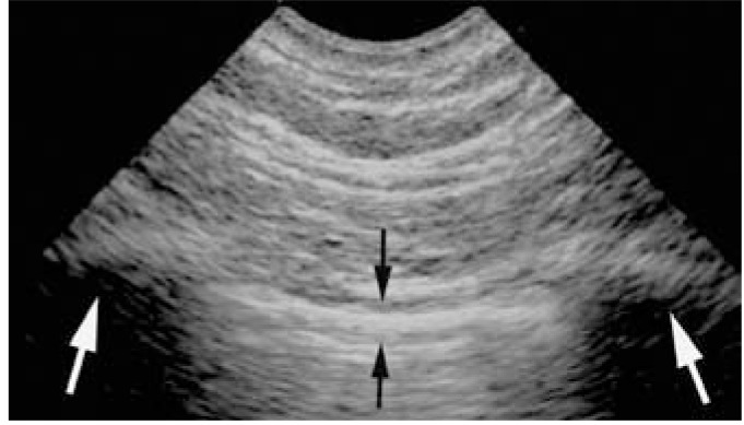
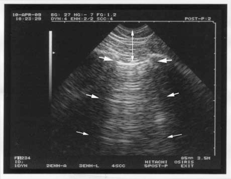
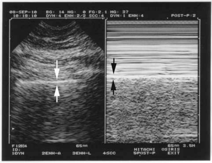
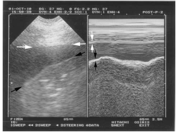
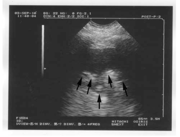
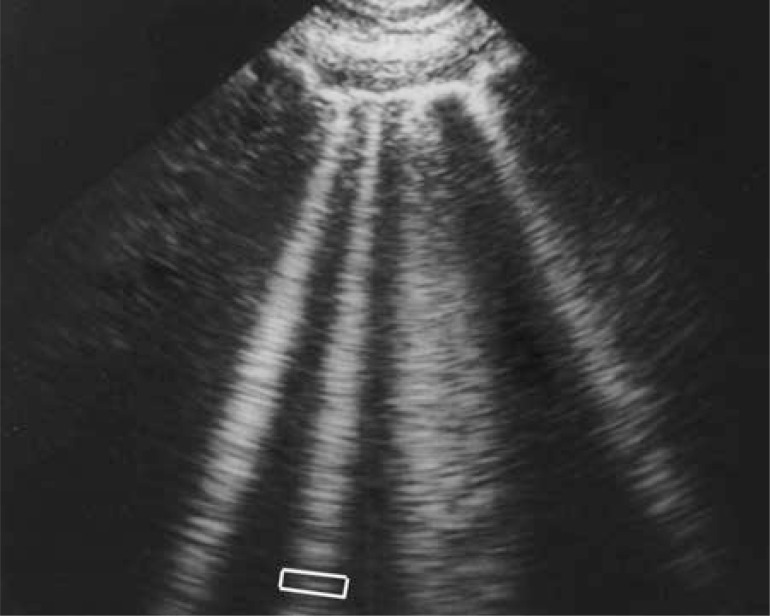
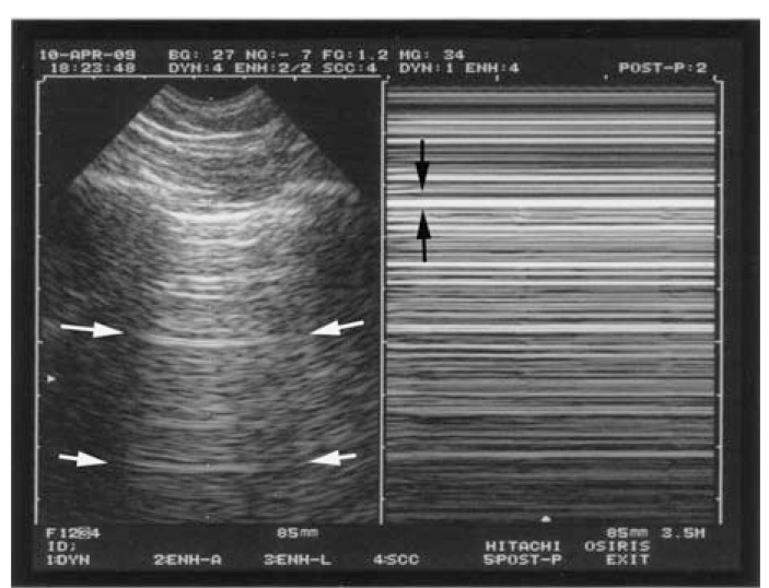
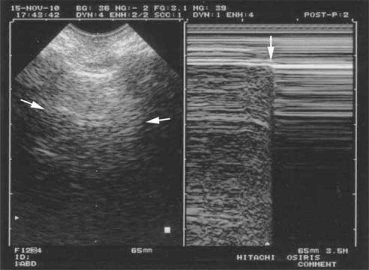
Critical ultrasound is a new tool for first-line physicians, including neonate intensivists. The consideration of the lung as one major target allows to redefine the priorities. Simple machines work better than up-to-date ones. We use a microconvex probe. Ten standardized signs allow a majority of uses: the bat sign (pleural line), lung sliding and the A-line (normal lung surface), the quad sign and sinusoid sign indicating pleural effusion regardless its echogenicity, the tissue-like sign and fractal sign indicating lung consolidation, the B-line artifact and lung rockets (indicating interstitial syndrome), abolished lung sliding with the stratosphere sign, suggesting pneumothorax, and the lung point, indicating pneumothorax. Other signs are used for more sophisticated applications (distinguishing atelectasis from pneumonia for instance...). All these disorders were assessed in the adult using CT as gold standard with sensitivity and specificity ranging from 90 to 100%, allowing to consider ultrasound as a reasonable bedside gold standard in the critically ill. The same signs are found, with no difference in the critically ill neonate. Fast protocols such as the BLUE-protocol are available, allowing immediate diagnosis of acute respiratory failure using seven standardized profiles. Pulmonary edema e.g. yields anterior lung rockets associated with lung sliding, making the B-profile. The FALLS-protocol, inserted in a Limited Investigation including a simple model of heart and vessels, assesses acute circulatory failure using lung artifacts. Interventional ultrasound (mainly, thoracocenthesis) provides maximal safety. Referrals to CT can be postponed. CEURF proposes personnalized bedside trainings since 1990. Lung ultrasound opens physicians to a visual medicine.


Figures

Fig. (1)
Fig. (1)
The bat sign. The pleural line, the first sign of standardized lung ultrasound. The white arrows indicate the shadow of the upper and lower ribs. The dark arrows indicate the exact level of the pleural line. This pattern is called the bat sign, since one can imagine the wings and body of a bat (a long-time user of ultrasound), provided longitudinal scans are performed. The bat pattern has the same proportions in adults and neonates.
Fig. (2)
Fig. (2)
The A-line. Arising from the pleural line (upper and larger arrows), two reflections of the pleural line are visible (middle and lower arrows). They are equidistant. The distance between two A-lines is equal to the skin-pleural line distance (vertical arrow). A-lines are the expression of air, i.e., normal alveolar air or free air of a pneumothorax.
Fig. (3)
Fig. (3)
The seashore sign (lung sliding). The normal pleural line shows a permanent movement which spreads homogeneously downwards. This results, in M-mode (right image), in a sandy pattern, arising exactly from the pleural line. Above the pleural line is a regular pattern, completely distinct from the sandy pattern seen below. The seashore sign is a simple way to display lung sliding on a frozen view. Note that both images (left, real-time, right, M-mode) are located at the very same level, a mandatory condition for a machine intended for use with lung ultrasound.
Fig. (4)
Fig. (4)
The quad sign and the sinusoid sign (pleural effusion). Left: real time, showing the quad sign. The pleural effusion is delineated by four regular borders: the pleural line (white arrows), the shadow of the ribs (not seen here), and mostly the lung line (black arrows), which demonstrates the lung surface (visceral pleura), always regular, and roughly parallel to the pleural line. Right: M-mode, showing the sinusoid sign. The lung line (black arrows) moves toward the pleural line (white arrows) on inspiration. The quad sign and sinusoid sign are universal, as opposed to the anechoic pattern of the effusion, which is applicable only to uncomplicated effusions. They are highly sensitive and quite specific.
Fig. (5)
Fig. (5)
The shred sign (alveolar syndrome). Real image arising from the pleural line. In spite of its anechoic tone (mimicking a pleural effusion according to the traditional definitions), the deep limit (arrows) is shredded. The shred sign is quite specific to lung consolidation.
Fig. (6)
Fig. (6)
Lung rockets (interstitial syndrome). This sign is highly relevant in acute lung ultrasound in the critically ill. It shows here four or five B-lines. The B-line is a comet-tail artifact, arising from the pleural line, hyperechoic like the pleural line, spreading out without fading to the edge of the screen, well-defined, erasing the A-lines, and moving in concert with lung sliding. Three or more B-lines are called lung rockets, and are equivalent to interstitial syndrome. They are used to differentiate the different types of acute respiratory failure, and as help in managing acute circulatory failure. In the frame, one J-line (among many) is isolated, showing that the B-line is a vertical line shaped by numerous small horizontal lines.
Fig. (7)
Fig. (7)
The stratosphere sign (pneumothorax). Pneumothorax. On the left, this real-time image is frozen and cannot display abolished lung sliding, yet the use of M-mode (right image) clearly shows the absolute absence of movement at the level of the pleural line (black arrows): the stratosphere sign. Note again on the left image the A-line sign, indicating that only A-lines can be visualized in pneumothorax (white arrows). This again indicates air (see Fig. 2).
Fig. (8)
Fig. (8)
The lung point (pneumothorax). Left (real-time), only an ill-defined (but sufficient) A-line is visible. Right, M-mode shows that in a particular area of the chest wall, the lung is (before the vertical arrow) or is no longer (after the arrow) in contact with the pleural line in a cyclic rhythm. This sudden appearance, or disappearance, of lung signs, the lung point, is specific to pneumothorax.

References

    1. Dénier A. Les ultrasons, leur application au diagnostic. Presse Méd. 1946;22:307–8.
    1. Greenough A, Dimitriou G, Alvares BR, Karani J. Routine daily chest radiographs in ventilated, very low birth weight infants. Eur J Pediatr. 2001;160:147–9. - PubMed
    1. Spitzer AR, Greer JG, Antunes M, Szema KF, Gross GW. The clinical value of screening chest radiography in the neonate with lung disease. Clin Pediatr. 1993;32:514–9. - PubMed
    1. Hauser GJ, Pollack MM, Sivit CJ, Taylor GA, Bulas DI, Guion CJ. Routine chest radiographs in pediatric intensive care: a prospective study. Pediatrics. 1989;83:465–70. - PubMed
    1. Weinberger SE, Drazen JM. Harrison’s principles of internal medicine. Ed. 16. New York: McGraw-Hill; 2005. Diagnostic procedures in respiratory diseases; pp. 1505–8.

LinkOut - more resources