. 2012 Dec 18:1:e00090.
doi: 10.7554/eLife.00090.

Doxorubicin blocks proliferation of cancer cells through proteolytic activation of CREB3L1

Affiliations

Doxorubicin blocks proliferation of cancer cells through proteolytic activation of CREB3L1

Bray Denard et al. Elife. .

Abstract

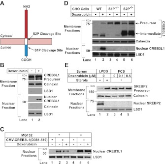
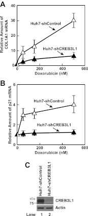
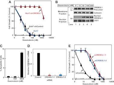
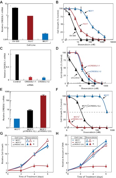
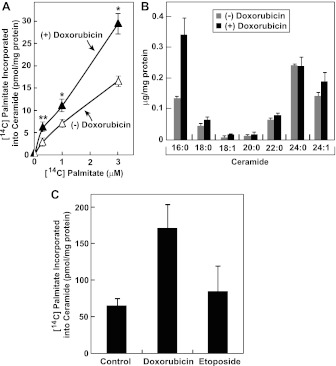
Doxorubicin is used extensively for chemotherapy of diverse types of cancer, yet the mechanism through which it inhibits proliferation of cancer cells remains unclear. Here we report that doxorubicin stimulates de novo synthesis of ceramide, which in turn activates CREB3L1, a transcription factor synthesized as a membrane-bound precursor. Doxorubicin stimulates proteolytic cleavage of CREB3L1 by Site-1 Protease and Site-2 Protease, allowing the NH(2)-terminal domain of CREB3L1 to enter the nucleus where it activates transcription of genes encoding inhibitors of the cell cycle, including p21. Knockdown of CREB3L1 mRNA in human hepatoma Huh7 cells and immortalized human fibroblast SV589 cells conferred increased resistance to doxorubicin, whereas overexpression of CREB3L1 in human breast cancer MCF-7 cells markedly enhanced the sensitivity of these cells to doxorubicin. These results suggest that measurement of CREB3L1 expression may be a useful biomarker in identifying cancer cells sensitive to doxorubicin.DOI:http://dx.doi.org/10.7554/eLife.00090.001.

Keywords: CREB3L1; Human; cancer; ceramide; doxorubicin.


Conflict of interest statement

The authors have declared that no competing interests exist.

Figures

Figure 1.
Figure 1.. Doxorubicin stimulates RIP of CREB3L1.
(A) Schematic diagram of CREB3L1. (B),(D) On Day 0, Huh7 cells (B) or wild type and mutant CHO cells (D) were seeded at 4 × 10⁵ cells per 60 mm dish. On day 1, cells were treated with 500 nM doxorubicin. On day 2, 24 hr after the treatment, the cells were separated into nuclear and membrane fractions, and analyzed by immunoblot with antibodies directed against CREB3L1, calnexin and LSD1. (C) On day 0, huh7 cells were seeded at 1 × 10⁵ cells per 60 mm dish. On day 1 they were transfected with pCMV-CREB3L1(Δ381-519) (0.1 µg per dish) as indicated. On day 2, they were treated with 500 nM doxorubicin as indicated. On day 3, 24 hr after the treatment, the cells were treated with 10 µM MG132 for 2 hr as indicated. Nuclear fraction of the cells was then analyzed by immunoblot analysis with antibody reacting against CREB3L1 and LSD1. (E) On day 0, CHO-7 cells were seeded at 2 × 10⁵ cells per 60 mm dish. On day 1, some cells were changed into sterol-depleting medium (medium A containing 50 µM compactin, 50 µM mevalonate, and 5% lipoprotein deficient serum [LPDS]) with or without supplementation of sterols (1 µg/ml 25-hydroxycholesterol and 10 µg/ml cholesterol). Other cells were changed into normal medium (medium A supplemented with 5% fetal calf serum [FCS]) containing the indicated concentrations of doxorubicin. On day 2, 24 hr after the treatment, the cells were separated into nuclear and membrane fractions, and analyzed by immunoblot with antibodies directed against SREBP2, calnexin and LSD1. DOI: http://dx.doi.org/10.7554/eLife.00090.003
Figure 2.
Figure 2.. Doxorubicin induces transcription of genes activated by CREB3L1.
(A),(B) On day 0, indicated cells were seeded at 3 × 10⁵ cells per 60 mm dish. On day 1, the cells were treated with the indicated concentration of doxorubicin. On day 2, 24 hr after the treatment, some of the cells were harvested for quantification of p21 mRNA through RT-QPCR (B). On day 4, 72 hr after the treatment, the rest of the cells were harvested for quantification of collagen 1α1 (COL1A1) mRNA through RT-QPCR (A). (A),(B) The value of each mRNA in cells that were not treated with the drug is set to 1. (C) Immunoblot analysis of CREB3L1 in indicated cells. DOI: http://dx.doi.org/10.7554/eLife.00090.004
Figure 3.
Figure 3.. CREB3L1 is required for doxorubicin to suppress proliferation of Huh7 cells.
(A) On day 0, indicated cells were seeded at 1.5 × 10⁵ cells per 60 mm dish. On day 1, they were treated with the indicated concentrations of doxorubicin. On day 3, 48 hr after the treatment, the cells were quantified to determine cell proliferation. The number of cells just prior to the drug treatment and after treatment with no drug for 48 hr is set to 0% and 100%, respectively. (B) Huh7 cells treated with the indicated concentrations of doxorubicin were analyzed as described in Figure 1B. (C) On day 0, Huh7 cells were seeded at 4 × 10⁵ cells per 60 mm dish. On day 1, cells were treated with the indicated concentrations of doxorubicin. On day 3, 48 hr after the treatment, cells were harvested to determine the percentage of the cells that underwent apoptosis through TUNEL assay. (D),(E) On day 0, Huh7 cells were seeded at 1 × 10⁵ cells per 60 mm dish. On day 1, the cells were transfected with indicted siRNAs. On day 2, the cells were treated with indicated concentrations of doxorubicin. On day 4, 48 hr after the treatment, some of the cells were harvested for quantification of CREB3L1 mRNA by RT-QPCR (D), while the others were used for determination of cell proliferation as described in Figure 3A (E). (A),(C),(D),(E) Results are reported as mean ± S.E.M. of three independent experiments. DOI: http://dx.doi.org/10.7554/eLife.00090.005
Figure 4.
Figure 4.. Sensitivity of cancer cells to doxorubicin is correlated to their expression of CREB3L1.
(A),(E) RT-QPCR quantification of CREB3L1 mRNA in indicated cells with its value in Huh7 (A) or MCF-7 cells (E) set to 1. (B),(F) Effect of doxorubicin on proliferation of the indicated cells was determined as described in Figure 3A. (C),(D) SV-589 cells were treated and analyzed as described in Figure 3D,E. (G),(H) On day 0, indicated cells were seeded at 1.5 × 10⁵ cells per 60 mm dish. On day 1, the cells were treated with or without 15 nM doxorubicin. After incubation for the indicated period of time, cell proliferation was determined by direct counting of the cells (G) or by measurement of the amount of cellular DNA (H). (G),(H) The number of cells just before doxorubicin treatment at time 0 is set to one. (AH) Results are reported as mean ± S.E.M. of three independent experiments. DOI: http://dx.doi.org/10.7554/eLife.00090.006
Figure 5.
Figure 5.. CREB3L1 activation is independent from DNA breaks.
(A) On day 0, indicated cells were seeded at 4 × 10⁵ cells per 60 mm dish. On day 1, cells were treated with 500 nM doxorubicin or 500 nM etoposide. On day 2, 24 hr after the treatment, the cells were harvested for immunoblot analysis with antibodies reacting against γH2AX or actin. (B) Huh7 cells were seeded and treated as described in (A). On day 2, cells were separated into nuclear and membrane fractions and analyzed by immunoblot analysis as described in Figure 1B. (C) The effect of etoposide on proliferation of the indicated cells was determined as described in Figure 3A. (D)–(G) On day 0, indicated cells were seeded at 1.5 × 10⁵ cells per 60 mm dish. On day 1, cells were treated with indicated concentrations of etoposide (D), doxorubicin (E), bleomycin (F), or paclitaxel (G). On day 3, 48 hr after the treatment, proliferation of the cells was determined by measurement of cellular DNA. The amount of DNA just prior to the drug treatment and after treatment with no drug for 48 hr is set to 0% and 100%, respectively. (C)–(G) Results are reported as mean ± S.E.M. of three independent experiments. DOI: http://dx.doi.org/10.7554/eLife.00090.007
Figure 6.
Figure 6.. Doxorubicin stimulates synthesis of ceramide.
(A) On day 0, Huh7 cells were seeded at 2 × 10⁵ per 60-mm dish. On day 1, the cells were treated with or without 500 nM doxorubicin. On day 2, 20 hr after the treatment, the cells were labeled with indicated concentrations of [14C]palmitate for additional 4 hr. Cell lipids were then extracted to determine the amount of [14C]palmitate incorporated into ceramide. *p=0.003; **p=0.02. (B) On day 0, Huh7 cells were seeded at 1.5 × 10⁵ per 60-mm dish. On day 1, the cells were treated with or without 500 nM doxorubicin. On day 2, 24 hr after the treatment, the cells were harvested for ceramide analysis via LC-MS as described in ‘Materials and methods’. The amount of ceramide with indicated amide-linked fatty acids was presented. (C) Huh7 cells were treated with 500 nM doxorubicin or 1 µM etoposide, labeled with 3 µM [14C]palmitate, and analyzed as described in Figure 6A. (A)–(C) Results are reported as mean ± S.E.M. of triplicate incubations from a representative experiment. DOI: http://dx.doi.org/10.7554/eLife.00090.008
Figure 7.
Figure 7.. Doxorubicin-induced synthesis of ceramide stimulates cleavage of CREB3L1.
(A),(C) On day 0, Huh7 cells were seeded at 4 × 10⁵ per 60-mm dish. On day 1, the cells were treated with indicated concentrations of myriocin (A) or fumonisin B1 (C) for 2 hr, followed by co-incubation with 200 nM doxorubicin. On day 2, 24 hr after the doxorubicin treatment, cells were analyzed for cleavage of CREB3L1 by immunoblot analysis as described in Figure 1B. (B) Huh7 cells treated with or without 30 µM myriocin for 2 hr followed by co-treatment with doxorubicin were analyzed as described in Figure 3A. (D) Huh7 cells treated with 10 µM C6-ceramide for 3 hr were analyzed as described in Figure 6B. Results are reported as mean ± S.E.M. of triplicate incubations from a representative experiment. (E) Huh7 cells treated with indicated concentration of C6-ceramide for 24 hr were analyzed as described in Figure 1B. (F) Indicated cells treated with indicated concentration of C6-ceramide for 48 hr were analyzed as described in Figure 3A. (B),(F) Results are reported as mean ± S.E.M. of three independent experiments. DOI: http://dx.doi.org/10.7554/eLife.00090.009

Comment in

  • How does doxorubicin work?
    Patel AG, Kaufmann SH. Patel AG, et al. Elife. 2012 Dec 18;1:e00387. doi: 10.7554/eLife.00387. Elife. 2012. PMID: 23256047 Free PMC article.

References

    1. Adams CM, Reitz J, De Brabander JK, Feramisco JD, Li L, Brown MS, et al. 2004. Cholesterol and 25-hydroxycholesterol inhibit activation of SREBPs by different mechanisms, both involving SCAP and Insigs. J Biol Chem 279:52772–80 - PubMed
    1. Bose R, Verheij M, Haimovitz-Friedman A, Scotto K, Fuks Z, Kolesnick R. 1995. Ceramide synthase mediates daunorubicin-induced apoptosis: An alternative mechanism for generating death signals. Cell 82:405–14 - PubMed
    1. Brown MS, Goldstein JL. 2009. Cholesterol feedback: from Schoenheimer's bottle to Scap's MELADL. J Lipid Res 50:S15–27 - PMC - PubMed
    1. Brown MS, Ye J, Rawson RB, Goldstein JL. 2000. Regulated intramembrane proteolysis: A control mechanism conserved from bacteria to humans. Cell 100:391–8 - PubMed
    1. DeBose-Boyd RA, Brown MS, Li WP, Nohturfft A, Goldstein JL, Espenshade PJ. 1999. Transport-dependent proteolysis of SREBP: relocation of site-1 protease from Golgi to ER obviates the need for SREBP transport to Golgi. Cell 99:703–12 - PubMed

Publication types

MeSH terms