Comparative Study
. 2013 Jan 8;110(2):E132-40.
doi: 10.1073/pnas.1213624110. Epub 2012 Dec 19.

In vivo translation rates can substantially delay the cotranslational folding of the Escherichia coli cytosolic proteome

Affiliations
Comparative Study

In vivo translation rates can substantially delay the cotranslational folding of the Escherichia coli cytosolic proteome

Prajwal Ciryam et al. Proc Natl Acad Sci U S A. .

Abstract

A question of fundamental importance concerning protein folding in vivo is whether the kinetics of translation or the thermodynamics of the ribosome nascent chain (RNC) complex is the major determinant of cotranslational folding behavior. This is because translation rates can reduce the probability of cotranslational folding below that associated with arrested ribosomes, whose behavior is determined by the equilibrium thermodynamics of the RNC complex. Here, we combine a chemical kinetic equation with genomic and proteomic data to predict domain folding probabilities as a function of nascent chain length for Escherichia coli cytosolic proteins synthesized on both arrested and continuously translating ribosomes. Our results indicate that, at in vivo translation rates, about one-third of the Escherichia coli cytosolic proteins exhibit cotranslational folding, with at least one domain in each of these proteins folding into its stable native structure before the full-length protein is released from the ribosome. The majority of these cotranslational folding domains are influenced by translation kinetics which reduces their probability of cotranslational folding and consequently increases the nascent chain length at which they fold into their native structures. For about 20% of all cytosolic proteins this delay in folding can exceed the length of the completely synthesized protein, causing one or more of their domains to switch from co- to posttranslational folding solely as a result of the in vivo translation rates. These kinetic effects arise from the difference in time scales of folding and amino-acid addition, and they represent a source of metastability in Escherichia coli's proteome.


Conflict of interest statement

The authors declare no conflict of interest.

Figures

Fig. 1.
Fig. 1.
A systems approach for predicting the cotranslational folding behavior of E. coli’s cytosolic proteome. (A) 50S ribosomal subunit with ribosomal protein and RNA molecules shown, respectively, in red and yellow. The protein G domain (blue) is shown emerging from the exit tunnel, and it is tethered to the P-site tRNA by an unstructured poly-glycine linker (white). (B) Cotranslational folding curves (PF) for a protein G domain synthesized at finite translation rates (red triangles, τA = 1.3 ms) and at an infinitely slow translation rate (black X symbols). Data were taken from the coarse-grained molecular dynamics simulations reported by O’Brien et al. (7). The deviation between the PF(i) curves is characterized by ΔLm, the separation in nascent chain lengths at which the native state becomes stable, that is when PF(i) > 0.5. The simulation results for τA = 60 ms are shown as blue circles; at this translation speed, ΔLm = 0. (C) ΔLm can be thought of as the additional number of residues (brown spheres) required for a domain to achieve its stable folded structure at finite translation rates (Lower) compared with infinitely slow translation (Upper). Structures were taken from coarse-grained simulations of protein G reported by O’Brien et al. (7). (D) Illustration of the work flow of our systems approach.
Fig. 2.
Fig. 2.
Cotranslational folding of cytosolic protein domains in E. coli. (A) Determination of a scaling relationship to model cotranslational domain folding/unfolding kinetics. The mean folding (green) and unfolding (magenta) times of protein G as a function of nascent chain length calculated from coarse-grained simulations (7) were fitted by Eqs. 3 and 4 (dashed lines). (B) Examples of cotranslational folding curves calculated for four different protein domains in E. coli at in vivo (red, Eq. 1) and infinitely slow (blue, Eq. 2) translation rates. The domains correspond, respectively, to (ASNC_ECOLI, domain 1; Upper Left), (3MG2_ECOLI, domain 1; Upper Right), (ILVC_ECOLI, domain 1; Lower Left), and (ENO, domain 1; Lower Right) in Dataset S1. (Upper Left) Note that the red and blue lines are superimposed. (C) Structural characterization of domains that fold cotranslationally and posttranslationally in E. coli cells that are dividing every 150 min at 37 °C. (Upper) Probability density function (PDF) vs. domain length. (Lower) Probability of different domain classifications in terms of mostly α (α), mostly β (β), or mixed α/β secondary structure. (D) As in C, except the data are from protein domains that fold cotranslationally with ΔLm = 0 and those that fold with ΔLm values greater than 41 residues. The noncontiguous distribution for the ΔLm > 41 distribution arises from the small number of domains used in its construction (n = 41 data points).
Fig. 3.
Fig. 3.
Extent to which kinetic effects are exhibited during in vivo cotranslational folding and its correlation with the separation in time scales. (A) Probability distribution of ΔLm values for domains that exhibit cotranslational folding in E. coli doubling every 150 min at 37 °C (n = 422). The cumulative distribution function (CDF) is shown as a solid red line. The arrows and numbers indicate (from left to right) the ΔLm values at which the CDF equals 0.5, 0.8, 0.9, and 0.95, respectively. (B) ΔLm for cotranslationally folding proteins as a function of the ratio of τF,m to τA,m at nascent chain length m at which the midpoint of domain folding stability occurs at an infinitely slow translation rate.
Fig. 4.
Fig. 4.
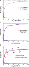
Probability distribution of ΔLm values (histograms) and cumulative distributions (solid lines) at two different E. coli growth rates listed in the legend as the doubling time (A), weighted by the protein expression level data denoted by BLT_WT in Dataset S1 (B), and for proteins that exhibit cooperative and noncooperative domain folding (C). In C, the ΔLm values are shown for probabilities corresponding to 0.8, 0.9, and 0.95 for the noncooperative dataset, whereas the cooperative dataset includes an additional ΔLm value reported at a probability of 0.5.
Fig. 5.
Fig. 5.
Slow translating synonymous codon mutations and their affect on the deviation of cotranslational folding from quasiequilibrium. The probability distribution of the change in ΔLm values for cotranslational folding domains on converting each codon in the WT mRNA transcripts to its corresponding slowest translating synonymous codon is shown. Cotranslational folders that have ΔLm = 0 for the WT mRNA are not included in this analysis because their ΔΔLm could never be anything other than 0. The CDF is shown in blue. The arrows and numbers indicate (from left to right) the ΔLm values at which the CDF equals 0.05, 0.10, 0.20, and 0.5, respectively.
Fig. P1.
Fig. P1.
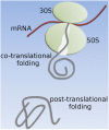
Schematic representation of the 30S and 50S subunits of the RNC complex in E. coli. The cotranslational folding of the nascent chain (gray) can be shifted to posttranslational folding due solely to the translation rates that occur naturally in E. coli.

References

    1. Nicola AV, Chen W, Helenius A. Co-translational folding of an alphavirus capsid protein in the cytosol of living cells. Nat Cell Biol. 1999;1(6):341–345. - PubMed
    1. Netzer WJ, Hartl FU. Recombination of protein domains facilitated by co-translational folding in eukaryotes. Nature. 1997;388(6640):343–349. - PubMed
    1. Agashe VR, et al. Function of trigger factor and DnaK in multidomain protein folding: Increase in yield at the expense of folding speed. Cell. 2004;117(2):199–209. - PubMed
    1. Elcock AH. Molecular simulations of cotranslational protein folding: Fragment stabilities, folding cooperativity, and trapping in the ribosome. PLOS Comput Biol. 2006;2(7):e98. - PMC - PubMed
    1. Komar AA, Lesnik T, Reiss C. Synonymous codon substitutions affect ribosome traffic and protein folding during in vitro translation. FEBS Lett. 1999;462(3):387–391. - PubMed

Publication types

LinkOut - more resources