Review
. 2012 Dec 20;65(1):1-46.
doi: 10.1124/pr.112.006809. Print 2013 Jan.

A modern understanding of the traditional and nontraditional biological functions of angiotensin-converting enzyme

Affiliations
Review

A modern understanding of the traditional and nontraditional biological functions of angiotensin-converting enzyme

Kenneth E Bernstein et al. Pharmacol Rev. .

Erratum in

  • Pharmacol Rev. 2013;65(1):544

Abstract

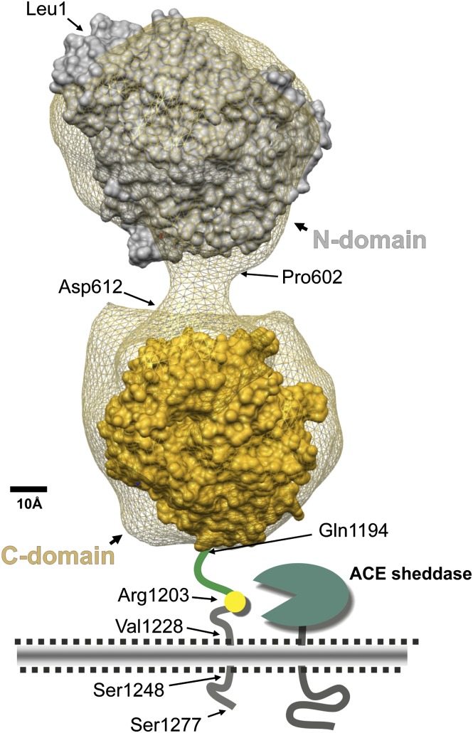
Angiotensin-converting enzyme (ACE) is a zinc-dependent peptidase responsible for converting angiotensin I into the vasoconstrictor angiotensin II. However, ACE is a relatively nonspecific peptidase that is capable of cleaving a wide range of substrates. Because of this, ACE and its peptide substrates and products affect many physiologic processes, including blood pressure control, hematopoiesis, reproduction, renal development, renal function, and the immune response. The defining feature of ACE is that it is composed of two homologous and independently catalytic domains, the result of an ancient gene duplication, and ACE-like genes are widely distributed in nature. The two ACE catalytic domains contribute to the wide substrate diversity of ACE and, by extension, the physiologic impact of the enzyme. Several studies suggest that the two catalytic domains have different biologic functions. Recently, the X-ray crystal structure of ACE has elucidated some of the structural differences between the two ACE domains. This is important now that ACE domain-specific inhibitors have been synthesized and characterized. Once widely available, these reagents will undoubtedly be powerful tools for probing the physiologic actions of each ACE domain. In turn, this knowledge should allow clinicians to envision new therapies for diseases not currently treated with ACE inhibitors.


Figures

Fig. 1.
Fig. 1.
Model of human somatic ACE. This model is a three-dimensional reconstruction of the electron microscopic appearance of porcine somatic ACE (the net) combined with available human X-ray crystal structures (Chen et al., 2010; Danilov et al., 2011). It shows the ACE N-domain (Leu1–Pro601), linker region (Pro602–Asp612), C-domain (Leu613–Pro1193), stalk region (Gln1194–Arg1227), transmembrane segment (Val1228–Ser1248), and intracellular domain (Gln1249–Ser1277) (Acharya et al., 2003; Corradi et al., 2006). We also indicate the membrane-bound ACE sheddase and Arg1203, the C-terminal residue of soluble ACE.
Fig. 2.
Fig. 2.
Response of ACE 10/10 mice to challenge with melanoma. B16-F10 melanoma cells were injected into the skin of ACE 10/10, ACE 10/ACE wild-type heterozygous (HZ), and ACE wild-type (WT) mice. Two weeks later, the mice were sacrificed and the tumor volume was measured. Data points from individual mice are shown, as well as group means and S.E.M. ACE 10/10 mice consistently showed much smaller tumors than wild-type mice (Shen et al., 2007).
Fig. 3.
Fig. 3.
Phylogenetic tree of ACE. (A) Human ACE amino acid sequence was used to search several publically available databases. Retrieved sequences were aligned for homology and plotted in tree form using JACOP software (http://myhits.isb-sib.ch/cgi-bin/jacop). The degree of homology is reflected in the position of each sequence, with more homologous sequences clustered together. The retrieved ACE homologs consist of proteins with two (blue), one (green), or no putative zinc binding-sites (black). Human ACE is indicated with an arrow. (B) The top panels show a diagonal dot plot that was prepared using Dotlet software (http://myhits.isb-sib.ch/cgi-bin/dotlet). The amino acid sequence of human ACE is positioned along the vertical axis, and is compared with the amino acid sequence indicated along the horizontal axis. The program used a sliding window of 39 amino acids with a grayscale dot that varies from 100% (white dot) to 0% (black dot). Comparing human ACE against itself produces a 45° diagonal of identity, as well as two other parallel segments confirming the amino acid sequence homology between the N- and C-domains. Comparison of the human sequence to sequences from five species from diverse phyla demonstrates the wide prevalence of an ACE-like protein containing two homologous domains. The bottom part of the figure shows, in schematic form, the evolutionary relationship between the six species. The DNA sequences used in this figure are as follows: no active domain: Brugia malayi (lymphatic filariasis roundworm): XP_001897661.1, Caenorhabditis briggsae: XP_002644637.1, and C. elegans: NP_001024453.1; one active domain: Acromyrmex echinatior (Panamanian leafcutter ant): EGI64832.1, Aedes aegypti (yellow fever mosquito): XP_001659916.1, Ornithorhynchus anatinus (platypus): XP_001515597.1, Pongo abelii (Sumatran orangutan): NP_001124604.1, Monodelphis domestica (gray short-tailed opossum): XP_001376640.2, Ailuropoda melanoleuca (giant panda): XP_002922087.1, Sorangium cellulosum So ce 56 (bacteria): YP_001615731.1, Anaeromyxobacter dehalogenans 2CP-C (bacteria): YP_465231.1, Stigmatella aurantiaca DW4/3-1 (bacteria): YP_003953730.1; Myxococcus fulvus HW-1 (bacteria): YP_004668148.1, Myxococcus xanthus DK 1622 (bacteria): YP_631771.1, Gloeobacter violaceus PCC 7421 (cyanobacteria): NP_926089.1, Shewanella baltica OS678 (bacteria): YP_005273382.1; Candidatus solibacter usitatus Ellin6076: YP_826088.1, Saccoglossus kowalevskii (acorn worm): XP_002734849.1, Crassostrea gigas (Pacific oyster): AEV53960.1, Hydra magnipapillata (hydra): XM_002162385.1, Strongylocentrotus purpuratus (purple sea urchin): XP_001183772.1, Ixodes scapularis (black-legged tick): XP_002435481.1, Pediculus humanus corporis (human body louse): XP_002431342.1, Tribolium castaneum (red flour beetle): NP_001164243.1, Pontastacus leptodactylus (narrow-clawed crayfish): CAX48990.1, Drosophila melanogaster (fruit fly): NP_477046.1, Acyrthosiphon pisum (pea aphid): NP_001129384.1, S. littoralis (African cotton leafworm): ABW34729.1, Apis mellifera (honey bee): XP_393561.3, Locusta migratoria (migratory locust): AAR85358.1, T. tessulatum (duck leech): AAS57725.1, and Trichinella spiralis (trichinosis nematode parasite): XP_003379675.1; two active domains: A. darlingi (American mosquito): EFR22959.1, C. quinquefasciatus (southern house mosquito): XP_001845716.1, A. gambiae str. PEST: XP_313865.3, Heterocephalus glaber (naked mole-rat): EHB07361.1, Mesocricetus auratus (golden hamster): BAD98304.1, Macaca mulatta (rhesus monkey): XP_002800616.1, Homo sapiens (human): NP_000780.1, Pan troglodytes (chimpanzee): NP_001008995.1, Mus musculus (house mouse): NP_997507.1, Rattus norvegicus (Norway rat): NP_036676.1, E. caballus (horse): XP_001495639.3, Oryctolagus cuniculus (rabbit): NP_001075864.1, Cavia porcellus (domestic guinea pig): XP_003465969.1, Canis lupus familiaris (dog): XP_003639297.1, Loxodonta africana (African savanna elephant): XP_003414368.1, Bos taurus (cattle): 449408, Sus scrofa (pig): ABL73884.1, Tetraodon nigroviridis (green spotted pufferfish): CAG04404.1, Danio rerio (zebrafish): XP_694336.5, Dicentrarchus labrax (European seabass): CBN81253.1, Oreochromis niloticus (Nile tilapia): XP_003438793.1, Xenopus (Silurana) tropicalis (Western clawed frog): NP_001116882.1, Anolis carolinensis (green anole): XP_003222396.1, Taeniopygia guttata (zebra finch): XP_002191587.1, Gallus gallus (chicken): NP_001161204.1, Meleagris gallopavo (turkey): XP_003213080.1, C. intestinalis (sea squirt): XP_002123029.1, Branchiostoma floridae (Florida lancelet): XM_002594635.1, N. vectensis (starlet sea anemone): XP_001627759.1, T. adhaerens (Placozoa): XP_002111333.1, and D. pulex (common water flea): EFX86779.1.
Fig. 3.
Fig. 3.
Phylogenetic tree of ACE. (A) Human ACE amino acid sequence was used to search several publically available databases. Retrieved sequences were aligned for homology and plotted in tree form using JACOP software (http://myhits.isb-sib.ch/cgi-bin/jacop). The degree of homology is reflected in the position of each sequence, with more homologous sequences clustered together. The retrieved ACE homologs consist of proteins with two (blue), one (green), or no putative zinc binding-sites (black). Human ACE is indicated with an arrow. (B) The top panels show a diagonal dot plot that was prepared using Dotlet software (http://myhits.isb-sib.ch/cgi-bin/dotlet). The amino acid sequence of human ACE is positioned along the vertical axis, and is compared with the amino acid sequence indicated along the horizontal axis. The program used a sliding window of 39 amino acids with a grayscale dot that varies from 100% (white dot) to 0% (black dot). Comparing human ACE against itself produces a 45° diagonal of identity, as well as two other parallel segments confirming the amino acid sequence homology between the N- and C-domains. Comparison of the human sequence to sequences from five species from diverse phyla demonstrates the wide prevalence of an ACE-like protein containing two homologous domains. The bottom part of the figure shows, in schematic form, the evolutionary relationship between the six species. The DNA sequences used in this figure are as follows: no active domain: Brugia malayi (lymphatic filariasis roundworm): XP_001897661.1, Caenorhabditis briggsae: XP_002644637.1, and C. elegans: NP_001024453.1; one active domain: Acromyrmex echinatior (Panamanian leafcutter ant): EGI64832.1, Aedes aegypti (yellow fever mosquito): XP_001659916.1, Ornithorhynchus anatinus (platypus): XP_001515597.1, Pongo abelii (Sumatran orangutan): NP_001124604.1, Monodelphis domestica (gray short-tailed opossum): XP_001376640.2, Ailuropoda melanoleuca (giant panda): XP_002922087.1, Sorangium cellulosum So ce 56 (bacteria): YP_001615731.1, Anaeromyxobacter dehalogenans 2CP-C (bacteria): YP_465231.1, Stigmatella aurantiaca DW4/3-1 (bacteria): YP_003953730.1; Myxococcus fulvus HW-1 (bacteria): YP_004668148.1, Myxococcus xanthus DK 1622 (bacteria): YP_631771.1, Gloeobacter violaceus PCC 7421 (cyanobacteria): NP_926089.1, Shewanella baltica OS678 (bacteria): YP_005273382.1; Candidatus solibacter usitatus Ellin6076: YP_826088.1, Saccoglossus kowalevskii (acorn worm): XP_002734849.1, Crassostrea gigas (Pacific oyster): AEV53960.1, Hydra magnipapillata (hydra): XM_002162385.1, Strongylocentrotus purpuratus (purple sea urchin): XP_001183772.1, Ixodes scapularis (black-legged tick): XP_002435481.1, Pediculus humanus corporis (human body louse): XP_002431342.1, Tribolium castaneum (red flour beetle): NP_001164243.1, Pontastacus leptodactylus (narrow-clawed crayfish): CAX48990.1, Drosophila melanogaster (fruit fly): NP_477046.1, Acyrthosiphon pisum (pea aphid): NP_001129384.1, S. littoralis (African cotton leafworm): ABW34729.1, Apis mellifera (honey bee): XP_393561.3, Locusta migratoria (migratory locust): AAR85358.1, T. tessulatum (duck leech): AAS57725.1, and Trichinella spiralis (trichinosis nematode parasite): XP_003379675.1; two active domains: A. darlingi (American mosquito): EFR22959.1, C. quinquefasciatus (southern house mosquito): XP_001845716.1, A. gambiae str. PEST: XP_313865.3, Heterocephalus glaber (naked mole-rat): EHB07361.1, Mesocricetus auratus (golden hamster): BAD98304.1, Macaca mulatta (rhesus monkey): XP_002800616.1, Homo sapiens (human): NP_000780.1, Pan troglodytes (chimpanzee): NP_001008995.1, Mus musculus (house mouse): NP_997507.1, Rattus norvegicus (Norway rat): NP_036676.1, E. caballus (horse): XP_001495639.3, Oryctolagus cuniculus (rabbit): NP_001075864.1, Cavia porcellus (domestic guinea pig): XP_003465969.1, Canis lupus familiaris (dog): XP_003639297.1, Loxodonta africana (African savanna elephant): XP_003414368.1, Bos taurus (cattle): 449408, Sus scrofa (pig): ABL73884.1, Tetraodon nigroviridis (green spotted pufferfish): CAG04404.1, Danio rerio (zebrafish): XP_694336.5, Dicentrarchus labrax (European seabass): CBN81253.1, Oreochromis niloticus (Nile tilapia): XP_003438793.1, Xenopus (Silurana) tropicalis (Western clawed frog): NP_001116882.1, Anolis carolinensis (green anole): XP_003222396.1, Taeniopygia guttata (zebra finch): XP_002191587.1, Gallus gallus (chicken): NP_001161204.1, Meleagris gallopavo (turkey): XP_003213080.1, C. intestinalis (sea squirt): XP_002123029.1, Branchiostoma floridae (Florida lancelet): XM_002594635.1, N. vectensis (starlet sea anemone): XP_001627759.1, T. adhaerens (Placozoa): XP_002111333.1, and D. pulex (common water flea): EFX86779.1.
Fig. 4.
Fig. 4.
TNFα production by peritoneal macrophages. Peritoneal macrophages were isolated from ACE knockout (ACE KO), wild-type (WT), ACE 10/10 (10/10), N-KO, and C-KO mice. The cells were cultured in vitro overnight with lipopolysaccharide, and the level of TNFα within the culture media was measured. Data points from individual mice are shown, as well as group means and S.E.M. Large amounts of TNFα were made by N-KO macrophages, implying fundamental biochemical differences between these macrophages and equivalent cells from wild-type mice (Ong et al., 2012).
Fig. 5.
Fig. 5.
Model comparing carboxypeptidase A and ACE. In this schematic, the active sites of carboxypeptidase A and ACE are compared. The pockets in the enzyme represent side chain binding sites within the active sites of the enzymes. In ACE, these pockets are often labeled S1, S1′, and S2′ based on their position relative to the zinc molecule. The + indicate positive charges and X-H represents a potential hydrogen bond. Interactions stabilizing substrates or inhibitors are indicated with dots. The substrate amino bond hydrolyzed by the enzymes is indicated with a bent line. Initial thinking about ACE inhibitors was derived from work showing that benzylsuccinic acid was an inhibitor of carboxypeptidase A (Byers and Wolfenden, 1973). A major advance was the use of a sulfhydryl group to bind to the zinc molecule (Ondetti and Cushmen, 1981). The figure also shows the structure of the ACE inhibitor lisinopril, which has a high affinity for ACE due to interaction with the S1 pocket, in addition to the zinc atom and other structural features of ACE. Adapted with permission from Cushman et al. (1977). Copyright (1978) American Chemical Society.
Fig. 6.
Fig. 6.
Binding of lisinopril to testis ACE. As compared with the schematic in Fig. 5, the crystal structure of human testis ACE–binding lisinopril shows the detailed interactions of individual ACE amino acids with the inhibitor (Natesh et al., 2003). Amino acid numbers are those of testis ACE. Stabilizing interactions are indicated by dashes. The S1-, S1′-, and S2′-binding pockets of ACE are indicated. The carboxyalkyl carboxylate of lisinopril (indicated with an asterisk) substitutes for the amide carbonyl in an ACE substrate. What would be the scissile amide nitrogen in an ACE substrate is indicated by an arrow. Adapted with permission from Macmillan Publishers Ltd: Natesh et al. (2003).

References

    1. AbdAlla S, Lother H, Langer A, el Faramawy Y, Quitterer U. (2004) Factor XIIIA transglutaminase crosslinks AT1 receptor dimers of monocytes at the onset of atherosclerosis. Cell 119:343–354 - PubMed
    1. Acharya KR, Sturrock ED, Riordan JF, Ehlers MR. (2003) Ace revisited: a new target for structure-based drug design. Nat Rev Drug Discov 2:891–902 - PMC - PubMed
    1. Agerholm-Larsen B, Nordestgaard BG, Tybjaerg-Hansen A. (2000) ACE gene polymorphism in cardiovascular disease: meta-analyses of small and large studies in whites. Arterioscler Thromb Vasc Biol 20:484–492 - PubMed
    1. Agerholm-Larsen B, Nordestgaard BG, Steffensen R, Sørensen TI, Jensen G, Tybjaerg-Hansen A. (1997) ACE gene polymorphism: ischemic heart disease and longevity in 10,150 individuals. A case-referent and retrospective cohort study based on the Copenhagen City Heart Study. Circulation 95:2358–2367 - PubMed
    1. Ahmed A, Fonarow GC, Zhang Y, Sanders PW, Allman RM, Arnett DK, Feller MA, Love TE, Aban IB, Levesque R, et al. (2012) Renin-angiotensin inhibition in systolic heart failure and chronic kidney disease. Am J Med 125:399–410 - PMC - PubMed

Publication types

MeSH terms

LinkOut - more resources