Review
. 2013 Jan;29(1):1-13.
doi: 10.1016/j.kjms.2012.08.001. Epub 2012 Oct 12.

New insights into desmosome regulation and pemphigus blistering as a desmosome-remodeling disease

Affiliations
Review

New insights into desmosome regulation and pemphigus blistering as a desmosome-remodeling disease

Yasuo Kitajima. Kaohsiung J Med Sci. 2013 Jan.

Abstract

Desmosomes in keratinocytes are the most important intercellular adhering junctions that provide structural strength for the epidermis. These junctions are connected directly with desmosomal cadherin proteins. Desmosomal cadherins are divided into four desmogleins (Dsgs), Dsg1-4, and three desmocollins (Dscs), Dsc1-3, all of which are involved in desmosomal adhesion by homo- and/or heterophilic binding between Dsgs and Dscs in a Ca(2+)-dependent manner. Cadherins are present on the cell surface and anchor keratin intermediate filaments (KIFs) to their inner cytoplasmic surface to generate an intracellular KIF-skeletal scaffold through several associate proteins, including plakoglobin, plakophillin, and desmoplakins. As such, the desmosomal contacts between adjacent cells generate an intercellular KIF scaffold throughout the whole epidermal sheet. However, despite these critical roles in maintaining epidermal adhesion and integrity, desmosomes are not static structures. Rather, they are dynamic units that undergo regular remodeling, i.e., assembly and disassembly, to allow for cell migration within the epidermis in response to outside-in signaling during epidermal differentiation. Recently, two cell-cell adhesion states controlled by desmosomes have been recognized, including "stable hyperadhesion (Ca(2+)-independent)" and "dynamic weak-adhesion (Ca(2+)-dependent)" conditions. These conditions are mutually reversible through cell signaling events involving protein kinase C (PKC) and epidermal growth factor receptor. Pemphigus vulgaris (PV) is an autoimmune bullous disease caused by anti-Dsg3 antibodies. Binding of these antibodies to Dsg3 causes endocytosis of Dsg3 from the cell surface and results in the specific depletion of Dsg3 from desmosomes, an event linked to acantholysis in the epidermis. This binding of anti-Dsg3 antibody to Dsg3 in epidermal keratinocytes activates PKC, to generate the "weak-adhesion (Ca(2+)-dependent)" state of desmosomes. The weak-adhesion desmosomes appear to be the susceptible desmosomal state and a prerequisite for Dsg3 depletion from desmosomes, pivotal and specific events leading to PV blistering. These observations allow us to propose a concept for pemphigus blistering disorders as a "desmosome-remodeling impairment disease" involving a mechanism of Dsg3 nonassembly and depletion from desmosomes through PV immunoglobulin G-activated intracellular signaling events.


Figures

Figure 1
Figure 1
KIF networks and desmosomes binding with KIF. Epidermal keratinocytes are connected with desmosomes anchored by individual cell cytoskeletons of KIFs. (A) Hematoxylin–eosin‐stained section of the epidermis. (B) Cultured human keratinocytes stained with antikeratin antibody, showing a keratin cytoskeletal network structure. (C) Electron microscopy of cultured human keratinocytes. KIF = keratin intermediate filament.
Figure 2
Figure 2
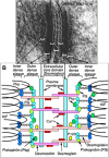
(A) Electron microscopy and (B) a molecular schema of desmosome. KIF = keratin intermediate filament; PG = plakoglobin; Pkp = plakophilin.
Figure 3
Figure 3
Human keratinocytes stained with antikeratin, anti‐Dsg3 and anti‐Dpk grown in (A) low and (B) high Ca2+ medium. When keratinocytes are grown in low Ca2+ medium, they grow independently without forming cell–cell contacts, whereas in Ca2+ switching up to normal, they move laterally and form cell–cell contact and colonies. In low Ca2+ medium, Dsg3 and Dpk are randomly distributed in the cell or on the cell surface. Many of these dots are colocalized, even though they do not appear to localize at cell–cell contact and do not form desmosome (lower part of A). After Ca2+ switch up, Dsg3 and Dpk are localized at cell–cell contacts, generating a linear punctate pattern. Dpk = desmoplakin; Dsg3 = desmoglein 3.
Figure 4
Figure 4
Human keratinocytes stained with anti‐Dsg3 pemphigus vulgaris IgG and with secondary gold‐particle‐labeled antihuman IgG in (A) low and (B) high Ca2+ medium. (A) Dsg3 aggregates on the plasma membrane without any attachment plaques inside the cells (upper) and also with attachment plaques bound with keratin intermediate filaments (lower) are formed in low Ca2+ medium. (B) In high (normal) Ca2+ medium, complete desmosomes, but almost no half‐desmosomes, are observed. Dsg3 = desmoglein 3; IgG = immunoglobulin G; KIF = keratin intermediate filament.
Figure 5
Figure 5
Schematic explanation of Ca2+‐sensitive low‐affinity and Ca2+‐resistant hyperadhesive desmosomes. These two types of desmosomes are convertible to each other by PKC, wound healing (EGFR activation), and Ca2+ switching. (A) In low Ca2+ medium, no desmosomes are formed (Dsg3 random distribution) (lower). (B) The Ca2+‐sensitive low‐affinity (dynamic) desmosomes have no electron‐dense midline in the core domain of desmosomes (upper insert), while (C) the Ca2+‐resistant hyperadhesive desmosomes demonstrate it (upper insert), as observed by electron microscopy. Mitotic keratinocytes demonstrate coarse spines of radial KIF at cell–cell contacts, which may have Ca2+‐sensitive low‐affinity (dynamic) desmosomes, whereas others may have stable desmosomes. Dsg3 = desmoglein 3; EGFR = epidermal growth factor receptor; KIF = keratin intermediate filament; PKC = protein kinase C.
Figure 6
Figure 6
Ulltrastructural features of Ca2+‐induced desmosome formation following adherens junction formation. (A) Low Ca2+‐grown cells show very small number of adherens. (B) Fifteen minutes after calcium switch‐up, many adherens junctions are formed, (C) then 30 minutes after the calcium switch, half‐desmosomes appear to get closer, just to be going to couple. (D) At 120 minutes after calcium switch‐up, almost complete desmosomes are formed, suggesting that adherens junction formation precedes desmosome formation. AD =adherens junction; DS =desmosome; KIF = keratin intermediate filament.
Figure 7
Figure 7
(A) PV‐IgG molecules bound to Dsg3 on half‐desmosomes in low Ca2+ medium do not inhibit coupling with each other to form complete desmosomes after Ca2+ switch to high. Keratinocytes are treated with PV‐IgG in the low Ca2+ medium for 30 minutes, followed by washing and staining with antihuman IgG FITC (for immunofluorescence microscopy) or gold‐particle (for immunoelectron microscopy)‐labeled IgG (goat), and then Ca2+ concentration of medium is switched up to high. This PV‐IgG bound with (B, C) gold‐particle‐labeled antihuman IgG on the surface of half‐desmosomes does not inhibit the (D) formation of complete desmosomes, which (E) sandwich the PV‐IgG and gold particles. FITC = fluorescein‐4‐isothiocyanate; IgG = immunoglobulin G; PV = pemphigus vulgaris.
Figure 8
Figure 8
Dsg3 molecules, which once bound with PV‐IgG in desmosomes within 2 minutes, disappear from desmosome within the following 5 hours after 30 minutes. (A) Keratinocytes are first treated with PV‐IgG for 2 minutes and washed with culture medium, and then incubated for further 5 hours without any antibody. (B) At the 30‐minute time point, some of Dsg3 bound with PV‐IgG are translocated from cell surface into the cytoplasm, generating a dotted pattern in the cells, and the rest are retained at cell–cell contacts, which are supposed to be desmosomes, as shown with co‐localized Dpks. (C, D) Only Dsg3 bound with PV‐IgG at cell–cell contacts disappears from the contacts by the time point at 5 hours, but not Dpks and Dscs, from desmosomes. Dpk = desmoplakin; Dsc = desmocollin; Dsg3 = desmoglein 3; IgG = immunoglobulin G; PV = pemphigus vulgaris.
Figure 9
Figure 9
Western blots of Tr‐X 100‐soluble and Tr‐X 100‐insoluble fractions from DJM‐1 cells which are treated with PV‐IgG and AK23 (a pathogenic anti‐Dsg3 monoclonal antibody) after 30 minutes and 24 hours. (A, B) In the Tr‐X 100‐soluble membrane fractions at 30 minutes and 24 hours after treatments with PV‐IgG and AK23, Dsg3 decrease down to 9% and 58%, respectively. (C) In the Tr‐X 100‐insoluble cytoskeleton‐desmosome fractions, 30‐minute treatment is not enough to reach the same reduction result of Dsg3 as that of the Tr‐X 100‐soluble fraction. (D) However, 24‐hour treatment produces the same maximum plateau reduction of Dsg3 as that of the Tr‐X 100‐soluble fraction. Dsg3 = desmoglein 3; IgG = immunoglobulin G; PV = pemphigus vulgaris; Tr‐X = Triton‐X.
Figure 10
Figure 10
Ultrastructural fate of desmoglein 3 after PV‐IgG binding in cultured keratinocytes treated with PV‐IgG. Keratinocytes are treated with PV‐IgG for 2 minutes, followed by washing and incubation with antihuman gold‐particle IgG for another 2 minutes. The aggregated PV‐IgG formed on the cell membrane during the first 2 minutes of incubation are internalized within 15 minutes and degraded within 60 minutes. IgG = immunoglobulin G; PV = pemphigus vulgaris.
Figure 11
Figure 11
Putative mechanisms for the formation of Dsg3‐depleted desmosomes with decreased adhesion strength and blistering in PV. Binding of anti‐Dsg3 antibodies contained in PV‐IgG to free Dsg3 proteins on the interdesmosomal cell surface leads to their internalization into endosomes, and Dsg3 proteins integrated in the core domain of desmosomes are excluded from these sites and possibly internalized into endosomes. Shortage of free Dsg3 on the cell membrane due to its endocytosis and exclusion of Dsg3 from desmosome core domains result in the formation of Dsg3‐depleted desmosomes, the adhesion strength of which is decreased. The processes are supposed to be controlled by a variety of signaling pathways. Dsg3 = desmoglein 3; IgG = immunoglobulin G; PV = pemphigus vulgaris.
Figure 12
Figure 12
Two‐step hypothesis of acantholysis mechanism in PV: Generation of dynamic (low‐affinity) Ca2+‐sensitive desmosomes and Dsg3 depletion. In cultured keratinocytes, PV‐IgG activates PKC and EGFR, which alter the Ca2+‐independent hyperadhesive desmosome to Ca2+‐sensitive dynamic desmosomes or even weaken the adhesive strength, leading keratinocytes to become more sensitive to blistering processes, i.e., nonassembly and depletion of Dsg3 in desmosomes. Dsg3 = desmoglein 3; EGFR = epidermal growth factor receptor; IgG = immunoglobulin G; PKC = protein kinase C; PV = pemphigus vulgaris.

References

    1. Kitajima Y.. Mechanisms of desmosome assembly and disassembly. Clin Exp Dermatol. 2002; 27: 684–690. - PubMed
    1. He W., Cowin P., Stokes D.L.. Untangling desmosomal knots with electron tomography. Science. 2003; 302: 109–113. - PubMed
    1. Kottke M.D., Margaret D., Kottke E.D., Andrew P.K.. The desmosome: cell science lessons from human diseases. J Cell Sci. 2006; 119: 797–806. - PubMed
    1. North A.J., Bardsley W.G., Hyam J., Bornslaeger E.A., Cordingley H.C., Trinnaman B., et al. Molecular map of the desmosomal plaque. J Cell Sci. 1999; 112: 4325–4336. - PubMed
    1. Jonca N., Guerrin M., Hadjiolova K., Caubet C., Gallinaro H., Simon M., et al. Corneodesmosin, a component of epidermal corneocyte desmosomes, displays homophilic adhesive properties. J Biol Chem. 2001; 277: 5024–5029. - PubMed

MeSH terms