. 2013 Jan;123(1):509-16.
doi: 10.1172/JCI65252. Epub 2012 Dec 21.

Thyroid hormone is required for hypothalamic neurons regulating cardiovascular functions

Affiliations

Thyroid hormone is required for hypothalamic neurons regulating cardiovascular functions

Jens Mittag et al. J Clin Invest. 2013 Jan.

Abstract

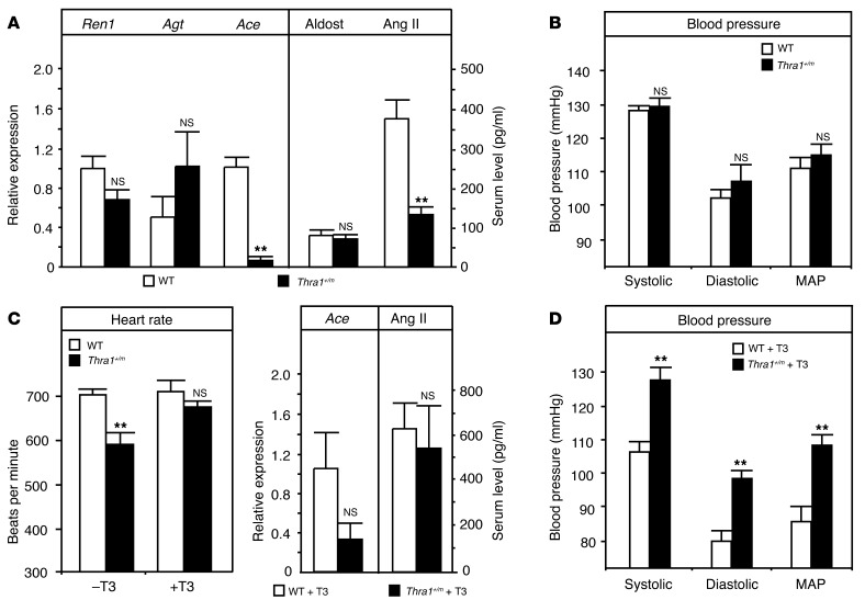
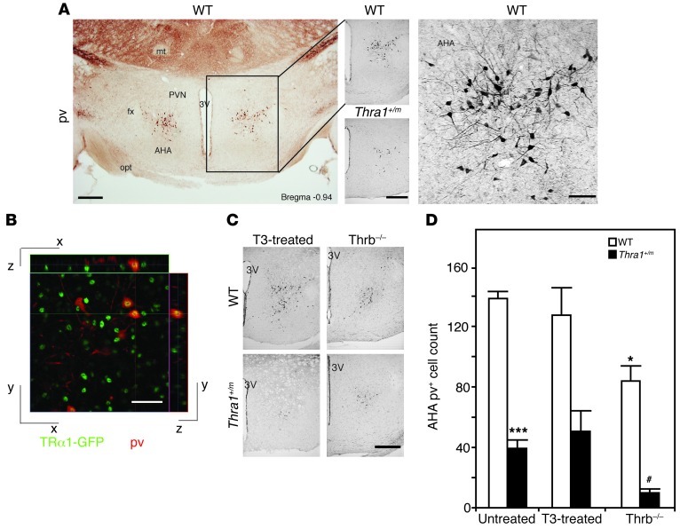
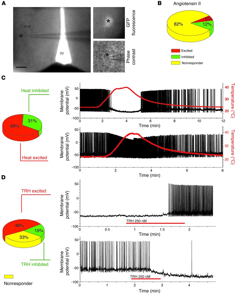
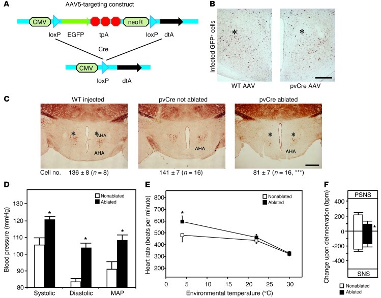
Thyroid hormone is well known for its profound direct effects on cardiovascular function and metabolism. Recent evidence, however, suggests that the hormone also regulates these systems indirectly through the central nervous system. While some of the molecular mechanisms underlying the hormone's central control of metabolism have been identified, its actions in the central cardiovascular control have remained enigmatic. Here, we describe a previously unknown population of parvalbuminergic neurons in the anterior hypothalamus that requires thyroid hormone receptor signaling for proper development. Specific stereotaxic ablation of these cells in the mouse resulted in hypertension and temperature-dependent tachycardia, indicating a role in the central autonomic control of blood pressure and heart rate. Moreover, the neurons exhibited intrinsic temperature sensitivity in patch-clamping experiments, providing a new connection between cardiovascular function and core temperature. Thus, the data identify what we believe to be a novel hypothalamic cell population potentially important for understanding hypertension and indicate developmental hypothyroidism as an epigenetic risk factor for cardiovascular disorders. Furthermore, the findings may be beneficial for treatment of the recently identified patients that have a mutation in thyroid hormone receptor α1.


Figures

Figure 1
Figure 1. Regulation of blood pressure in Thra1+/m mice before and after treatment with T3.
(A) mRNA expression of renal renin (Ren1), hepatic angiotensinogen (Agt), and pulmonary Ace as well as serum aldosterone (Aldost) and angiotensin II (Ang II) levels in wild-type and Thra1+/m mice. (B) Systolic, diastolic, and mean arterial blood pressure (MAP) in wild-type and Thra1+/m mice. (C) Heart rate in wild-type and Thra1+/m mice before and after T3 treatment as well as pulmonary Ace mRNA expression and serum angiotensin II levels in T3-treated animals. (D) Systolic, diastolic, and mean arterial blood pressure in T3-treated wild-type and Thra1+/m mice. All values are mean ± SEM; n = 5. **P < 0.01. NS, not significant.
Figure 2
Figure 2. Reduced number of pv cells in the anterior hypothalamus of Thra1+/m mice.
(A) Immunohistochemistry for pv in the anterior hypothalamus, as overview (left; scale bar: 250 μm) and high magnification (right; scale bar: 50 μm) in wild-type and Thra1+/m mice (middle; scale bar: 250 μm). fx, fornix; mt, mamillothalamic tract; PVN, paraventricular nucleus of the hypothalamus; 3V, 3rd ventricle; opt, optic tract. (B) Double immunohistochemistry for GFP (green) and pv (red) in the AHA of a mouse strain expressing a chimeric TRα1-GFP protein. Yellow indicates overlapping staining. Scale bar: 25 mm. (C) pv neurons in T3-treated wild-type and Thra1+/m mice or crossings with hyperthyroid Thrb–/– mice. Scale bar: 250 μm. (D) Quantification of pv neurons in the AHA of the different animal models. All values are mean ± SEM; n = 4–9. *P < 0.05 to untreated wild type; ***P < 0.001 to untreated wild type; #P < 0.05 to untreated Thra1+/m.
Figure 3
Figure 3. Electrophysiological responses of pv+ cells in the AHA.
(A) Differential interference contrast (DIC) micrograph showing a recorded AHA pv+ neuron (indicated by an asterisk) (left; scale bar: 200 μm) and higher-magnification images of the same GFP-positive neuron under fluorescence and DIC (right; 500-fold magnification). (B) Response of AHA pv+ neurons to angiotensin II (82% no response; n = 14 out of 17). (C) Temperature responsiveness of the AHA pv+ cells to heat (31% inhibited, n = 5 out of 16, and 69% excited, n = 11 out of 16) in patch-clamp recordings on hypothalamic sections of transgenic pvGFP mice. (D) Response of AHA pv+ neurons to TRH (48% excited, n = 10 out of 21; 19% inhibited, n = 4 out of 21; and 33% nonresponsive, n = 7 out of 21) (the neuron in the top panel was held below threshold to prevent action potential firing; no holding current was applied in the other experiments).
Figure 4
Figure 4. Effect of the in vivo ablation of AHA pv cells in pvCre mice.
(A) AAV construct before and after Cre recombination. CMV, cytomegalovirus promotor; loxP, Cre recombination site; tpA, triple polyadenylation site; neoR, neomycin resistance gene; dtA, diphtheria toxin A. (B) Immunohistochemistry for EGFP at the site of the injection (indicated by asterisks) showing AAV-infected cells (scale bar: 250 μm). (C) Immunohistochemistry for pv in AAV-injected wild-type, nonablated pvCre, or AAV-injected ablated pvCre mice (the overall ablation efficiency is shown in the cell count at the bottom; ***P < 0.001 to nonablated, unpaired 2-tailed Student’s t test; the respective groups for the subsequent cardiac and metabolic analyses had cell counts of 81 ± 13 in the ablated animals vs. 142 ± 10 in the nonablated animals; n = 6, P = 0.002; scale bar: 500 μm). Asterisks indicate the site of injection. (D) Systolic, diastolic, and mean arterial blood pressure in mice with reduced numbers of pv+ cells in the AHA (black bars) and controls (white bars; *P < 0.05 for ablated vs. nonablated, unpaired 2-tailed Student’s t test). (E) Heart rates in these mice (*P < 0.05 for ablated vs. nonablated at 4°C, 2-way ANOVA). (F) Change in heart rate upon pharmacological deinnervation of the parasympathetic nervous system (PSNS) (scopolamine methyl bromide) or the sympathetic nervous system (SNS) (timolol) in mice with reduced numbers of pv+ cells in the AHA (black bars) and controls (white bars; *P < 0.05 for ablation, 2-way ANOVA). All values are mean ± SEM.

Comment in

References

    1. Magnus-Levy A. Uber den respiratorischen Gaswechsel unter dem Einfluss der Thyroidea sowie unter verschiedenen pathologischen Zustanden. Berlin Klin Wochenschr. 1895;34:650–652.
    1. Klein I, Ojamaa K. Thyroid hormone and the cardiovascular system. N Engl J Med. 2001;344(7):501–509. doi: 10.1056/NEJM200102153440707. - DOI - PubMed
    1. Kim B. Thyroid hormone as a determinant of energy expenditure and the basal metabolic rate. Thyroid. 2008;18(2):141–144. doi: 10.1089/thy.2007.0266. - DOI - PubMed
    1. Kahaly GJ, Dillmann WH. Thyroid hormone action in the heart. Endocr Rev. 2005;26(5):704–728. doi: 10.1210/er.2003-0033. - DOI - PubMed
    1. Silva JE. Thermogenic mechanisms and their hormonal regulation. Physiol Rev. 2006;86(2):435–464. doi: 10.1152/physrev.00009.2005. - DOI - PubMed

Publication types

MeSH terms

Substances