

Review
doi: 10.1039/c2cs35416k.
Epub 2012 Dec 21.
Chemical probing of glycans in cells and organisms
Affiliations
- PMID: 23257905
- PMCID: PMC3641795
- DOI: 10.1039/c2cs35416k
Item in Clipboard
Review
Chemical probing of glycans in cells and organisms
Chem Soc Rev.
.
Abstract
Among the four major building blocks of life, glycans play essential roles in numerous physiological and pathological processes. Due to their non-templated biosynthesis, advances towards elucidating the molecular details of glycan functions are relatively slow compared with the pace of protein and nucleic acid research. Over the past 30 years, chemical tools have emerged as powerful allies to genetics and molecular biology in the study of glycans in their native environment. This tutorial review will provide an overview of the recent technological developments in the field, as well as the progress in the application of these techniques to probe glycans in cells and organisms.
Figures
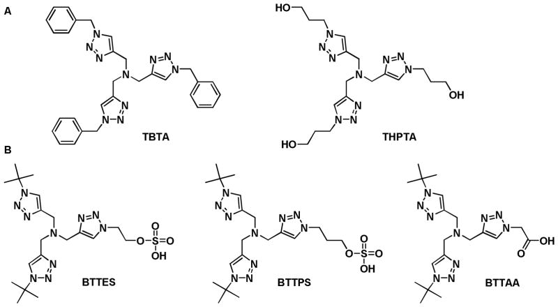
The first (A) and the second generation (B) tris(triazolylmethyl)amine-based ligands that accelerate CuAAC.
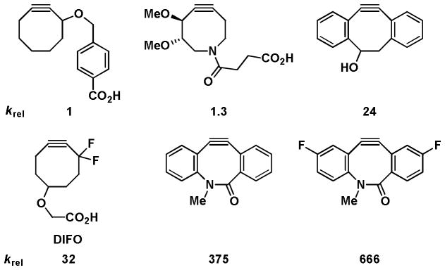
Relative reactivity of cyclooctyne and biarylazacyclooctynone probes.
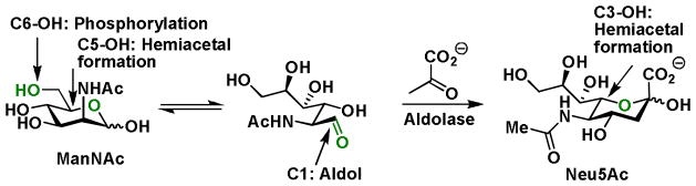
Functional groups in ManNAc participating in enzymatic transformations and hemiacetal formation.
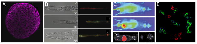
(A) A fluorescent image of Alexa Fluor 647-labeled fucosides in a zebrafish embryo (10 hpf). (B) Fluorescent images of old (green) and newly synthesized O-glycans (red) in C. elegans at different developmental stages (right panels), corresponding differential interference contrast (DIC) images (left panels) (adapted from reference 32). (C) Far-red fluorescence imaging of sialylated glycans in a mouse tumor model. Upper image shows signals from a vehicle control, lower image shows NA649 signal from a metabolically labeled mouse co-localizing with flanked tumor. (D) SPECT and X-ray CT fusion images of the same tumor model as in (C) labeled with DOTA-111In-conjugated neutravidin (adapted from reference 33). (E) Fluorescence images of S. cerevisiae with cell surface N-linked glycans labeled with Alexa Fluor 488 (green) or 647 (red) dye (adapted from reference 34).
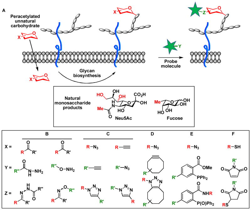
(A) Labeling cell-surface sialylated and fucosylated glycans using metabolic oligosaccharide engineering and bioorthogonal chemistry. Positions in sialic acid and fucose that are amenable for chemical tagging are highlighted in red. (B) condensation of ketones with aminooxy and hydrazide compounds. (C) CuAAC. (D) Copper-free click chemistry. (E) The Staudinger ligation. (F) Thiol-maleimide coupling reaction.
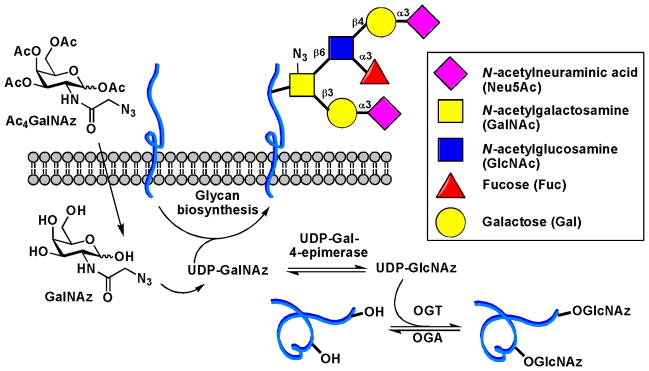
Metabolic labeling of mucin-type O-glycans and O-GlcNAcylated proteins
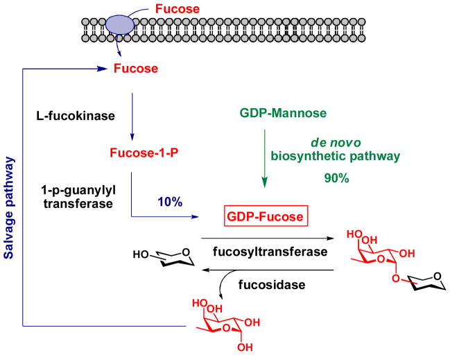
GDP-Fucose de novo biosynthetic pathway and salvage pathway in vertebrates.
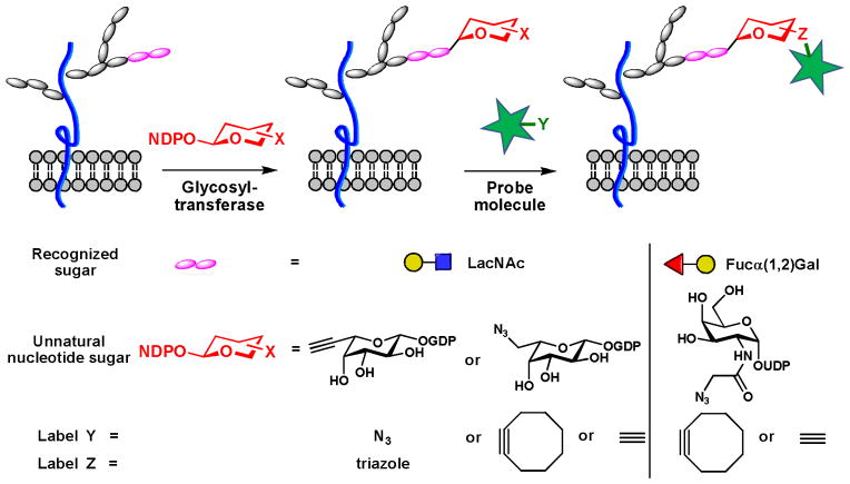
Chemoenzymatic labeling of cell-surface polysaccharides.
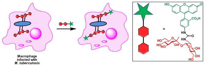
Detection of M. tuberculosis in infected macrophages.
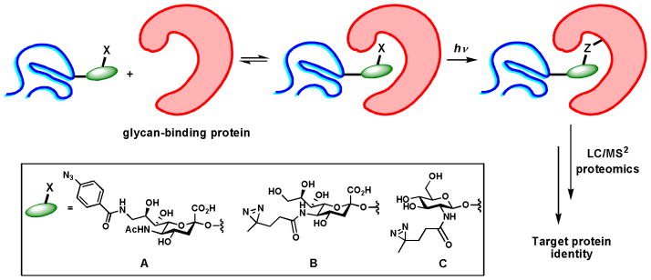
Identification of glycan-binding protein partners via photo crosslinking. (A, B) Unnatural sialic acid analogs used to capture sialic acid binding proteins. (C) The unnatural GlcNAc analog used to capture proteins that bind to O-GlcNAc-modified proteins.
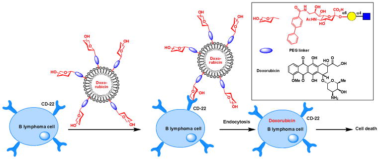
Using glycans as targeting epitopes to deliver drugs selectively to B lymphoma cells in mice models.
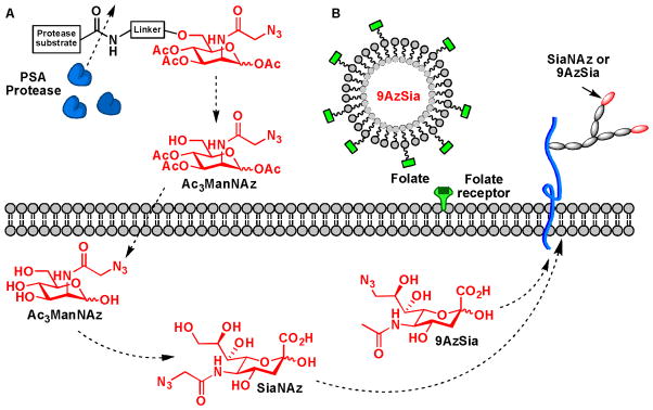
Targeted delivery of unnatural monosaccharide metabolites. (A) An uncaging strategy for targeted delivery of ManNAz to cancer cells. (B) Using folic acid as the targeting epitope.
References
Publication types
MeSH terms
Substances
Grants and funding
LinkOut - more resources
Full Text Sources
Other Literature Sources
Miscellaneous