. 2013 Jan;8(1):162-75.
doi: 10.1038/nprot.2012.150. Epub 2012 Dec 20.

Directed cardiomyocyte differentiation from human pluripotent stem cells by modulating Wnt/β-catenin signaling under fully defined conditions

Affiliations

Directed cardiomyocyte differentiation from human pluripotent stem cells by modulating Wnt/β-catenin signaling under fully defined conditions

Xiaojun Lian et al. Nat Protoc. 2013 Jan.

Abstract

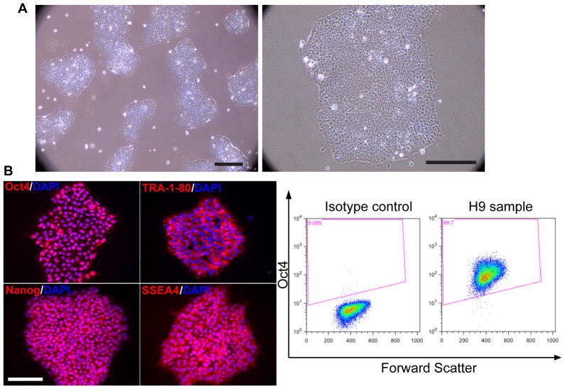
The protocol described here efficiently directs human pluripotent stem cells (hPSCs) to functional cardiomyocytes in a completely defined, growth factor- and serum-free system by temporal modulation of regulators of canonical Wnt signaling. Appropriate temporal application of a glycogen synthase kinase 3 (GSK3) inhibitor combined with the expression of β-catenin shRNA or a chemical Wnt inhibitor is sufficient to produce a high yield (0.8-1.3 million cardiomyocytes per cm(2)) of virtually pure (80-98%) functional cardiomyocytes in 14 d from multiple hPSC lines without cell sorting or selection. Qualitative (immunostaining) and quantitative (flow cytometry) characterization of differentiated cells is described to assess the expression of cardiac transcription factors and myofilament proteins. Flow cytometry of BrdU incorporation or Ki67 expression in conjunction with cardiac sarcomere myosin protein expression can be used to determine the proliferative capacity of hPSC-derived cardiomyocytes. Functional human cardiomyocytes differentiated via these protocols may constitute a potential cell source for heart disease modeling, drug screening and cell-based therapeutic applications.


Conflict of interest statement

COMPETING FINANCIAL INTERESTS

T.J.K. is a founder and consultant for Cellular Dynamics International, a company that uses human stem cells for drug testing. All the other authors declare no competing financial interests.

Figures

Figure 1
Figure 1
Analysis of undifferentiated hPSCs. (a) H9 hESCs were cultured on Matrigel-coated 6-well plates in mTeSR1 for 2 days. Bright field images of the typical morphology of undifferentiated H9 colonies are shown. Scale bar = 100 μm. (b) Immunofluorescent staining for Oct4, Nanog, TRA-1-80 and SSEA4 was performed on undifferentiated H9 cells. Scale bar = 100 μm. (c) Flow cytometry analysis of Oct4 expression in H9 hESCs cultivated on Matrigel-coated plates in mTeSR1.
Figure 2
Figure 2
Analysis of cardiomyocyte progenitor marker expression. (a) 19-9-11 iPSCs on Matrigel-coated 6-well plates were treated with 12 μM CHIR99021 for 24 hours. After 24 hours, flow cytometry analysis of brachyury expression was performed. The green histogram represents brachyury expression and the red histogram is an isotype control. (b) 19-9-11 iPSCs were cultured on Matrigel-coated 6-well plates in mTeSR1 for 4 days before exposure to 12 μM CH on day 0 and 5 μM IWP2 in RPMI/B27-insulin on day 3. At day 5, differentiated cells were singularized and replated on gelatin coated coverslips. At day 6, Isl1 expression was assessed by immunostaining and nucleus were stained with DAPI. Scale bar = 50 μm.
Figure 3
Figure 3
Schematic of protocol to differentiate of cardiomyocytes from hPSCs with small molecule modulators of canonical Wnt signaling. Bright field images of the typical morphology of day −3, day 0, day 1, day 5, day 8, day 15 cells from 19-9-11 are shown at 10X and 20X magnifications. Scale bar = 100 μm.
Figure 4
Figure 4
Structural characterization of cardiomyocytes generated from hPSCs via small molecule modulation of Wnt signaling (a–b) Cardiomyocytes were generated from 19-9-11 iPSCs using the protocol described in Fig. 3, with 12 μM CH treatment at day 0 and 5 μM IWP2 treatment at day 3. At day 20, cells were individualized and replated on 0.1% gelatin coated coverslips. Immunostaining for α-actinin (green) and MLC2a (red) shows sarcomere organization. Nuclei were stained with DAPI (a) Scale bar = 50 μm. (b) Scale bar = 20 μm.
Figure 5
Figure 5
Quantitative analysis of cardiomyocytes differentiated from hPSCs via small molecule modulation of Wnt signaling (a–b) Cardiomyocytes were generated from 19-9-11 iPSCs using the protocol described in Fig. 3, with 12 μM CH treatment at day 0 and 5 μM IWP2 treatment at day 3. At day 20, cells were analyzed for (a) cTnT/SMA and cTnT/MLC2a expression, and (b) MF20/Ki67 and MF20/BrdU incorporation by flow cytometry.

References

    1. Kehat I, et al. Human embryonic stem cells can differentiate into myocytes with structural and functional properties of cardiomyocytes. J Clin Invest. 2001;108:407–414. - PMC - PubMed
    1. Graichen R, et al. Enhanced cardiomyogenesis of human embryonic stem cells by a small molecular inhibitor of p38 MAPK. Differentiation. 2008;76:357–370. - PubMed
    1. Yang L, et al. Human cardiovascular progenitor cells develop from a KDR+ embryonic-stem-cell-derived population. Nature. 2008;453:524–528. - PubMed
    1. Kattman SJ, et al. Stage-specific optimization of activin/nodal and BMP signaling promotes cardiac differentiation of mouse and human pluripotent stem cell lines. Cell Stem Cell. 2011;8:228–240. - PubMed
    1. Mohr JC, et al. The microwell control of embryoid body size in order to regulate cardiac differentiation of human embryonic stem cells. Biomaterials. 2010;31:1885–1893. - PMC - PubMed

Publication types