Review
. 2013 Jan;14(1):25-37.
doi: 10.1038/nrm3494.

Microtubule attachment and spindle assembly checkpoint signalling at the kinetochore

Affiliations
Review

Microtubule attachment and spindle assembly checkpoint signalling at the kinetochore

Emily A Foley et al. Nat Rev Mol Cell Biol. 2013 Jan.

Abstract

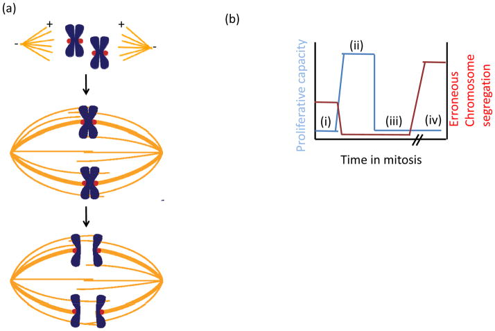
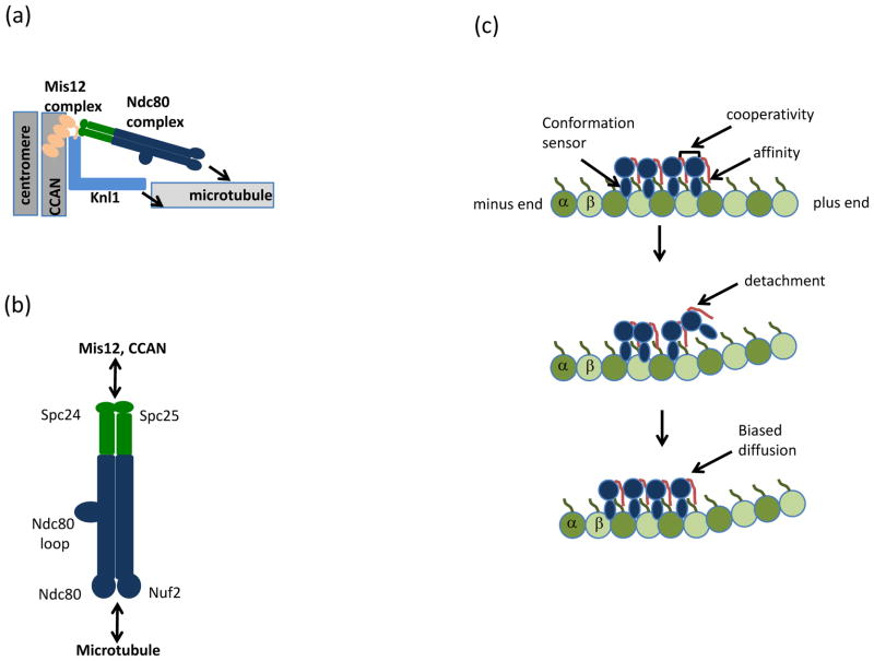
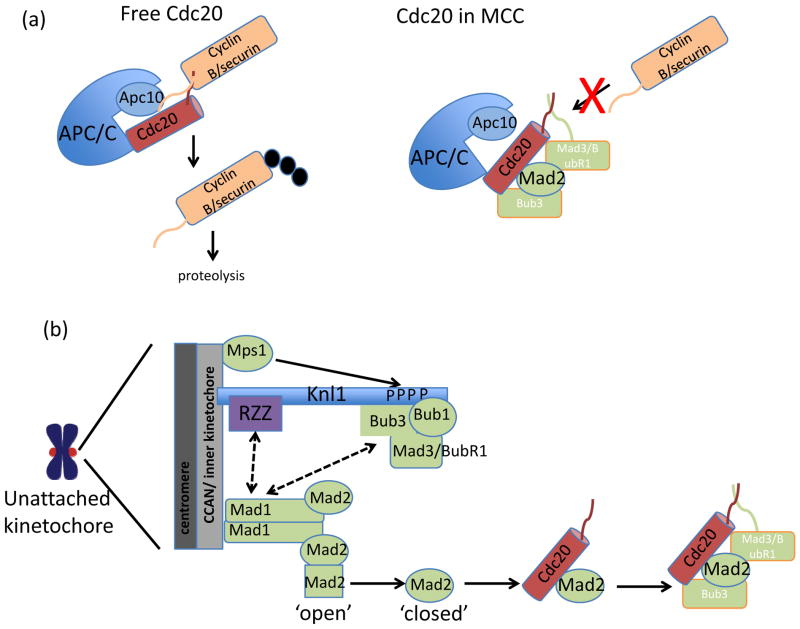
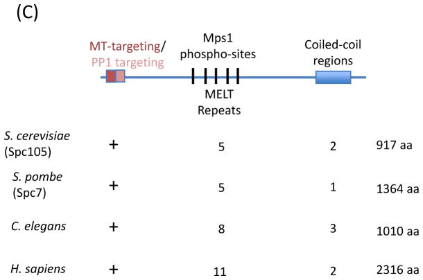
In eukaryotes, chromosome segregation during cell division is facilitated by the kinetochore, a multiprotein structure that is assembled on centromeric DNA. The kinetochore attaches chromosomes to spindle microtubules, modulates the stability of these attachments and relays the microtubule-binding status to the spindle assembly checkpoint (SAC), a cell cycle surveillance pathway that delays chromosome segregation in response to unattached kinetochores. Recent studies are shaping current thinking on how each of these kinetochore-centred processes is achieved, and how their integration ensures faithful chromosome segregation, focusing on the essential roles of kinase-phosphatase signalling and the microtubule-binding KMN protein network.


Figures

Figure 1
Figure 1. Speed and fidelity in cell division
(a) Schematic of chromosome segregation. (top) At the start of mitosis, a bipolar spindle composed of microtubules (orange) assembles with plus-ends (+) oriented towards the spindle equator and minus-ends (−) clustered at the spindle pole. At the spindle poles, centrosomes (not shown) are a major site of microtubule nucleation. (middle) Pairs of replicated chromosomes (blue) attach to the spindle via kinetochores (red). Error-free chromosome segregation depends on each sister attaching to microtubules from opposite spindle poles (bi-orientation). (bottom) Once all chromosomes have bi-oriented, sister chromatids are segregated to opposite spindle poles. (b) Schematic plot of proliferative capacity after cell division (blue) and relative chromosome segregation errors (red) as a function of time spent in mitosis. (i) When the spindle assembly checkpoint (SAC) is inactive, exit from mitosis occurs rapidly, independent of kinetochore-microtubule attachment, increasing the frequency of erroneous chromosome segregation and hence decreasing proliferative capacity. (ii) In an unperturbed cell cycle, proper chromosome-spindle attachments are established quickly allowing for timely exit from mitosis. Meanwhile, the SAC ensures anaphase occurs only when all kinetochores have attached to the spindle. (iii) Modest increases in the time spent in mitosis, which can arise from SAC activity, decrease proliferative capacity. This can occur despite the absence of segregation defects, due to activation of stress-response pathways. (iv) When the SAC cannot be satisfied, for example, in the presence of anti-mitotic drugs, cells arrest in mitosis, followed by cell cycle arrest or apoptosis.
Figure 2
Figure 2. Architectural organization of the KMN network
(a) Schematic of the Knl1-Mis12 complex-Ndc80 complex (KMN) network at the kinetochore. The four-subunit Mis-segregation 12 (Mis12) complex bridges Kinetochore null 1 (Knl1) and the Nuclear division cycle 80 (Ndc80) complex to the Conserved Centromere-Associated Network (CCAN) and underlying centromeric DNA. Arrows indicate microtubule binding activities in the Ndc80 complex and Knl1. (b) The Ndc80 complex, a dumbbell shaped heterotetramer, consists of a pair of microtubule-binding calponin-homology domains within the Ndc80 protein and Nuclear filamentous 2 (Nuf2) subunits (blue). The kinetochore domain is distal to the microtubule-binding domain and resides in the Spindle pole component 24 (Spc24) and Spc25 heterodimer (green). The subcomplexes interact via coiled-coil domains, which in the Ndc80 protein is interrupted by a loop that mediates protein-protein interactions. (c) Model for Ndc80 protein (blue) association with tubulin (green). Modified with permission from. The Ndc80 protein has the unusual property of recognizing the region between α and β tubulin subunits, in contrast to most microtubule-binding proteins which bind microtubules once per tubulin dimer. Recognition of both intra- and inter-tubulin interfaces is thought to allow the Ndc80 complex to preferentially bind straight (top) versus the curled (middle) tubulin interfaces at disassembling microtubule tips. During detachment of the Ndc80 protein (middle) from curved microtubule structures, cooperativity between Ndc80 complexes (mediated by the Ndc80 tail, red) allows persistent microtubule and diffusion towards the microtubule minus-end (bottom). The positively charged Ndc80 tail also increases the affinity of the Ndc80 complex for microtubules via interaction with the negatively charged tubulin E-hook (green line) present in α and β tubulin subunits.
Figure 3
Figure 3. Molecular mechanism of kinetochore-based SAC activation
(a) Chromosome segregation and exit from mitosis are triggered by the Anaphase Promoting Complex/Cyclosome (APC/C). Cell division cycle 20 (Cdc20), an activator of APC/C, forms a docking site with Apc10 that recognizes destruction box sequences in cyclin B and securin and promotes ubiquitylation (black circles) of these substrates. The SAC promotes the formation of a Cdc20 inhibitory complex known as Mitotic Checkpoint Complex (MCC). In the MCC, Cdc20 interaction with Apc10 is disrupted, and Mitotic arrest deficient 3 (Mad3)/Bub1-related 1 (BubR1) binds degron recognition sites in Cdc20, preventing ubiquitylation of cyclin B and securin. (b) Schematic of SAC activation at the kinetochore. On unattached kinetochores, Monopolar spindle 1 (Mps1) phosphorylates Knl1. Phosphorylated Knl1 binds Budding uninhibited by benomyl 1 (Bub1) and Bub3, and recruits Mad3/BubR1. In metazoans, Knl1 also recruits the Rod-ZW10-Zwilch (RZZ) complex. Together, these proteins recruit to the kinetochore heterodimer of Mad1 and ‘closed’ Mad2. The Mad1-Mad2 complex catalyzes the conversion of soluble ‘open’ Mad2 to ‘closed’ Mad2, which can associate with Cdc20 in the cytoplasm. Mad2-Cdc20 complexes are then bound by Mad3/BubR1 and Bub3 to form the MCC. (c) Knl1 schematic. Features of Knl1 that contribute to SAC signaling include the N-terminal basic patch that mediates microtubule binding (red) and protein phosphatase 1 (PP1) binding motif (pink), multiple MELT (M [D/E][I/L/V/M][S/T]) repeats (black lines), which are phosphorylated by Mps1, and coiled-coil domains (blue box) which mediate kinetochore targeting. The presence and/or number of these features in different systems are indicated below. Solid arrows indicate direct interactions; dashed arrows indicate unknown mechanism of recruitment.
Figure 4
Figure 4. Molecular mechanisms of kinetochore-microtubule attachment
(a–b) Dynamic regulation of kinetochore-microtubule binding affinity by phospho-regulation. (a) Schematic plot of microtubule binding affinity versus phosphorylation at unattached kinetochores. (b) Molecular effects of altering the kinase/phosphatase balance on KMN network microtubule binding. KMN network diagrammed as in Fig. 2. (1) If Aurora B kinase is inhibited and phosphorylation is low, microtubule-binding affinity is high and erroneous attachments cannot be eliminated. (2) Partial phosphorylation allows attachments that are sufficiently dynamic to allow stabilization of attachments that result in bi-orientation and release of improper attachments. (3) If B56-protein phosphatase 2A (PP2A) is inhibited, phosphorylation marks generated by Aurora B kinase increase on the KMN network, preventing stable kinetochore-microtubule binding. (c) Speculative model of phospho-regulation of kinetochore (grey)-microtubule (black) binding through outer-kinetochore phosphorylation by Aurora B and B56-PP2A. (i) On unattached kinetochores, which lack tension, centromeric Aurora B kinase is close to its outer kinetochore substrates and phosphorylation is high, despite the localization of B56-PP2A phosphatase at kinetochores. (ii) During prometaphase, lateral interactions produce intermediate tension, increasing both inter- and intra-kinetochore stretching which increases the distance between Aurora B and outer kinetochore substrates, while B56-PP2A localization to relative to the outer kinetochore is unchanged. The net result in decreased phosphorylation and (iii) stabilization of initial microtubule tip interactions. (iv) At metaphase, full microtubule occupancy results in the loss of B56-PP2A from the kinetochore, and limited access of Aurora B to its substrates allowing for stable attachments. (d) Association of the Ndc80 complex with the Duo1 and Mps1 interacting factor 1 (Dam1) complex and the Spindle and Kinetochore-Associated (SKA) complex biases and strengthens Ndc80 complex localization to the microtubule plus-end, enabling the formation of persistent kinetochore-microtubule interactions. (d) Tension, arising from bi-orientation, stabilizes attachments both by increasing the lifetime of kinetochore-microtubule interactions and by promoting microtubule polymerization. As diagrammed here, force-dependent stabilization of kinetochore-microtubule interactions depends on the Ndc80 complex, but other microtubule-binding proteins at the kinetochore may also contribute.
Figure 5
Figure 5. Molecular mechanisms of SAC extinction at the kinetochore
(a) SAC extinction at the kinetochore proceeds through PP1 targeting to Knl1, microtubule binding by Knl1, and, in metazoans, dynein-dependent stripping of kinetochore proteins including Mad1, Mad2, and Spindly, which binds to the RZZ complex. The substrates of PP1 at the kinetochore are not known but possible candidates include the MELT domains in Knl1 that are phosphorylated by Mps1. Together, these mechanisms lead to loss of SAC regulators from attached kinetochores but the interdependencies and molecular mechanisms of kinetochore eviction during SAC extinction remain largely unclear.

Similar articles

Cited by

References

    1. McAinsh AD, Meraldi P. The CCAN complex: linking centromere specification to control of kinetochore-microtubule dynamics. Semin Cell Dev Biol. 2011;22:946–52. - PubMed
    1. Cheeseman IM, Chappie JS, Wilson-Kubalek EM, Desai A. The conserved KMN network constitutes the core microtubule-binding site of the kinetochore. Cell. 2006;127:983–97. A seminal paper that identifies the KMN network as a conserved essential microtubule-binding activity at the kinetochore. - PubMed
    1. Gordon DJ, Resio B, Pellman D. Causes and consequences of aneuploidy in cancer. Nat Rev Genet. 2012;13:189–203. - PubMed
    1. Lengauer C, Kinzler KW, Vogelstein B. Genetic instabilities in human cancers. Nature. 1998;396:643–9. - PubMed
    1. Basu J, et al. Mutations in the essential spindle checkpoint gene bub1 cause chromosome missegregation and fail to block apoptosis in Drosophila. J Cell Biol. 1999;146:13–28. - PMC - PubMed

Publication types