. 2012 Dec;10(12):1555-66.
doi: 10.1158/1541-7786.MCR-12-0314.

PRSS3/mesotrypsin is a therapeutic target for metastatic prostate cancer

Affiliations

PRSS3/mesotrypsin is a therapeutic target for metastatic prostate cancer

Alexandra Hockla et al. Mol Cancer Res. 2012 Dec.

Abstract

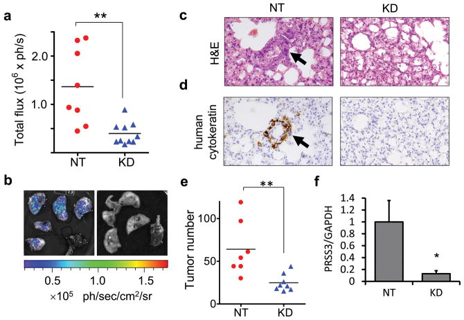
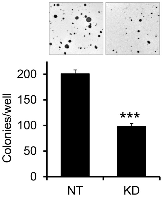
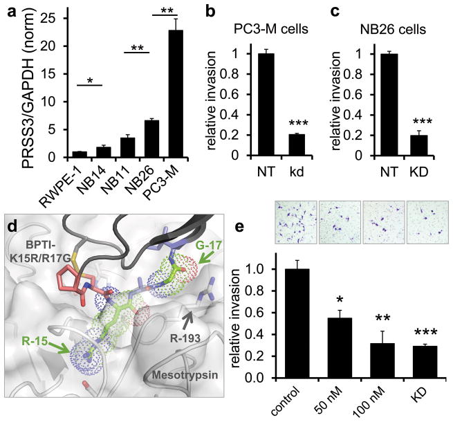
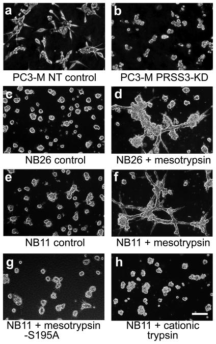
PRSS3/mesotrypsin is an atypical isoform of trypsin that has been associated with breast, lung, and pancreatic cancer cell malignancy. In analyses of open source transcriptional microarray data, we find that PRSS3 expression is upregulated in metastatic prostate cancer tissue, and that expression of PRSS3 in primary prostate tumors is prognostic of systemic progression following prostatectomy. Using a mouse orthotopic model with bioluminescent imaging, we show that PRSS3/mesotrypsin is critical for prostate cancer metastasis. Silencing of PRSS3 inhibits anchorage-independent growth of prostate cancer cells in soft agar assays, and suppresses invasiveness in Matrigel transwell assays and three-dimensional (3D) cell culture models. We further show that treatment with recombinant mesotrypsin directly promotes an invasive cellular phenotype in prostate cancer cells and find that these effects are specific and require the proteolytic activity of mesotrypsin, because neither cationic trypsin nor a mesotrypsin mutant lacking activity can drive the invasive phenotype. Finally, we show that a newly developed, potent inhibitor of mesotrypsin activity can suppress prostate cancer cell invasion to a similar extent as PRSS3 gene silencing. This study defines mesotrypsin as an important mediator of prostate cancer progression and metastasis, and suggests that inhibition of mesotrypsin activity may provide a novel modality for prostate cancer treatment.


Conflict of interest statement

Conflict of Interest Statement: The authors have no potential conflicts of interests to disclose.

Figures

Figure 1
Figure 1
PRSS3 expression in human prostate tumors. (a,b) PRSS3 expression correlates with increasing cancer progression. (a) Metastatic tumor tissue (n=5) showed significantly higher levels of PRSS3 expression than either benign prostate (n=5; p<0.001) or clinically localized cancers (n=5; p<0.005). The difference in PRSS3 expression between benign tissue and localized tumors was not significant in this dataset. Transcriptional microarray data were from (22). (b) Normal prostate (n=25), primary prostate tumors (n=65), and metastatic tumors (n=24 samples from 4 patients) showed a trend of increasing PRSS3 expression, with a significant difference between normal prostate and metastatic tissue (p=0.006). The difference in PRSS3 expression between primary tumors and metastases was not significant in this dataset. Transcriptional microarray data were from (23). (c) High PRSS3 expression in resected prostate tumors was significantly associated with early cancer recurrence following prostatectomy when comparing above-median (n=11) to below-median expressers (n=11) with respect to PRSS3 transcript levels (p=0.013). Transcriptional microarrays and associated survival data were from (24).
Figure 2
Figure 2
Bioluminescence imaging facilitates detection of tumor growth and metastasis in mouse orthotopic xenograft model. (a) PC3-M cells transduced with lentiviruses for firefly luciferase and GFP expression showed high transduction efficiency by comparing phase contrast (left) and fluorescence microscopy (right). Scale bar=50μm. (b) A luciferase assay of cell dilutions detected as few as 19 cells. (c) When PC3-M cells transduced with luciferase virus were implanted orthotopically (3 × 10⁴ cells), tumor growth could be followed in vivo by bioluminescence imaging. (d) Ex vivo bioluminescence imaging of organs and tissues was used to identify metastases in lung lobes, liver, kidneys, diaphragm, and spleen.
Figure 3
Figure 3
PRSS3 silencing slows prostate tumor growth in orthotopic xenografts. (a) Transduction with shRNA lentivirus targeting human PRSS3 resulted in effective knockdown in PC3-M cells (KD) relative to cells transduced with non-target control virus (NT), as assessed by qRT/PCR (bars, SEM; n=3). (b) PRSS3 KD cell lysates show reduced expression of mesotrypsinogen (32 kDa) relative to NT cell lysates on a Western blot; the blot was stripped and reprobed for actin (44 kDa) as a loading control. (c) In mice implanted with cells with knockdown of PRSS3 (KD) vs. nontarget control cells (NT), both superinfected with luciferase virus, PRSS3 knockdown mice (n=10) showed little change in tumor growth relative to the control mice (n=8), as assessed by in vivo bioluminescence imaging. (d) Tumors from three PRSS3 knockdown mice harvested after 2 weeks showed persistent suppression of PRSS3 expression relative to a tumor from a control mouse (NT) (bars, SEM; n=3). (e) Ex vivo imaging of resected prostates with PRSS3 knockdown primary tumors (n=10) showed reduced luminescence relative to controls (n=8), although the difference was not statistically significant. (f) Resected tumor-bearing prostates from the PRSS3 knockdown group (n=10) weighed significantly less than resected prostates from the control mice (n=8). Bars, SEM. *, p < 0.01; **, p < 0.005.
Figure 4
Figure 4
PRSS3 silencing inhibits prostate to lung metastasis in orthotopic xenografts. (a) Mice implanted with PC3-M cells in which PRSS3 was knocked down with viral shRNA (KD; n=10) showed significantly reduced pulmonary metastasis at two weeks post-implantation, as assessed by ex vivo imaging at necropsy, relative to control mice (NT; n=8) (**, p=0.0021). (b) Lungs from a mouse implanted with nontarget control virus-transduced PC3-M cells (NT) demonstrate flux measurement near the group median with extensive metastasis in all lobes (left), by contrast with lungs from a representative mouse implanted with PC3-M cells in which PRSS3 was knocked down with viral shRNA (KD), which show no evidence of metastasis (right). (c,d) Lung section from a mouse implanted with NT PC3-M cells (left panels) showed the presence of many small metastases such as that indicated by the black arrow in panels showing H&E staining (c) and immunohistochemical staining for human cytokeratins (d). Lung section from a mouse implanted with PRSS3 KD PC3-M cells (right panels) illustrates normal lung morphology (c) and background staining for human cytokeratins (d). (e) Histological and immunohistochemical analyses found a significantly lower mean number of lung metastases per section in the PRSS3 knockdown group (KD; n=8) compared to the control group (NT; n=7) (**, p=0.0022). (f) Lung tissue bearing metastases from mice with PRSS3 KD tumors (n=5) showed significantly lower expression of human PRSS3 when normalized to human GAPDH, relative to lung tissue from mice bearing NT control tumors (n=5) (bars, SEM; *, p=0.043).
Figure 5
Figure 5
PRSS3 silencing inhibits anchorage independent growth of prostate cancer cells. PC3-M cells transduced with shRNA virus targeting PRSS3 (KD) or with a nontarget control virus (NT) were plated in soft agar and grown for 2 weeks. Cells in which PRSS3 expression was suppressed showed significantly fewer colonies per well. Photographs of representative fields are shown above graphical results. ***, p<0.0001; bars, SEM; n=4 biological replicates.
Fig. 6
Fig. 6
PRSS3 silencing and pharmacological inhibition of mesotrypsin inhibit invasion of prostate cancer cells. (a) Endogenous PRSS3 expression, assessed by qRT/PCR, increases with progression in the RWPE-1 derived cell series, and is highly upregulated in the aggressive metastatic PC3-M cell line (bars, SEM; n=3). (b,c) Knockdown of PRSS3 in PC3-M cells (b) or NB26 cells (c) significantly reduced invasion in Matrigel transwell assays (bars, SEM; n=4). (d) Structure of mesotrypsin complex with prototype mesotrypsin inhibitor BPTI-K15R/R17G double mutant, highlighting (in green) inhibitor residues mutated to improve steric complementarity toward mesotrypsin active site residue Arg-193. (e) In Matrigel transwell invasion assays, PC3-M NT cells assayed in the presence of 50 or 100 nM BPTI-K15R/R17G showed significantly reduced invasion; 100 nM inhibitor gave results similar to PRSS3 knockdown. Photographs of representative fields from invasion filters are shown above graphical results (bars, SEM; n=4 biological replicates). *, p<0.05; **, p<0.01; ***, p<0.0005.
Figure 7
Figure 7
Endogenous and recombinant mesotrypsin stimulate invasive 3D growth morphology. (a,b) PC3-M cells transduced with nontarget control virus (a) or with PRSS3-targeted shRNA virus (b) were grown in Matrigel in serum-free media for 3 days and then photographed. (c,d) NB26 cells were grown in Matrigel and treated with (c) buffer only or (d) 100 nM active recombinant human mesotrypsin for 5 days and then photographed, (e,f,g,h) NB11 cells were grown in Matrigel and treated with (e) buffer only, (f) 100 nM active recombinant human mesotrypsin, (g) 100 nM catalytically inactive mesotrypsin-S195A mutant, or (h) 100 nM active recombinant human cationic trypsin for 5 days and then photographed. Scale bar=100 μm.

Comment in

References

    1. Siegel R, Naishadham D, Jemal A. Cancer statistics, 2012. CA Cancer J Clin. 2012;62:10–29. - PubMed
    1. Shah RB, Mehra R, Chinnaiyan AM, Shen R, Ghosh D, Zhou M, et al. Androgen-independent prostate cancer is a heterogeneous group of diseases: lessons from a rapid autopsy program. Cancer Res. 2004;64:9209–16. - PubMed
    1. Freedland SJ, Humphreys EB, Mangold LA, Eisenberger M, Dorey FJ, Walsh PC, et al. Risk of Prostate Cancer–Specific Mortality Following Biochemical Recurrence After Radical Prostatectomy. JAMA. 2005;294:433–9. - PubMed
    1. Arya M, Bott SR, Shergill IS, Ahmed HU, Williamson M, Patel HR. The metastatic cascade in prostate cancer. Surg Oncol. 2006;15:117–28. - PubMed
    1. Nguyen DX, Bos PD, Massague J. Metastasis: from dissemination to organ-specific colonization. Nat Rev Cancer. 2009;9:274–84. - PubMed

Publication types