

TGF expression and macrophage accumulation in atherosclerotic renal artery stenosis
- PMID: 23258796
- PMCID: PMC3613956
- DOI: 10.2215/CJN.06460612
TGF expression and macrophage accumulation in atherosclerotic renal artery stenosis
Abstract
Background and objectives: Atherosclerotic renal artery stenosis (ARAS) reduces renal blood flow and is a potential cause of chronic kidney injury, yet little is known regarding inflammatory pathways in this disorder in human participants. This study aimed to examine the hypothesis that reduced renal blood flow (RBF) in ARAS would be associated with tissue TGF-β activation and inflammatory cell accumulation.
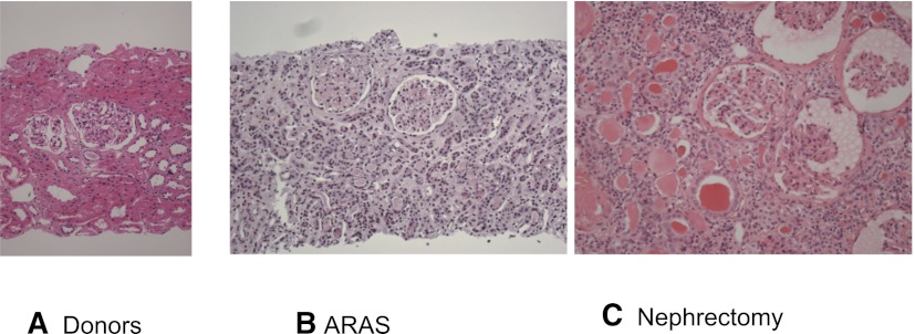
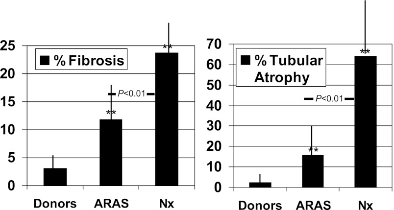
Design, setting, participants, & measurements: This cross-sectional study of ARAS of varying severity compared transjugular biopsy specimens in patients with ARAS (n=12, recruited between 2008 and 2012) with tissue from healthy kidney donors (n=15) and nephrectomy specimens from individuals with total vascular occlusion (n=65). ARAS patients were studied under controlled conditions to measure RBF by multidetector computed tomography and tissue oxygenation by blood oxygen level-dependent magnetic resonance imaging.
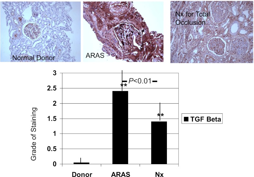
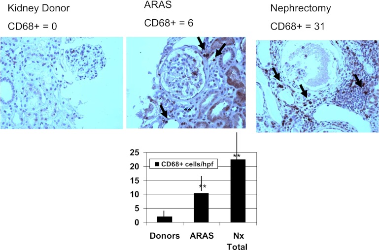
Results: Compared with the nonstenotic contralateral kidneys, RBF was reduced in poststenotic kidneys (242±149 versus 365+174 ml/min; P<0.01) as was single-kidney GFR (28±17 versus 41±19 ml/min; P<0.01), whereas cortical and medullary oxygenation were relatively preserved. Tissue TGF-β immunoreactivity was higher in ARAS patients compared with those with both normal kidneys and those with total occlusion (mean score 2.4±0.7 versus 1.5+1.1 in the nephrectomy group and versus 0±0 in donors; P<0.01). By contrast, the number of CD68+ macrophages was higher with greater disease severity (from 2.2±2.7 in normal to 22.4±18 cells/high-power field in nephrectomy samples; P<0.001).
Conclusions: The results of this study indicate robust stimulation of TGF-β associated with macrophage infiltration within the human kidney with vascular occlusive disease.
Figures
References
-
- Wheatley K, Ives N, Gray R, Kalra PA, Moss JG, Baigent C, Carr S, Chalmers N, Eadington D, Hamilton G, Lipkin G, Nicholson A, Scoble J, ASTRAL Investigators : Revascularization versus medical therapy for renal-artery stenosis. N Engl J Med 361: 1953–1962, 2009 - PubMed
-
- Korsakas S, Mohaupt MG, Dinkel HP, Mahler F, Do DD, Voegele J, Baumgartner I: Delay of dialysis in end-stage renal failure: Prospective study on percutaneous renal artery interventions. Kidney Int 65: 251–258, 2004 - PubMed
-
- Guo H, Kalra PA, Gilbertson DT, Liu J, Chen SC, Collins AJ, Foley RN: Atherosclerotic renovascular disease in older US patients starting dialysis, 1996 to 2001. Circulation 115: 50–58, 2007 - PubMed
-
- Greco BA, Breyer JA: The natural history of renal artery stenosis: Who should be evaluated for suspected ischemic nephropathy? Semin Nephrol 16: 2–11, 1996 - PubMed
Publication types
MeSH terms
Substances
Grants and funding
LinkOut - more resources
Full Text Sources
Other Literature Sources
Medical
