. 2012:2012:761843.
doi: 10.1155/2012/761843. Epub 2012 Nov 20.

Characterization of Porcine Ventral Mesencephalic Precursor Cells following Long-Term Propagation in 3D Culture

Affiliations

Characterization of Porcine Ventral Mesencephalic Precursor Cells following Long-Term Propagation in 3D Culture

Pia S Jensen et al. Stem Cells Int. 2012.

Abstract

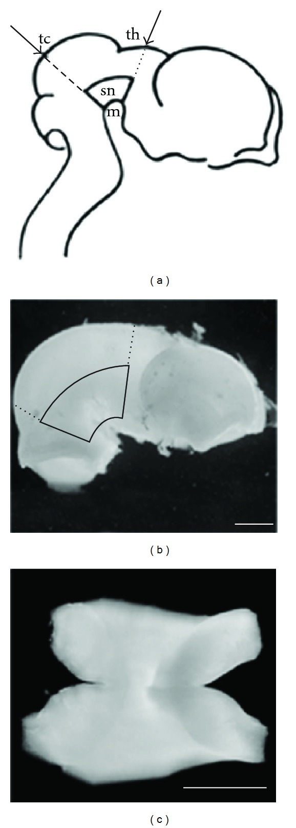
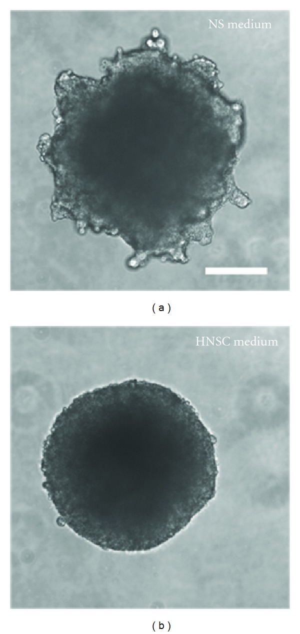
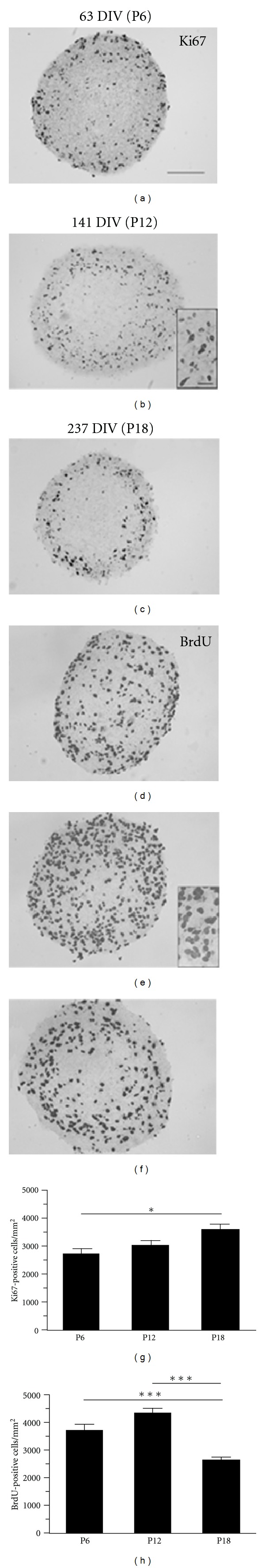
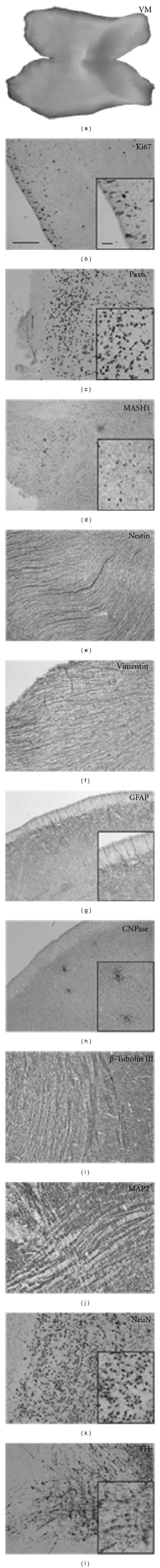
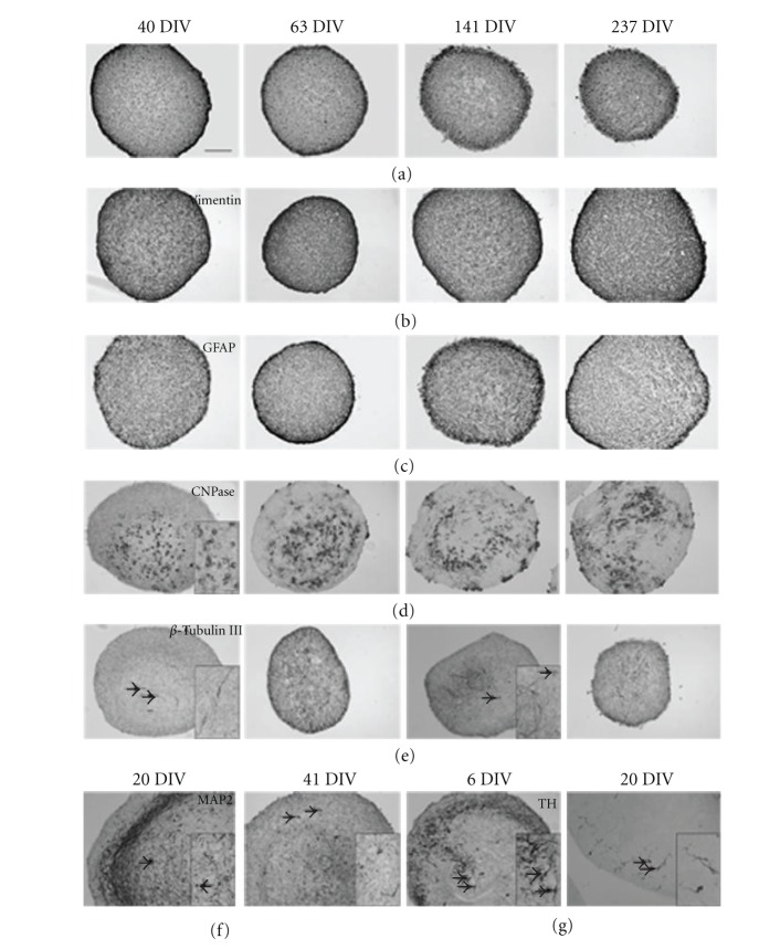
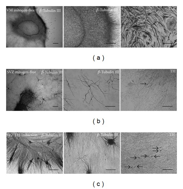
The potential use of predifferentiated neural precursor cells for treatment of a neurological disorder like Parkinson's disease combines stem cell research with previous experimental and clinical transplantation of developing dopaminergic neurons. One current obstacle is, however, the lack of ability to generate dopaminergic neurons after long-term in vitro propagation of the cells. The domestic pig is considered a useful nonprimate large animal model in neuroscience, because of a better resemblance of the larger gyrencephalic pig brain to the human brain than the commonly used brains of smaller rodents. In the present study, porcine embryonic (28-30 days), ventral mesencephalic precursor cells were isolated and propagated as free-floating neural tissue spheres in medium containing epidermal growth factor and fibroblast growth factor 2. For passaging, the tissue spheres were cut into quarters, avoiding mechanical or enzymatic dissociation in order to minimize cellular trauma and preserve intercellular contacts. Spheres were propagated for up to 237 days with analysis of cellular content and differentiation at various time points. Our study provides the first demonstration that porcine ventral mesencephalic precursor cells can be long-term propagated as neural tissue spheres, thereby providing an experimental 3D in vitro model for studies of neural precursor cells, their niche, and differentiation capacity.


Figures

Figure 1
Figure 1
Dissection of porcine embryonic (E28–30) ventral mesencephalon (VM). (a) Lateral perspective showing the angle of the first two cuts of the dissection, with imaginary lines (arrows) intersecting the dorsal surface of the tectum (tc) and the thalamus (th) (from Dunnett and Bjorklund, 1992). (b) Brain of a 28-day-old embryo with the dissection lines from (a) marked. The area encircled by solid lines represents the VM. (c) The ventral surface of the isolated VM explant. Scale bars: 500 μm.
Figure 2
Figure 2
Long-term propagated porcine ventral mesencephalic (VM) neural tissue spheres (NTS). The NTS were propagated in either (a) neurosphere- (NS-) proliferation medium or (b) human neural stem cell- (HNSC-) proliferation medium. Scale bar: 200 μm.
Figure 3
Figure 3
Proliferation in long-term propagated porcine ventral mesencephalic (VM) neural tissue spheres (NTS) visualized by Ki67 and BrdU immunostaining of histological sections. Photomicrographs of NTS showing expression of Ki67 at (a) 63 days in vitro (DIV) (passage (P) 6), (b) 141 DIV (P12), (c) 237 DIV (P18) and BrdU labelling at (d) 63 DIV, (e) 141 DIV, and (f) 237 DIV. Scale bars: 100 μm (overview) and 20 μm (insert). Quantification of Ki67 (g) and BrdU (h) expressing cells showed significantly more Ki67-positive cells at P18 as compared to P6, whereas there were significantly more BrdU-positive cells at P6 and P12 as compared to P18. Data are expressed as mean ± SEM (n = 15–19; *P < 0.05, ***P < 0.001).
Figure 4
Figure 4
Expression of stem cell markers and markers of glial and neuronal cells in primary (noncultured) porcine ventral mesencephalon (VM) obtained at embryonic day 28–30. Photomicrographs showing an intact VM (a), and immunoexpression of the proliferation marker Ki67 (b), the proneural markers Pax6 (c) and MASH1 (d), the neural stem cell marker nestin (e), the glial markers vimentin (f) and GFAP (g), the oligodendrocyte marker CNPase (h), the neuronal markers β Tubulin III (i), MAP2 (j) and NeuN (k), and the dopaminergic marker TH (l). Scale bars: 100 μm (overview) and 20 μm (insert).
Figure 5
Figure 5
Neural stem cell, glial, and neuronal markers in long-term propagated embryonic porcine ventral mesencephalic, neural tissue-spheres (NTS), stained after 40, 63, 141, and 237 days in vitro (DIV). Photomicrographs showing immunoexpression of nestin (a), vimentin (b), GFAP (c), CNPase (d), and β Tubulin III (e) in NTS. The neuronal marker MAP2 and the dopaminergic marker TH were not expressed in the long-term propagated NTS, but younger NTS cultures showed MAP2 expression (f) in NTS after 20 and 41 DIV and TH expression (g) after 6 and 21 DIV. Arrows mark neuronal cell soma magnified in inserts. DIV = days in vitro. Scale bars = 100 μm (overview) and 20 μm (insert).
Figure 6
Figure 6
Differentiation of long-term propagated embryonic porcine neural tissue spheres (NTS). Differentiation of VM NTS (51 and 170 days of age) induced by mitogen withdrawal did after 25 days induce a high number of β Tubulin III positive cells in the central core of the differentiated NTS (a), but no TH-positive cells were identified. Instead, the differentiated cells primarily expressed the astroglial marker GFAP and displayed a characteristic astroglial morphology. To verify the differentiation potential of the protocols used, 51-day-old NTS cultures derived from the embryonic porcine subventricular zone (SVZ) were included in the differentiation study as a positive control. Differentiation of SVZ NTS for 25 days by mitogen withdrawal (b) or by addition of the TH-inducing factors a-FGF, forskolin, TPA, and db-cAMP (c), known to stimulate TH expression in mammalian forebrain cultures, both resulted in β Tubulin III- and TH-positive cells. The β Tubulin III-positive cells had a characteristic neuronal morphology and were located in clusters/clones in the zone of migrating cells and in the central core of the differentiated SVZ NTS. The TH-positive cells were more numerous and appeared more morphologically immature by having shorter processes (c) compared to the cultures differentiated under mitogen-free conditions (b). Arrows mark soma of TH-positive cells. Scale bars = 200 μm and 100 μm in microphotographs of β Tubulin III expression at low and high magnification, respectively. 100 μm in microphotographs of GFAP and TH expression.

Similar articles

Cited by

References

    1. Dauer W, Przedborski S. Parkinson’s disease: mechanisms and models. Neuron. 2003;39(6):889–909. - PubMed
    1. Freed CR, Breeze RE, Rosenberg NL, et al. Therapeutic effects of human fetal dopamine cells transplanted in a patient with Parkinson’s disease. Progress in Brain Research. 1990;82:715–721. - PubMed
    1. Freed CR, Greene PE, Breeze RE, et al. Transplantation of embryonic dopamine neurons for severe Parkinson’s disease. The New England Journal of Medicine. 2001;344(10):710–719. - PubMed
    1. Brundin P, Pogarell O, Hagell P, et al. Bilateral caudate and putamen grafts of embryonic mesencephalic tissue treated with lazaroids in Parkinson’s disease. Brain. 2000;123(7):1380–1390. - PubMed
    1. Björklund A, Dunnett SB, Brundin P, et al. Neural transplantation for the treatment of Parkinson’s disease. The Lancet Neurology. 2003;2(7):437–445. - PubMed

LinkOut - more resources