. 2012 Jul;3(1):s125-s131.
doi: 10.1007/s12975-012-0174-9.

Gr1+ Macrophages and Dendritic Cells Dominate the Inflammatory Infiltrate 12 Hours After Experimental Intracerebral Hemorrhage

Affiliations

Gr1+ Macrophages and Dendritic Cells Dominate the Inflammatory Infiltrate 12 Hours After Experimental Intracerebral Hemorrhage

Matthew D Hammond et al. Transl Stroke Res. 2012 Jul.

Abstract

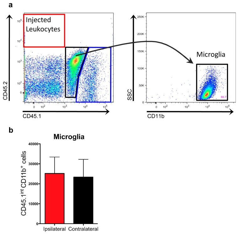
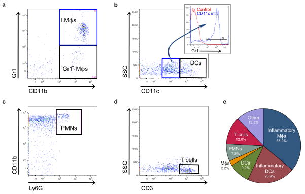
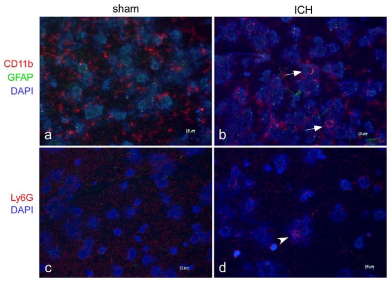
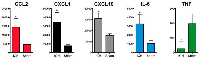
Intracerebral hemorrhage (ICH) is a devastating disease lacking an effective treatment. While the initial injury occurs within minutes, an inflammatory response contributes to ongoing tissue damage over hours to days. Relatively little is known about leukocyte trafficking into the brain in the hours after ICH onset. Understanding these events may lead to identification of new therapeutic targets. Using the blood injection mouse model of ICH, the numbers of leukocytes in the ipsilateral and contralateral brain were quantified by flow cytometry 12 hours after surgery. Perihematomal inflammation was confirmed by histology and chemokines and cytokines in the brain quantified by multiplex ELISA. Few neutrophils were detected in the brain 12 hours after ICH. The majority of leukocytes consisted of inflammatory macrophages (CD45.1(hi)CD3(-)Ly6G(-)CD11c(-)CD11b(+)Gr1(+) cells) and inflammatory dendritic cells (CD45.1(hi)CD3(-)Ly6G(-)CD11c(int)CD11b(+)Gr1(+) cells). Microglia numbers did not differ between the hemispheres. These results indicate that blood-derived monocyte populations traffic into brain early after ICH and outnumber neutrophils at 12 hours.


Figures

Fig. 1
Fig. 1
Microglia and blood-derived leukocytes can be distinguished from injected leukocytes. a Representative plots from ipsilateral brain samples showing few donor CD45.2+ cells at 12 hours. The CD45.1int population (black gate, center) is 99.7% CD11b+ and are classified as microglia. The blue gate (lower right corner) represents the host-derived CD45.1hi leukocyte population. b Total microglia numbers are not different between hemispheres. n=5
Fig. 2
Fig. 2
Representative plots showing leukocyte populations present after ICH. a Inflammatory macrophages (I.Mϕs) and Gr1 macrophages (Gr1 Mϕs) are both present in brain, but the inflammatory macrophages are much more numerous. This plot is gated on CD45.1hiCD3Ly6GCD11c cells. b Plot showing CD11cint (blue gate, center) and CD11chi dendritic cell populations (DCs, black gate, right) is gated on CD45.1hiCD3Ly6G cells. The majority of CD11cint cells express Gr1 (inset) and are termed inflammatory dendritic cells. c Few neutrophils (PMNs) are found in brain at 12 hours. The plot is gated on CD45.1hiCD3. d T cells are found in the ipsilateral hemisphere by 12 hours. The plot is gated on CD45.1hi cells. e Pie chart showing the distribution of leukocyte populations expressed as the mean percentage of infiltrating cells from 5 mice. “Other” indicates CD45.1hi cells that do not fall into any of the aforementioned gating strategies
Fig. 3
Fig. 3
Immunofluorescence staining demonstrates local inflammation. Representative perihematomal sections from 12 hours after ICH or sham surgery are shown in a–d. a–b Staining for CD11b, a marker for myeloid cells, demonstrates most CD11b+ cells after sham surgery (a) have branched cytoplasmic processes consistent with resting microglia. b Arrows indicate larger, spherical CD11b+ cells consistent with macrophages at the border of the ICH that were not found after sham surgery. Astrocytic processes, identified by GFAP+ staining, were difficult to identify after sham surgery but became more evident after ICH. c–d Neutrophils, identified by staining with Ly6G, could not be found after sham surgery and were found in small numbers around the ICH at 12 hours (arrowhead). Scale bar indicates 10 μm
Fig. 4
Fig. 4
ICH induces elevations of several chemokines and cytokines within 12 hours. The chemokines CCL2, CXCL1, and CXCL10 are significantly higher in ipsilateral ICH (n=4) brain versus ipsilateral sham (n=3) brain. The pro-inflammatory cytokine IL-6 is also elevated, but TNF is lower in ipsilateral ICH brain. All values are shown in pg/g brain protein. * p<0.05 by t-test after Holm-Bonferroni correction for multiple hypotheses

References

    1. Qureshi AI, Mendelow AD, Hanley DF. Intracerebral haemorrhage. Lancet. 2009;373:1632–1644. - PMC - PubMed
    1. Wang J, Dore S. Inflammation after intracerebral hemorrhage. J Cereb Blood Flow Metab. 2007;27:894–908. - PubMed
    1. Kane PJ, Modha P, Strachan RD, Cook S, Chambers IR, Clayton CB, Mendelow AD. The effect of immunosuppression on the development of cerebral oedema in an experimental model of intracerebral haemorrhage: Whole body and regional irradiation. J Neurol Neurosurg Psychiatry. 1992;55:781–786. - PMC - PubMed
    1. Lee ST, Chu K, Jung KH, Kim SJ, Kim DH, Kang KM, Hong NH, Kim JH, Ban JJ, Park HK, Kim SU, Park CG, Lee SK, Kim M, Roh JK. Anti-inflammatory mechanism of intravascular neural stem cell transplantation in haemorrhagic stroke. Brain. 2008;131:616–629. - PubMed
    1. Sansing LH, Kasner SE, McCullough L, Agarwal P, Welsh FA, KK Autologous blood injection to model spontaneous intracerebral hemorrhage in mice. Journal of Visualized Experiments. 2011 http://www.jove.com/index/Details.stp?ID=2618. - PMC - PubMed