. 2012 Dec 19;3(12):1063-72.
doi: 10.1021/cn300145z. Epub 2012 Sep 13.

Rotenone inhibits autophagic flux prior to inducing cell death

Affiliations

Rotenone inhibits autophagic flux prior to inducing cell death

Burton J Mader et al. ACS Chem Neurosci. .

Abstract

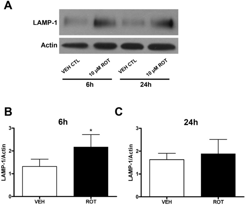
Rotenone, which selectively inhibits mitochondrial complex I, induces oxidative stress, α-synuclein accumulation, and dopaminergic neuron death, principal pathological features of Parkinson's disease. The autophagy-lysosome pathway degrades damaged proteins and organelles for the intracellular maintenance of nutrient and energy balance. While it is known that rotenone causes autophagic vacuole accumulation, the mechanism by which this effect occurs has not been thoroughly investigated. Treatment of differentiated SH-SY5Y cells with rotenone (10 μM) induced the accumulation of autophagic vacuoles at 6 h and 24 h as indicated by Western blot analysis for microtubule associated protein-light chain 3-II (MAP-LC3-II). Assessment of autophagic flux at these time points indicated that autophagic vacuole accumulation resulted from a decrease in their effective lysosomal degradation, which was substantiated by increased levels of autophagy substrates p62 and α-synuclein. Inhibition of lysosomal degradation may be explained by the observed decrease in cellular ATP levels, which in turn may have caused the observed concomitant increase in acidic vesicle pH. The early (6 h) effects of rotenone on cellular energetics and autophagy-lysosome pathway function preceded the induction of cell death and apoptosis. These findings indicate that the classical mitochondrial toxin rotenone has a pronounced effect on macroautophagy completion that may contribute to its neurotoxic potential.


Figures

Figure 1
Figure 1
Rotenone induced SH-SY5Y cell death and caspase-3-like activity are concentration- and time-dependent. Rotenone induced a concentration-dependent decrease in cell viability (A) and increase in caspase-3-like activity (B), observed 24 h after treatment. 10 μM rotenone induced a time-dependent decrease in SH-SY5Y cell viability (C) that was accompanied by an increase in caspase-3-like activity (D) at 48 and 72 h of treatment. Complete inhibition of rotenone-induced caspase-3-like activity (not shown) with the broad caspase inhibitor Boc-FMK (Boc-Asp-FMK) does not attenuate the decrease in cell viability after 48 h treatment with 10 μM rotenone (E). Rotenone (10 μM) induced a significant decrease in cellular ATP levels (μM/well) at 6 and 12 h after treatment compared to CTL (F). Results represent mean ± standard deviation, and experiments were repeated independently at least three times with similar results. *p < 0.05 vs 0 μM rotenone CTL (A, B, E, F); *p < 0.05 vs 0 h rotenone (C, D).
Figure 2
Figure 2
Rotenone causes accumulation of autophagic vacuoles by blocking their effective degradation. (A) Representative Western blot of LC3-II (14 kDa) and actin (42 kDa) loading control for SH-SY5Y lysates collected 6 and 24 h following treatment with 10 μM rotenone (ROT) in the presence or absence of 100 nM bafilomycin A1 (BafA1). Rotenone caused a significant increase in LC3-II immunoreactivity at both 6 and 24 h following treatment (A–C). To measure autophagic flux, 100nM BafA1 was added for the last 2 h of rotenone treatment prior to preparing lysates. Quantification of LC3-II/actin ratios for each treatment is expressed graphically as fold CTL for 6 h rotenone (B) and 24 h rotenone (C). Treatment with 10 μM rotenone induced a significant increase in AV accumulation, an effect that was not significantly greater upon treatment with 100 nM BafA1. Representative Western blot indicates that rotenone increase levels of the autophagy substrate p62 at both 6 and 24 h after treatment (D). Side bar in (D) indicates higher p62-immunoreactive species following 6 and 24 h treatment with rotenone that suggests its enhanced ubiquitination. Blots were stripped and reprobed for actin. Immunoreactivity for p62 (normalized to actin) is quantified graphically in (E) and is expressed as fold VEH CTL. Results = mean ± standard deviation from three to five independent experiments. *p < 0.05 vs VEH CTL.
Figure 3
Figure 3
Rotenone increases levels of α-synuclein. (A) Representative Western blot analysis of whole cell lysates indicates an increase in high molecular weight species of α-synuclein (>50 kDa) following 48 h treatment with 10 μM rotenone. (B) Quantification of high-molecular weight species of α-synuclein indicates a significant increase vs vehicle control. Results = mean ± standard deviation obtained from three independent experiments. *p < 0.05 vs VEH CTL.
Figure 4
Figure 4
Rotenone causes an increase in acidic vesicle pH. (A) Representative histogram of alterations in acidic vesicle pH in SH-SY5Y cells after treatment with DMSO vehicle (VEH, 24 h, orange line), rotenone (ROT, 10 μM, 24 h, blue line), or bafilomycin A1 (BafA, 100 nM, 4 h, pink line) as determined by flow cytometry. Effects of rotenone treatment are expressed as a leftward shift in fluorescence in the cell population when compared to vehicle control, suggesting a loss of lysosomal/acidic vesicle pH. (B) Quantification of LTR mean fluorescence intensity (MFI) indicates a >30% decrease following 6 and 24 h treatment with rotenone. (C) Phase contrast and fluorescence microscopy were used to image the rotenone-induced attenuation of LTR fluorescence at 24 h after treatment. Results = mean ± standard deviation from five independent experiments. *p < 0.05 vs VEH CTL.
Figure 5
Figure 5
Rotenone does not induce lysosomal membrane permeabilization (LMP). Confocal microscopy images obtained via double label immunocytochemistry for the soluble lysosomal enzyme cathepsin D (red, Cy3) and the lysosomal membrane protein LAMP-1 (green, FITC) following treatment for 24 h with DMSO vehicle (top row), 10 μM rotenone (ROT, middle row), or 50 μM chloroquine (CQ, bottom row). Punctate immunoreactivity for cathepsin D that colocalized to regions of the cell exhibiting intense LAMP-1 immunoreactivity was apparent in vehicle and rotenone-treated cells. Diffuse staining for cathepsin D was observed in chloroquine-treated cells that did not localize intracellularly to that of LAMP-1, suggesting the onset of LMP. Images are representative of LMP assessment from three independent experiments.
Figure 6
Figure 6
Rotenone increases levels of LAMP-1. Representative Western blot (A) for LAMP-1 (∼110 kDa) along with actin (42 kDa) loading control for lysates obtained from SH-SY5Y cells treated with vehicle control (VEH CTL) or 10 μM rotenone (ROT) for 6 or 24 h. Quantification of LAMP-1 signal averaged from four independent experiments indicated that levels of LAMP-1 were significantly greater at 6 h following rotenone. *p < 0.05 vs vehicle CTL.
Figure 7
Figure 7
Rotenone exposure does not alter TFEB levels. (A) Representative Western blot analysis for TFEB (53 kDa) and GAPDH (37 kDa) loading control following 6 and 24 h treatment with vehicle control (CTL) or 10 μM rotenone (ROT). (B) Quantification of TFEB/GAPDH ratios following 6 and 24 h treatment of rotenone are expressed graphically as fold CTL. Results = mean ± standard deviation obtained from six independent experiments.

References

    1. Olanow C. W.; Stern M. B.; Sethi K. (2009) The scientific and clinical basis for the treatment of Parkinson disease (2009). Neurology 72, S1–136. - PubMed
    1. Dauer W.; Przedborski S. (2003) Parkinson’s disease: mechanisms and models. Neuron 39, 889–909. - PubMed
    1. Klionsky D. J.; Abeliovich H.; Agostinis P.; Agrawal D. K.; Aliev G.; Askew D. S.; Baba M.; Baehrecke E. H.; Bahr B. A.; Ballabio A.; Bamber B. A.; Bassham D. C.; Bergamini E.; Bi X.; Biard-Piechaczyk M.; Blum J. S.; Bredesen D. E.; Brodsky J. L.; Brumell J. H.; Brunk U. T.; Bursch W.; Camougrand N.; Cebollero E.; Cecconi F.; Chen Y.; Chin L. S.; Choi A.; Chu C. T.; Chung J.; Clarke P. G.; Clark R. S.; Clarke S. G.; Clave C.; Cleveland J. L.; Codogno P.; Colombo M. I.; Coto-Montes A.; Cregg J. M.; Cuervo A. M.; Debnath J.; Demarchi F.; Dennis P. B.; Dennis P. A.; Deretic V.; Devenish R. J.; Di Sano F.; Dice J. F.; Difiglia M.; Dinesh-Kumar S.; Distelhorst C. W.; Djavaheri-Mergny M.; Dorsey F. C.; Droge W.; Dron M.; Dunn W. A. Jr.; Duszenko M.; Eissa N. T.; Elazar Z.; Esclatine A.; Eskelinen E. L.; Fesus L.; Finley K. D.; Fuentes J. M.; Fueyo J.; Fujisaki K.; Galliot B.; Gao F. B.; Gewirtz D. A.; Gibson S. B.; Gohla A.; Goldberg A. L.; Gonzalez R.; Gonzalez-Estevez C.; Gorski S.; Gottlieb R. A.; Haussinger D.; He Y. W.; Heidenreich K.; Hill J. A.; Hoyer-Hansen M.; Hu X.; Huang W. P.; Iwasaki A.; Jaattela M.; Jackson W. T.; Jiang X.; Jin S.; Johansen T.; Jung J. U.; Kadowaki M.; Kang C.; Kelekar A.; Kessel D. H.; Kiel J. A.; Kim H. P.; Kimchi A.; Kinsella T. J.; Kiselyov K.; Kitamoto K.; Knecht E.; Komatsu M.; Kominami E.; Kondo S.; Kovacs A. L.; Kroemer G.; Kuan C. Y.; Kumar R.; Kundu M.; Landry J.; Laporte M.; Le W.; Lei H. Y.; Lenardo M. J.; Levine B.; Lieberman A.; Lim K. L.; Lin F. C.; Liou W.; Liu L. F.; Lopez-Berestein G.; Lopez-Otin C.; Lu B.; Macleod K. F.; Malorni W.; Martinet W.; Matsuoka K.; Mautner J.; Meijer A. J.; Melendez A.; Michels P.; Miotto G.; Mistiaen W. P.; Mizushima N.; Mograbi B.; Monastyrska I.; Moore M. N.; Moreira P. I.; Moriyasu Y.; Motyl T.; Munz C.; Murphy L. O.; Naqvi N. I.; Neufeld T. P.; Nishino I.; Nixon R. A.; Noda T.; Nurnberg B.; Ogawa M.; Oleinick N. L.; Olsen L. J.; Ozpolat B.; Paglin S.; Palmer G. E.; Papassideri I.; Parkes M.; Perlmutter D. H.; Perry G.; Piacentini M.; Pinkas-Kramarski R.; Prescott M.; Proikas-Cezanne T.; Raben N.; Rami A.; Reggiori F.; Rohrer B.; Rubinsztein D. C.; Ryan K. M.; Sadoshima J.; Sakagami H.; Sakai Y.; Sandri M.; Sasakawa C.; Sass M.; Schneider C.; Seglen P. O.; Seleverstov O.; Settleman J.; Shacka J. J.; Shapiro I. M.; Sibirny A.; Silva-Zacarin E. C.; Simon H. U.; Simone C.; Simonsen A.; Smith M. A.; Spanel-Borowski K.; Srinivas V.; Steeves M.; Stenmark H.; Stromhaug P. E.; Subauste C. S.; Sugimoto S.; Sulzer D.; Suzuki T.; Swanson M. S.; Tabas I.; Takeshita F.; Talbot N. J.; Talloczy Z.; Tanaka K.; Tanida I.; Taylor G. S.; Taylor J. P.; Terman A.; Tettamanti G.; Thompson C. B.; Thumm M.; Tolkovsky A. M.; Tooze S. A.; Truant R.; Tumanovska L. V.; Uchiyama Y.; Ueno T.; Uzcategui N. L.; van der Klei I.; Vaquero E. C.; Vellai T.; Vogel M. W.; Wang H. G.; Webster P.; Wiley J. W.; Xi Z.; Xiao G.; Yahalom J.; Yang J. M.; Yap G.; Yin X. M.; Yoshimori T.; Yu L.; Yue Z.; Yuzaki M.; Zabirnyk O.; Zheng X.; Zhu X.; Deter R. L. (2008) Guidelines for the use and interpretation of assays for monitoring autophagy in higher eukaryotes. Autophagy 4, 151–175. - PMC - PubMed
    1. Shacka J. J.; Roth K. A.; Zhang J. (2008) The autophagy-lysosomal degradation pathway: role in neurodegenerative disease and therapy. Front. Biosci. 13, 718–736. - PubMed
    1. Pivtoraiko V. N.; Stone S. L.; Roth K. A.; Shacka J. J. (2009) Oxidative stress and autophagy in the regulation of lysosome-dependent neuron death. Antioxid. Redox Signaling 11, 481–496. - PMC - PubMed

Publication types

MeSH terms

Substances