. 2012 Dec 15;1(12):1258-63.
doi: 10.1242/bio.20122816. Epub 2012 Oct 11.

Reelin-dependent ApoER2 downregulation uncouples newborn neurons from progenitor cells

Affiliations

Reelin-dependent ApoER2 downregulation uncouples newborn neurons from progenitor cells

F Javier Pérez-Martínez et al. Biol Open. .

Abstract

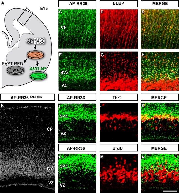
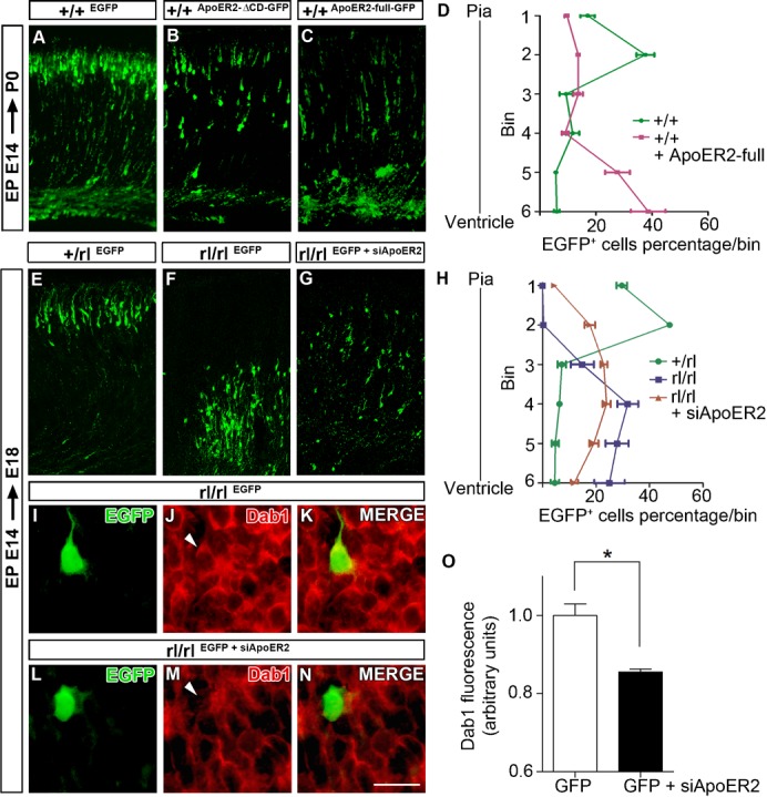
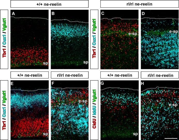
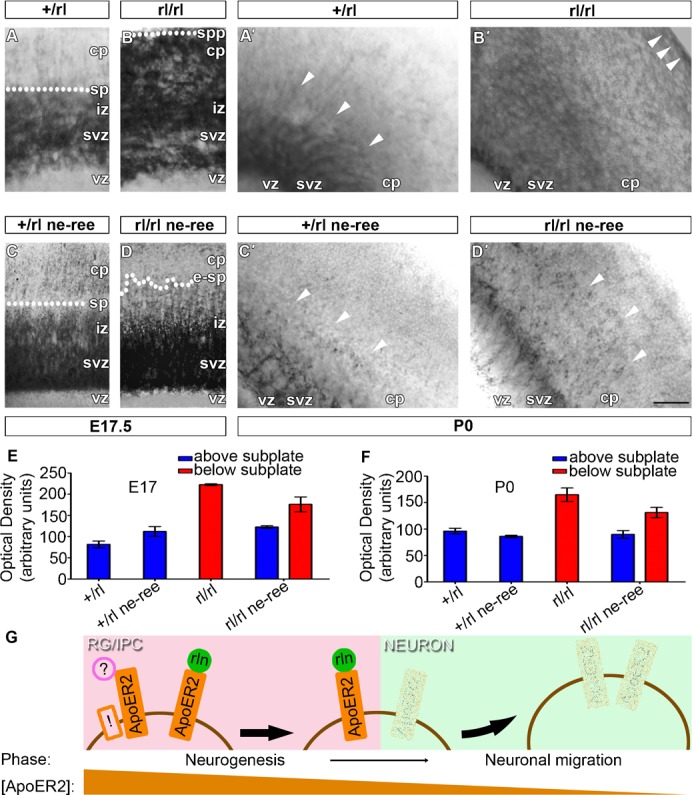
Reelin and its receptor machinery are well known to be required for the migration and positioning of neocortical projection neurons. More recently, reelin has been shown both necessary and sufficient to determine the rate of neocortical neurogenesis. The molecular links underlying its seemingly distinct proliferative and post-proliferative functions remain unknown. Here we reveal an enriched expression of functional reelin receptors, largely of Apolipoprotein E Receptor 2 (ApoER2), in radial glia basal processes and intermediate progenitor cells during mid/late cortical development. In vivo, ApoER2 overexpression inhibits neuronal migration. In contrast, precluding excessive levels of ApoER2 in reelin-deficient cortices, by either ApoER2 knock-down or the transgenic expression of reelin in neural progenitor cells, improves neuronal migration and positioning. Our study provides groundwork for the highly orchestrated clearance of neocortical neurons from their birth site, suggesting that a reelin-dependent ApoER2 downregulation mechanism uncouples newborn neurons from progenitor cells, thereby enabling neurons to migrate.

Keywords: ApoER2; Dab1; Mouse; Neurogenesis; Neuronal migration; Reelin.


Conflict of interest statement

Competing interests: The authors have no competing interests to declare.

Figures

Fig. 1.
Fig. 1.. Neocortical NPCs express functional ApoER2.
In situ binding with AP-RR36, an Alkaline Phosphatase-fusion probe of the receptor binding region of reelin, reveals the presence of functional reelin receptors, largely ApoER2, in the developing cortex at E15 (A,B). AP-RR36 staining is observed in several BLBP+ radial glia processes spanning the cortical wall (C–E) as well as in subpopulations of radial glia cell bodies in the basal most VZ (F–H). Most Tbr2+ IPCs in the SVZ (I–K) and some BrdU+ cycling progenitors (L–N) stain also with AP-RR36. Scale bar: 200 µm in B; 150 µm in C–E; 50 µm in F–N.
Fig. 2.
Fig. 2.. ApoER2 loading determines neuronal migratory behavior.
Overexpression of ApoER2 inhibits neuronal migration (A–D). P0 brains that had been transfected at E14 with an EGFP plasmid (control) (A) or with truncated (B) or full-length (C) ApoER2 plasmids are depicted. Note the alignment of a layer of neurons beneath the marginal zone in control cortex (A). Fewer neurons are aligned and many of them appear halted in their radial migratory trajectories in the brains transfected with ApoER2 plasmids (B,C). ApoER2 knock-down rescues neuronal migration and Dab1 levels in reeler (E–O). E18 cortices that had been transfected at E14 either with an EGFP plasmid (control) or with an EGFP plasmid plus an RNAi plasmid for ApoER2 (siApoER2) are depicted. A band of aligned neurons is visible beneath the marginal zone in EGFP-electroporated heterozygous cortices (E). In EGFP-electroporated reeler cortices neurons do not form an apparent band and appears confined at the bottom of the cortical wall (F). Partial rescue of neuronal migration is apparent in EGFP+siApoER2-electroporated reeler cortices (G). Dab1 levels are reduced in reeler cortical neurons after EGFP+siApoER2 electroporation (I–O). Scale bar: 150 µm in A–C,E–G; 15 µm in E–K,L–N.
Fig. 3.
Fig. 3.. Partial rescue of inside-out patterning in reeler by ectopic reelin in NPCs.
Immunohistochemical detection at P0 of Tbr1, Cux1 and VGlut1 on adjacent sections of ne-reelin (A,B) and reeler ne-reelin (C,D) cortices. In the WT background, the expression of the ne-reelin transgene does not alter cortical lamination. In the reeler background it does rescue cortical layering in the cell population above, but not that below, the ectopic subplate. Use of Fab blocking fragments confirmed these results allowing the combination of antibodies on the same section (E,F). Sequential injection of the nucleotide analogues CldU and IddU by E13 and E14, respectively, further demonstrate at P0 a rescue of the inside-out lamination pattern above the ectopic ne-reelin reeler subplate (G,H). Scale bar: 200 µm.
Fig. 4.
Fig. 4.. Partial rescue of ApoER2 downregulation in reeler by ectopic reelin in NPCs.
AP-RR36 in situ staining detects functional ApoER2 at E17 (A–D) and P0 (A′–D′) in heterozygous (control), reeler, heterozygous ne-reelin and reeler ne-reelin cortices. AP-RR36 staining is much more intense in the reeler CP. In the presence of the endogenous protein (heterozygous background) the ne-reelin transgene does not alter AP-RR36 staining. In the reeler background, ne-reelin induces a strong decrease of AP-RR36 staining above the ectopic subplate (circles and arrowheads indicate the approximate position of the subplate remnants). Semi-quantitative densitometric analysis of these effects (E,F). Summary of results and proposed model (G): Radial glia and intermediate progenitor cells (RG/IPC) are enriched with functional ApoER2. A reelin (ree)-dependent ApoER2 downregulation mechanism uncouples newborn neurons from RG/IPC, thereby enabling cells to migrate (with a plasma membrane ApoER2 concentration [ApoER2]-dependent switch from negative to positive effects). Alternative ApoeER2 ligands (?) or reelin co-receptors (!) might further depict reelin as a permissive rather than instructive signal for neuronal migration, Scale bar: 120 µm.

References

    1. Anthony T. E., Klein C., Fishell G., Heintz N. (2004). Radial glia serve as neuronal progenitors in all regions of the central nervous system. Neuron 41, 881–890 10.1016/S0896-6273(04)00140-0 - DOI - PubMed
    1. Arnaud L., Ballif B. A., Förster E., Cooper J. A. (2003). Fyn tyrosine kinase is a critical regulator of disabled-1 during brain development. Curr. Biol. 13, 9–17 10.1016/S0960-9822(02)01397-0 - DOI - PubMed
    1. Benhayon D., Magdaleno S., Curran T. (2003). Binding of purified Reelin to ApoER2 and VLDLR mediates tyrosine phosphorylation of Disabled-1. Brain Res. Mol. Brain Res. 112, 33–45 10.1016/S0169-328X(03)00032-9 - DOI - PubMed
    1. Blake S. M., Strasser V., Andrade N., Duit S., Hofbauer R., Schneider W. J., Nimpf J. (2008). Thrombospondin-1 binds to ApoER2 and VLDL receptor and functions in postnatal neuronal migration. EMBO J. 27, 3069–3080 10.1038/emboj.2008.223 - DOI - PMC - PubMed
    1. Bock H. H., Herz J. (2003). Reelin activates SRC family tyrosine kinases in neurons. Curr. Biol. 13, 18–26 10.1016/S0960-9822(02)01403-3 - DOI - PubMed