. 2012 Jun 6;5(Suppl 1):S7.
doi: 10.1186/1755-1536-5-S1-S7. eCollection 2012.

Molecular mechanisms of endothelial to mesenchymal cell transition (EndoMT) in experimentally induced fibrotic diseases

Affiliations

Molecular mechanisms of endothelial to mesenchymal cell transition (EndoMT) in experimentally induced fibrotic diseases

Sonsoles Piera-Velazquez et al. Fibrogenesis Tissue Repair. .

Abstract

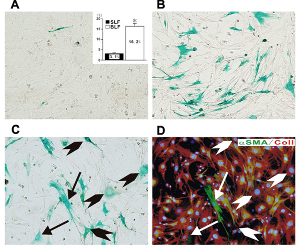
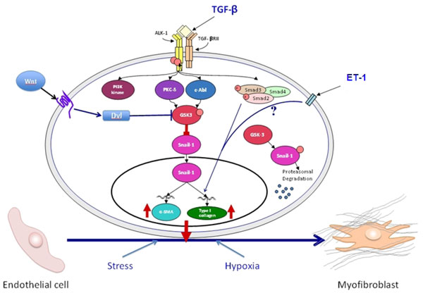
Several recent studies have demonstrated that endothelial to mesenchymal transition (EndoMT), a newly recognized type of cellular transdifferentiation may be an important source of myofibroblasts during the development of experimentally induced pulmonary, cardiac and kidney fibrosis. EndoMT is a complex biological process induced by members of the transforming growth factor (TGF-β) family of regulatory polypeptides in which endothelial cells adopt a mesenchymal or myofibroblastic phenotype acquiring motile and contractile properties and initiating expression of mesenchymal cell products such as α smooth muscle actin (α-SMA) and type I collagen. Although these experimental studies provide compelling evidence for the participation of EndoMT in the development of experimentally-induced fibrotic processes the precise role of EndoMT in the pathogenesis of human fibrotic disorders requires confirmation and validation from studies of human clinical pathologic conditions. Such confirmation should lead to a change in the paradigm of the origin of profibrogenic myofibroblasts involved in human fibrotic diseases. Further understanding of the molecular mechanisms and the regulatory pathways involved in EndoMT may lead to the development of novel therapeutic approaches for the incurable and often devastating fibrotic disorders.


Figures

Figure 1
Figure 1
Demonstration of endothelial cell-derived fibroblasts in fibroblast cultures established from lung parenchyma of mice with bleomycin induced pulmonary fibrosis. Fibroblast cultures were established from lungs from mice injected intratrachealy with either normal saline or bleomycin. When the cultures reached confluency they were stained with x-gal to identify the cells from endothelial lineage. Note the absence of x-gal staining cells in the cultures from saline injected control mice (A) in contrast with the marked abundance of x-gal staining fibroblasts in the cultures from bleomycin injected mice (B). The inset in A shows the percentage of x-gal positive cells in four separate samples of cultured fibroblasts from saline injected mice (SLF) compared to eight separate samples of fibroblasts cultured from bleomycin injected mice (BLF). Figures C and D show sequential staining of a fibroblast culture from bleomycin injected mice with x-gal (C) followed by immunocytochemistry for the mesenchymal cell markers type I collagen (red) and α- SMA (green). The arrows indicate cells positive for x-gal, type I collagen, and α- SMA, whereas the arrowheads indicate cells positive for x-gal and type I collagen. Reproduced from reference 26 with permission.
Figure 2
Figure 2
Schematic diagram showing the putative TGF-β signaling pathways involved in EndoMT. The diagram shows the numerous putative pathways that may participate in the EndoMT process and may be involved in the pathogenesis of human fibrotic disorders. One central pathway initiated following ligand-binding activation of the Smad-independent TGF-β pathway causes phosphorylation of GSK-3β mediated by PKC-δ and the cAbl non-receptor kinase. Phosphorylation of GSK-3β at serine 9 (ser9) causes its inhibition which then allows Snail1 to enter the nucleus. Nuclear accumulation of Snail1 results in marked stimulation of Snail1 expression which then leads to acquisition of the myofibroblast phenotype with stimulation of α-SMA. The inhibition of GSK-3β ser9 phosphorylation by specific inhibition of PKC-δ or c-Abl activity allows GSK-3β to phosphorylate Snail1 targeting it for proteosomal degradation and thus, effectively abolishes the acquisition of the myofibroblastic phenotype and the fibrotic response. Other pathways such as the ET-1, Wnt, hypoxia and cellular stress pathways may also participate although the molecular events have not been fully elucidated. Modified from Li and Jimenez [37].

References

    1. Hinz B, Gabbiani G. Fibrosis: recent advances in myofibroblast biology and new therapeutic perspectives. F1000 Biol Rep. 2010;11:2–78. - PMC - PubMed
    1. Neilson EG. Mechanisms of disease: Fibroblasts--a new look at an old problem. Nat Clin Pract Nephrol. 2006;2:101–8. doi: 10.1038/ncpneph0093. - DOI - PubMed
    1. Abraham DJ, Eckes B, Rajkumar V, Krieg T. New developments in fibroblast and myofibroblast biology: implications for fibrosis and scleroderma. Curr Rheumatol Rep. 2007;9:136–43. doi: 10.1007/s11926-007-0008-z. - DOI - PubMed
    1. Heinz B, Phan SH, Thannickal VJ, Galli A, Bochaton-Piallat ML, Gabbiani G. The myofibroblast: one function, multiple origins. Am J Pathol. 2007;170:1807–16. doi: 10.2353/ajpath.2007.070112. - DOI - PMC - PubMed
    1. Rosenbloom J, Castro SV, Jimenez SA. Narrative review: fibrotic diseases: cellular and molecular mechanisms and novel therapies. Ann Intern Med. 2010;152:159–66. - PubMed

LinkOut - more resources