. 2013 Jan;19(1):77-84.
doi: 10.3201/eid1901.121078.

Novel polyomavirus associated with brain tumors in free-ranging raccoons, western United States

Affiliations

Novel polyomavirus associated with brain tumors in free-ranging raccoons, western United States

Florante N Dela Cruz Jr et al. Emerg Infect Dis. 2013 Jan.

Abstract

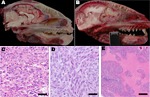
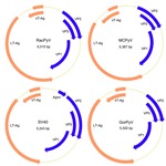
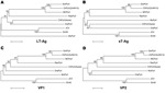
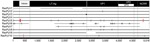
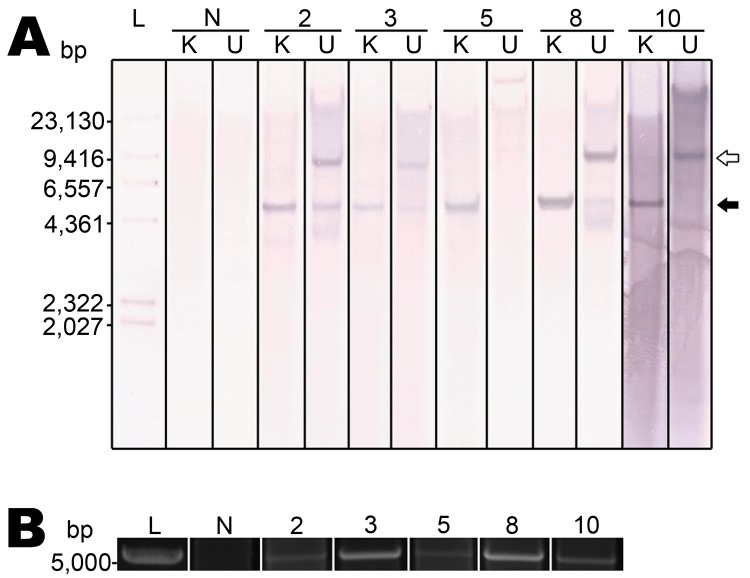
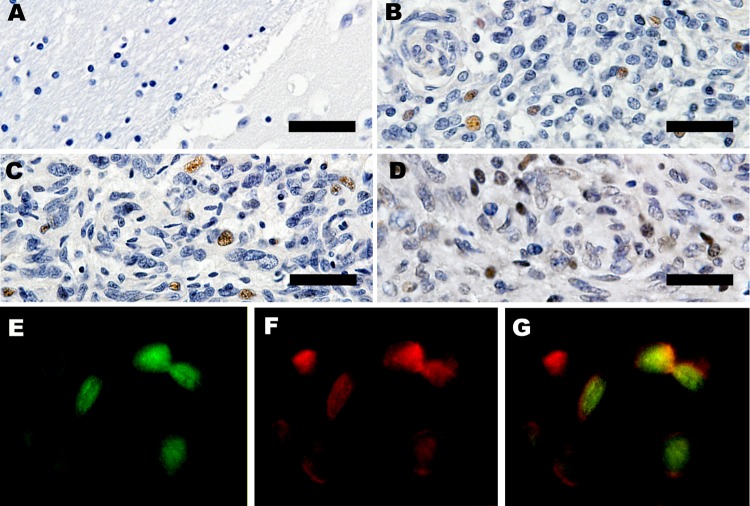
Tumors of any type are exceedingly rare in raccoons. High-grade brain tumors, consistently located in the frontal lobes and olfactory tracts, were detected in 10 raccoons during March 2010-May 2012 in California and Oregon, suggesting an emerging, infectious origin. We have identified a candidate etiologic agent, dubbed raccoon polyomavirus, that was present in the tumor tissue of all affected animals but not in tissues from 20 unaffected animals. Southern blot hybridization and rolling circle amplification showed the episomal viral genome in the tumors. The multifunctional nuclear protein large T-antigen was detectable by immunohistochemical analyses in a subset of neoplastic cells. Raccoon polyomavirus may contribute to the development of malignant brain tumors of raccoons.


Figures

Figure 1
Figure 1
Pathology of raccoon polyomavirus–associated tumors. A) Normal anatomy, head of unaffected raccoon, midsagittal section. An intact cribriform plate separates the ethmoid turbinates from the olfactory tract. B) Gross pathology, head of raccoon no. 9 (Rac9), left parasagittal section. The tumor obliterates the left olfactory tract and extends into the left frontal lobe to the level of the midbrain. The tumor compresses the brain and distorts the cerebellum. The raccoon head in length (crown to nose) is 16 cm. C) Histopathology (hematoxylin and eosin staining) for Rac5. Marked anisokaryosis and anisocytosis are evident. Original magnification ×40. D) Histopathology (hematoxylin and eosin staining) for Rac10. Shown is a region that has streams of elongated cells. Original magnification ×40. Scale bar = 40 μm. E) Histopathology (hematoxylin and eosin staining) for Rac3. Cell-dense sheets of neoplastic cells are interrupted by vast regions of necrosis. Original magnification ×20.
Figure 2
Figure 2
Genome organization of RacPyV. The entire dsDNA viral genome for RacPyV10 comprises 5,015 bp. The viral genome has a noncoding regulatory region and putative open reading frames for the late proteins VP1, VP2, and VP3 and the early proteins LT-Ag and sT-Ag. MCPyV and GorPyV, which are phylogenetic neighbors, and SV40 are presented for comparison. RacPyV, raccoon polyomavirus; LT-Ag, large T-antigen; sT-Ag, small T-antigen; VP, viral protein; MCPyV, Merkel cell polyomavirus; SV40, simian virus 40. GorPyV, gorilla polyomavirus.
Figure 3
Figure 3
Phylogenetic relationship of RacPyV with representative polyomavirus species. Phylogenetic trees were individually generated on the basis of amino acid sequences of LT-Ag (A), sT-Ag (B), VP1, and VP2 (C, D) by using the neighbor-joining method with p-distance and 1,000 bootstrap replications. RacPyV, raccoon polyomavirus; LT-Ag, large T-antigen; sT-Ag, small T-antigen; VP, viral protein; GorPyV, gorilla polyomavirus; ChPyV, chimpanzee polymavirus; MCPyV, Merkel cell polyomavirus; OraPyV, orangutan polyomavirus; JCV, JC virus; SV40, simian virus 40; MuPyV, murine polyomavirus. The bar represents 5% estimated phylogenetic divergence.
Figure 4
Figure 4
Partial genome sequences (ranging from 2,998 bp in raccoon polyomavirus 6 [RacPyV6] to 4,667 bp in RacPyV9) were obtained from 4 raccoons that either had undergone prolonged storage (RacPyV9) or were available only as formalin-fixed, paraffin-embedded tissue (dashed lines, RacPyVs 1, 6, and 7). Gaps in the sequences correspond to regions where amplification reactions failed. The genomes of RacPyVs 2, 3, 4, 5, 8, and 10 were sequenced in their entirety by using a primer walking method to complete the RacPyV circular genome. Open circles represent noncoding mutations; the closed circle represents a coding mutation. Horizontal bars indicate deletions. LT-Ag, large T-antigen; VP, viral protein; NCRR, noncoding regulatory region.
Figure 5
Figure 5
Raccoon polyomavirus (RacPyV) is episomal in raccoon brain tumors as detected by Southern blot hybridization and rolling circle amplification. A unique KpnI site in viral protein 1 (Technical Appendix Figure 2) conserved across the viral genomes was predicted to linearize circular RacPyV DNA. Genomic DNA was KpnI digested and probed with a 799-bp probe designed to hybridize to large T-antigen (Technical Appendix Figure 3). A) Southern blot hybridization. DNA digested with KpnI (K) and undigested DNA (U) and hybridized with RacPyV_LT_Probe1 shows identical banding patterns in each tumor. In KpnI-digested samples, a single band appears at ≈5 kb (closed arrow), which is the expected position for linearized RacPyV genome. B) RCA applied to RacPyV DNA by using random hexamers was digested with KpnI. Amplification of the circular RacPyV genome occurred in the same cohort of samples that was successful for the Southern blot hybridization. L, DNA ladder; N, DNA extracted from a raccoon that did not have a brain tumor (i.e., normal raccoon).
Figure 6
Figure 6
Expression of large T-antigen (LT-Ag) and p53 within a subset of tumors. A) Control, immunohistochemical analysis of frontal lobe of normal raccoon brain tissue. Astrocytes in this image and throughout the section were not immunoreactive for LT-Ag. Original magnification ×40. B, C) Immunohistochemical analysis for raccoon no. 2 (Rac2) and Rac3. LT-Ag is expressed within the nuclei of a subset of neoplastic astrocytes in 2 independent tumors. Original magnification ×40. D) Immunohistochemical analysis for Rac3. p53 is present within the nuclei of a subset of tumor cells. Original magnification ×40. Scale bars for panels A–D = 100 μm. E–G) Immunofluorescence of Rac4. p53 co-localizes with LT-Ag within nuclei of neoplastic cells. Original magnification ×60.

Similar articles

Cited by

References

    1. Johne R, Buck CB, Allander T, Atwood WJ, Garcea RL, Imperiale MJ, et al. Taxonomical developments in the family Polyomaviridae. Arch Virol. 2011;156:1627–34. 10.1007/s00705-011-1008-x - DOI - PMC - PubMed
    1. Colegrove KM, Wellehan JF Jr, Rivera R, Moore PF, Gulland FM, Lowenstine LJ, et al. Polyomavirus infection in a free-ranging California sea lion (Zalophus californianus) with intestinal T-cell lymphoma. J Vet Diagn Invest. 2010;22:628–32. 10.1177/104063871002200422 - DOI - PubMed
    1. Rollison DE. Epidemiologic studies of polyomaviruses and cancer: previous findings, methodologic challenges and future directions. Adv Exp Med Biol. 2006;577:342–56. 10.1007/0-387-32957-9_24 - DOI - PubMed
    1. Poulin DL, DeCaprio JA. Is there a role for SV40 in human cancer? J Clin Oncol. 2006;24:4356–65. 10.1200/JCO.2005.03.7101 - DOI - PubMed
    1. Chang Y, Moore PS. Merkel cell carcinoma: a virus-induced human cancer. Annu Rev Pathol. 2012;7:123–44. 10.1146/annurev-pathol-011110-130227 - DOI - PMC - PubMed

Publication types

MeSH terms

Substances

LinkOut - more resources