. 2012 Dec 19;103(12):2475-83.
doi: 10.1016/j.bpj.2012.11.008. Epub 2012 Dec 18.

Red blood cell membrane dynamics during malaria parasite egress

Affiliations

Red blood cell membrane dynamics during malaria parasite egress

Andrew Callan-Jones et al. Biophys J. .

Abstract

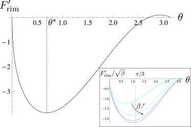
Precisely how malaria parasites exit from infected red blood cells to further spread the disease remains poorly understood. It has been shown recently, however, that these parasites exploit the elasticity of the cell membrane to enable their egress. Based on this work, showing that parasites modify the membrane's spontaneous curvature, initiating pore opening and outward membrane curling, we develop a model of the dynamics of the red blood cell membrane leading to complete parasite egress. As a result of the three-dimensional, axisymmetric nature of the problem, we find that the membrane dynamics involve two modes of elastic-energy release: 1), at short times after pore opening, the free edge of the membrane curls into a toroidal rim attached to a membrane cap of roughly fixed radius; and 2), at longer times, the rim radius is fixed, and lipids in the cap flow into the rim. We compare our model with the experimental data of Abkarian and co-workers and obtain an estimate of the induced spontaneous curvature and the membrane viscosity, which control the timescale of parasite release. Finally, eversion of the membrane cap, which liberates the remaining parasites, is driven by the spontaneous curvature and is found to be associated with a breaking of the axisymmetry of the membrane.


Figures

Figure 1
Figure 1
Merozoite egress from RBCs. Differential interference contrast microscopy reveals sequence of events leading to merozoite egress: a pore in the plasma membrane opens, allowing the first parasites to leave; next, the pore rim curls outward, causing further parasite escape; and finally, the remaining membrane cap everts, leading to complete egress.
Figure 2
Figure 2
Illustration of membrane dynamics. (a) Curling of the membrane, starting at the free edge, occurs at early times after pore formation; feeding of the membrane cap into the rim occurs at later times. (b) At a given moment, the RBC membrane can be described as a toroidal rim connected to a spherical cap. The arrows around the rim represent the direction of lipid flow in the membrane during feeding; the changing direction of the lipid flow gives rise to surface viscous dissipation.
Figure 3
Figure 3
The rim energy, Frim, as a function of pore opening angle. Frim is plotted in units of 2π2κR0/e and θ is in rads. The maximum in Frim near θ=π represents the nucleation barrier for pore opening; the minimum is a result of axisymmetry of the problem, and indicates that pure curling will stop near θ=θ. Parameter values: β=10 and γ˜=1. (Inset) Frim/β versus θ for β = 10, 100, 1000, and 10,000, illustrating the convergence of θ toward π/3.
Figure 4
Figure 4
Membrane dynamics during parasite egress. (a) The pore opening angle, θ, and cap radius, R, versus t. θ is shown for different values of the ratio of surface to bulk viscosities, ηs/η0, ranging from 0 to 500 μm, in increments of 100 μm. R is shown for ηs/η0=100μm. (Inset) Zoom of the early time behavior. θ is shown for ηs=0, revealing a smooth change from curling to feeding. R is shown for ηs/η0=100μm, showing a small decrease, expected initially during feeding based on the geometry. The other parameter values are c0=10μm−1, R0=3μm, e=0.05μm, ln=5, κ/η0=0.2μm3/ms, γ˜=1, and η/η0=100. (b) The cap depth, h, and number of turns in the rim, N, versus t. h is shown for different values of the surface/bulk viscosity ratio, ηs/η0, ranging from 0 to 500 μm, in increments of 100 μm. N is shown for ηs/η0=100μm. The other parameters are the same as in a.
Figure 5
Figure 5
Model fit to experimental results. The cap depth, h, versus t during parasite egress from RBCs. Data from Abkarian et al. (1) (circles) is fitted with the model developed in this work. The fit parameters are c0=13±4μm−1 and ηs/η0=1400±400μm. Deviation between the data and the model is expected for small h; the arrow indicates the expected shift in the model when the constraint of axisymmetry is relaxed (see text). The other, fixed parameters are: R0=3μm, e=0.05μm, ln=5, κ/η0=0.2μm3/ms, γ˜=1, and η/η0=100.
Figure 6
Figure 6
Final stage of parasite egress: eversion of the cap. As feeding ends, further elastic energy can be released by everting the cap, thus breaking the axisymmetry. For clarity, after eversion, the cap bends mainly along the x-direction. Eversion leads to a lateral displacement of the rim, denoted u.

References

    1. http://www.ncbi.nlm.nih.gov/pubmedhealth/PMH0001646/.
    1. Understanding Malaria. Information booklet. http://health.nih.gov/topic/Malaria.
    1. Abkarian M., Massiera G., Braun-Breton C. A novel mechanism for egress of malarial parasites from red blood cells. Blood. 2011;117:4118–4124. - PubMed
    1. Dvorak J.A., Miller L.H., Shiroishi T. Invasion of erythrocytes by malaria merozoites. Science. 1975;187:748–750. - PubMed
    1. Trager W. On the release of malaria merozoites. Trends Parasitol. 2002;18:60–61. - PubMed

Publication types

MeSH terms