. 2012 Dec 21;151(7):1608-16.
doi: 10.1016/j.cell.2012.11.018.

Foxa2 and H2A.Z mediate nucleosome depletion during embryonic stem cell differentiation

Affiliations

Foxa2 and H2A.Z mediate nucleosome depletion during embryonic stem cell differentiation

Zhaoyu Li et al. Cell. .

Abstract

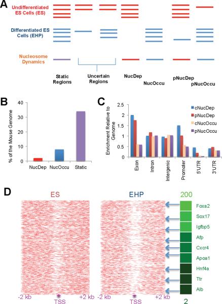
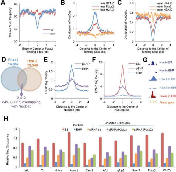
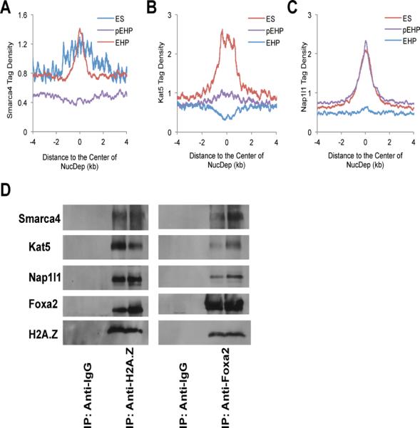
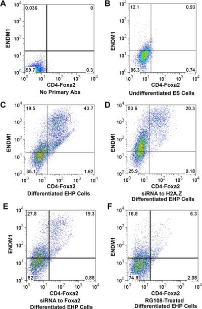
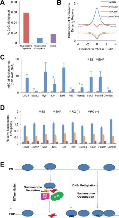
Nucleosome occupancy is fundamental for establishing chromatin architecture. However, little is known about the relationship between nucleosome dynamics and initial cell lineage specification. Here, we determine the mechanisms that control global nucleosome dynamics during embryonic stem (ES) cell differentiation into endoderm. Both nucleosome depletion and de novo occupation occur during the differentiation process, with higher overall nucleosome density after differentiation. The variant histone H2A.Z and the winged helix transcription factor Foxa2 both act to regulate nucleosome depletion and gene activation, thus promoting ES cell differentiation, whereas DNA methylation promotes nucleosome occupation and suppresses gene expression. Nucleosome depletion during ES cell differentiation is dependent on Nap1l1-coupled SWI/SNF and INO80 chromatin remodeling complexes. Thus, both epigenetic and genetic regulators cooperate to control nucleosome dynamics during ES cell fate decisions.


Figures

Figure 1
Figure 1
Nucleosome dynamics during embryonic stem cell differentiation (A) Schematic view of our computational analysis of nucleosome occupancy changes between undifferentiated ES (ES) and lineage-committed endodermic/hepatic progenitor (EHP) cells. Red and blue lines represent nucleosome occupancy in undifferentiated and differentiated ES cells, respectively. The nucleosome-bound and dynamic regions were identified computationally by the algorithm DANPOS (Figure S1B). Complete and partial nucleosome depletion regions (NucDep and pNucDep) and complete and partial nucleosome occupation regions (NucOccu and pNucOccu) were further defined following the DANPOS analysis. `Complete' means no sequencing tags found in either ES or EHP cells; `partial' means sequencing tags found in both cell types, but with at least a 4-fold difference. (B) Percentage of dynamic nucleosome regions in the whole genome during ES cell differentiation. (C) Distribution of nucleosome dynamic regions in the genome is normalized to the genomic distribution of all regions. 1 equals genomic distribution of each region. Genome, the whole mouse genome (mm8). (D) Nucleosome distribution near TSS of the 2,000 most activated (by mRNA level) genes during ES cell differentiation. Genes with increased expression after differentiation exhibit nucleosome depletion near their TSS. The gene list includes key hepatic differentiation markers, and the green shading indicates the degree of gene activation as measured by fold change of mRNA levels (up to 200) between EHP and ES cells. See also Figures S1 and S2.
Figure 2
Figure 2
H2A.Z and Foxa2 are required for nucleosome depletion during ES cell differentiation (A) Nucleosome distribution surrounding Foxa2 binding sites in ES and EHP cells. Note the decrease in nucleosome occupancy at Foxa2 sites in differentiated cells. (B and C) Genome-wide distribution of nucleosome depletion or occupation regions near H2A.Z, H2A.X and Foxa2 binding sites. (D) Co-binding regions of Foxa2 and H2A.Z overlap with nucleosome depletion regions. Foxa2 binding sites were pooled from EHP and pEHP (partially differentiated, see Figure S3A) cells; H2A.Z binding sites were pooled from ES and pEHP cells. (E and F) Foxa2 and H2A.Z tag density near Foxa2/H2A.Z-associated nucleosome depletion regions. (G) Co-localization of Foxa2/H2A.Z and nucleosome depletion regions in the intron of the Foxa1 gene. (H) Nucleosome depletion during ES-cell differentiation is dependent on Foxa2 and H2A.Z. Nucleosome occupancy was determined by qPCR at ten nucleosome depletion regions that are bound by both H2A.Z and Foxa2 in differentiated ES cells. Relative nucleoscome occupancy is shown for ES cells, sorted EHP cells, un-sorted EHP (siRNA (−), scramble siRNA control) cells, and un-sorted EHP cells transfected with siRNAs for H2afz (H2A.Z) or Foxa2. Error bars represent standard error of the mean. See also Figures S2–S5.
Figure 3
Figure 3
Foxa2/H2A.Z-driven nucleosome depletion complexes during ES cell differentiation. (A–C) Smarca4 (Brg1), Kat5 (Tip60), and Nap1l1, are enriched at nucleosome depletion regions. The tag density was normalized to 10 million sequencing tags for each sample; bin = 1 bp. (D) Immunoprecipitation (IP) experiments were performed with differentiated EHP cells using anti-H2A.Z or anti-Foxa2 antibodies and IP complexes were detected using western blotting with antibodies against Foxa2, H2A.Z, Smarca4, Kat5 and Nap1l1. Anti-IgG antibodies were used as negative IP controls. See also Figures S2–S5.
Figure 4
Figure 4
Nucleosome dynamics and ES cell differentiation. Flow cytometry analysis with dual cell surface markers of Foxa2/CD4 and ENDM1 for the assessment of the extent of differentiation (A) Control, differentiated ES cells sorted without incubation with primary antibodies. (B) Undifferentiated ES cells sorted with both antibodies. Less than 1% of the cells are double positive. (C) Differentiated ES cells, with more than 40% double-positive cells. (D) ES cells treated with siRNA to H2afz show decreased differentiation potential. (E) ES cells treated with siRNA to Foxa2 show decreased differentiation potential. (F) ES cells treated with 100 μM RG108, a DNA methyltransferase inhibitor, exhibit decreased differentiation. See also Figure S5.
Figure 5
Figure 5
DNA methylation promotes nucleosome occupation during ES cell differentiation (A) Cytosine DNA methylation profiles in undifferentiated ES cells were obtained from GEO (GSE11304), and compared to our nucleosome maps. The percentage of DNA methylation sites in ES cells is much higher at nucleosome-bound than at nucleosome-free regions. (B) Genome-wide distribution of dynamic nucleosome regions surrounding DNA methylation sites (m5C) in undifferentiated ES cells. (C) Nucleosomal DNA in ES and EHP cells was immunoprecipitated with an antibody against methylated cytosine (m5C), and DNA methylation was determined by qPCR at genomic regions near key pluripotency maker genes, which are silenced during ES cell differentiation. DNA at these loci is unmethylated in undifferentiated ES cells, but shows increased methylation after differentiation. The fold enrichment was normalized to genomic DNA. *, p<0.05; all others p< 0.01. Error bars represent standard error of the mean. (D) Nucleosome occupancy at nucleosome occupation regions of pluripotencey marker genes (the same regions assayed in Figure 5C) in ES cells, sorted EHP cells, un-sorted control EHP (RG(−)) cells and un-sorted EHP cells treated with RG108 (RG (+)), a DNA methyltransferase inhibitor. Nucleosome occupancy is severely blunted when DNA methylation is inhibited. Error bars represent standard error of the mean. (E) Schematic view of nucleosome dynamics during embryonic stem cell differentiation. mC, methylated cytosine. ES, undifferentiated embryonic stem cells; EHP, endodermic and hepatic progenitor cells. See also Figure S4.

Comment in

  • H2A.Z sets the stage in ESCs.
    Pandey R, Dou Y. Pandey R, et al. Cell Stem Cell. 2013 Feb 7;12(2):143-4. doi: 10.1016/j.stem.2013.01.012. Cell Stem Cell. 2013. PMID: 23395439

References

    1. Chodavarapu RK, Feng S, Bernatavichute YV, Chen PY, Stroud H, Yu Y, Hetzel JA, Kuo F, Kim J, Cokus SJ, et al. Relationship between nucleosome positioning and DNA methylation. Nature. 2010;466:388–392. - PMC - PubMed
    1. Clapier CR, Cairns BR. The biology of chromatin remodeling complexes. Annu Rev Biochem. 2009;78:273–304. - PubMed
    1. Gadue P, Gouon-Evans V, Cheng X, Wandzioch E, Zaret KS, Grompe M, Streeter PR, Keller GM. Generation of monoclonal antibodies specific for cell surface molecules expressed on early mouse endoderm. Stem Cells. 2009;27:2103–2113. - PMC - PubMed
    1. Gadue P, Huber TL, Nostro MC, Kattman S, Keller GM. Germ layer induction from embryonic stem cells. Exp Hematol. 2005;33:955–964. - PubMed
    1. Gadue P, Huber TL, Paddison PJ, Keller GM. Wnt and TGF-beta signaling are required for the induction of an in vitro model of primitive streak formation using embryonic stem cells. Proc Natl Acad Sci U S A. 2006;103:16806–16811. - PMC - PubMed

Publication types