. 2013 Feb;61(2):630-8.
doi: 10.1016/j.cyto.2012.11.014. Epub 2012 Dec 19.

Cloning and expression of feline colony stimulating factor receptor (CSF-1R) and analysis of the species specificity of stimulation by colony stimulating factor-1 (CSF-1) and interleukin-34 (IL-34)

Affiliations

Cloning and expression of feline colony stimulating factor receptor (CSF-1R) and analysis of the species specificity of stimulation by colony stimulating factor-1 (CSF-1) and interleukin-34 (IL-34)

Deborah J Gow et al. Cytokine. 2013 Feb.

Abstract

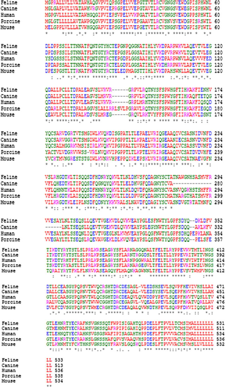
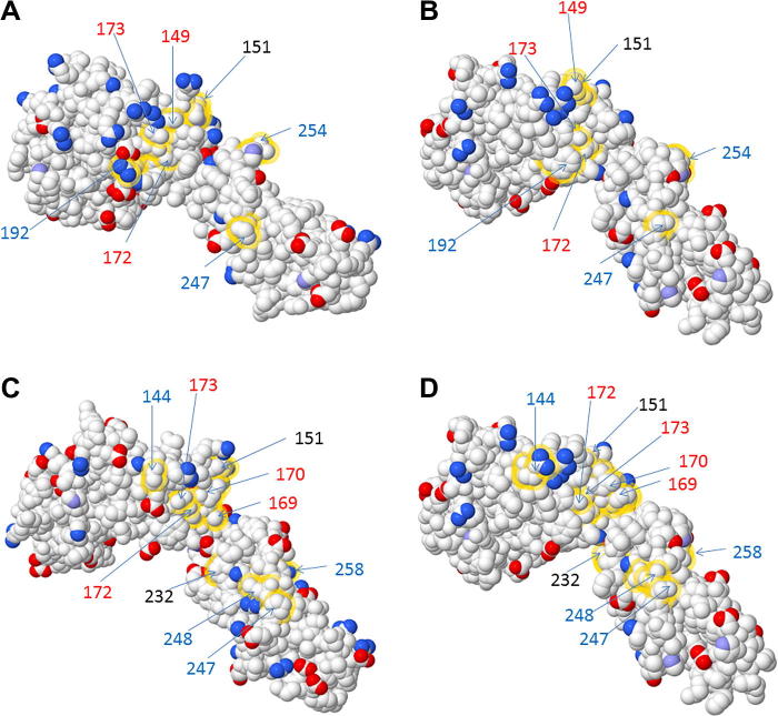
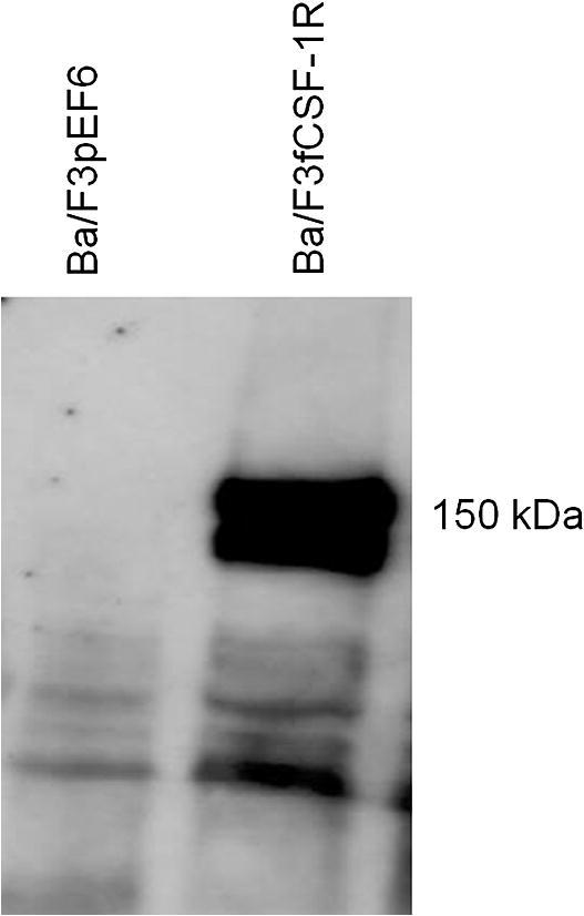
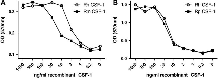
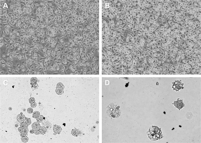
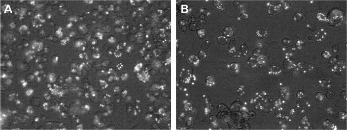
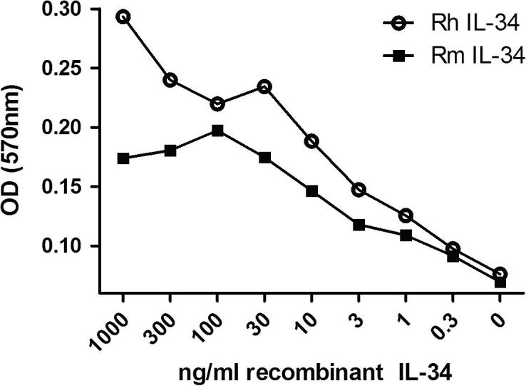
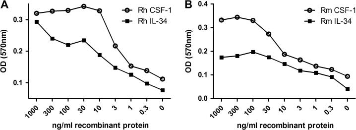
Colony stimulating factor (CSF-1) and its receptor, CSF-1R, have been previously well studied in humans and rodents to dissect the role they play in development of cells of the mononuclear phagocyte system. A second ligand for the CSF-1R, IL-34 has been described in several species. In this study, we have cloned and expressed the feline CSF-1R and examined the responsiveness to CSF-1 and IL-34 from a range of species. The results indicate that pig and human CSF-1 and human IL-34 are equally effective in cats, where both mouse CSF-1 and IL-34 are significantly less active. Recombinant human CSF-1 can be used to generate populations of feline bone marrow and monocyte derived macrophages that can be used to further dissect macrophage-specific gene expression in this species, and to compare it to data derived from mouse, human and pig. These results set the scene for therapeutic use of CSF-1 and IL-34 in cats.


Figures

Fig. 1
Fig. 1
Alignment of cloned feline CSF-1R extracellular domain with human, pig and mouse. Alignment was performed using Clustal W (http://www.ebi.ac.uk/Tools/msa/clustalw2/). The alignment clearly demonstrates the high level of homology that exists in the CSF-1R extracellular domain containing the IL-34 and CSF-1 binding sites. Of particular note is the high level of homology between both the feline and canine CSF-1R (88%) and feline and human CSF-1R (83%) while the homology between feline and the mouse and pig CSF-1R share 75%and 80% homology respectively. The red colour represents small hydrophobic amino acids, blue colour represents acidic amino acids, magenta denotes basic amino acids, and the green colour corresponds to hydrophilic or polar amino acids. Identical amino acids are represented by “∗”, conserved substitutions are represented by “:” and semi-conserved substitutions are represented by “.”.
Fig. 2
Fig. 2
3D models of non-conserved CSF-1 and IL-34 contact amino acids between human, mouse and cat CSF-1R. 3D models demonstrating the charged amino acid changes between human (A) and cat (B) CSF-1R and between mouse (C) and cat (D) CSF-1R. Models were generated using the PDB files for mouse CSF-1R (3EJJ) and human CSF-1R (4DKD chain X). The mouse PDB file (3EJJ) was used as a template to generate the cat CSF-1R. Published contact amino acids for both human and mouse CSF-1R binding sites for CSF-1 and IL-34 were analysed and non-conserved contact amino acids highlighted using FirstGlance (http://firstglance.jmol.org). Non-conserved amino acids for CSF-1 binding are represented by black numbers; red numbers indicate CSF-1 and IL-34 binding, while blue numbers indicates IL-34 binding. Positively charged atoms are represented by blue colour and negatively charged atoms by red colour. Medium blue coloured atoms denote partially charged atoms.
Fig. 3
Fig. 3
Western blot of transfected Ba/F3 cells expressing feline CSF-1R. A western blot was performed on Ba/F3 cells expressing feline CSF-1R. Cells were cultured in rhCSF-1 prior to collection of cell lysate. Cell lysate was collected from both Ba/F3 cells transfected with feline CSF-1R, but also Ba/F3 cell transfected with empty pEF6 vector as a negative control. A band at 150 kDa, corresponding to the predicted size is clearly visible for the feline CSF-1R, with no band identified for the negative control.
Fig. 4
Fig. 4
Activity of human, mouse and porcine recombinant CSF-1 on feline CSF-1R expressed in Ba/F3 cells. An MTT cell viability assay was used to assess the biological activity of recombinant human, mouse and porcine CSF-1 in the feline CSF-1R. (A) Both human and mouse CSF-1 are biologically active on the feline CSF-1R, with mouse CSF-1 demonstrating reduced activity compared to human CSF-1. (B) Comparing the effects of human and porcine CSF-1 on the feline CSF-1R demonstrates that these recombinant proteins are identical in their activation.
Fig. 5
Fig. 5
Feline monocyte derived and bone marrow derived macrophages. Feline peripheral blood monocytes and bone marrow cells progenitor cells were cultured with 10Units/ml CSF-1 for 8 days. For both the peripheral blood mononuclear cell (A), and bone marrow cell (B), cultures with rh-CSF-1, cells were adherent, had increased in size and granularity and divided, thus producing blood monocyte and bone marrow derived macrophages respectively. Cytospin analysis of the blood monocyte derived macrophages (C) and bone marrow derived macrophages (D) demonstrate that both populations display macrophage-like morphology.
Fig. 6
Fig. 6
Phagocytic activity of feline monocyte and bone marrow derived macrophages. Cells were cultured for 10 days in rh-CSF-1, prior to the addition of FITC-labelled Zymosan particles for 1 h. Both cell populations were highly phagocytic and ingested numerous particles of Zymosan after the 1 h incubation. (A) BMC and (B) PBMC.
Fig. 7
Fig. 7
Activity of recombinant human and mouse IL-34 on feline CSF-1R expressed in Ba/F3 cells. An MTT cell viability was used to assess the biological activity of human and mouse IL-34 on expressed feline CSF-1R. Both human and mouse IL-34 are biologically active on the feline CSF-1R in a dose dependant manner. The EC50 for mouse IL-34 on the feline CSF-1R was approximately 3-fold higher than for human IL-34.
Fig. 8
Fig. 8
Comparing the activity of human and mouse IL-34 and CSF-1 on expressed feline CSF-1R. An MTT cell viability assay was used to compare the biological activity of human and mouse IL-34 with human and mouse CSF-1. For both human (A) and mouse (B), IL-34 has demonstrated approximately half the biological activity compared to human and mouse CSF-1.

Similar articles

Cited by

References

    1. Hume D.A., Macdonald K.P. Therapeutic applications of macrophage colony-stimulating factor-1 (CSF-1) and antagonists of CSF-1 receptor (CSF-1R) signaling. Blood. 2012;119:1810–1820. - PubMed
    1. Gow D.J., Garceau V., Kapetanovic R., Sester D.P., Fici G.J., Shelly J.A. Cloning and Expression of Porcine Colony Stimulating Factor-1 (CSF-1) and Colony Stimulating Factor-1 Receptor (CSF-1R) and analysis of the species specificity of stimulation by CSF-1 and Interleukin 34. Cytokine. 2012 http://dx.doi.org/10.1016/j.cyto.2012.08.008. - PMC - PubMed
    1. Chitu V., Stanley E.R. Colony-stimulating factor-1 in immunity and inflammation. Curr Opin Immunol. 2006;18:39–48. - PubMed
    1. Bonifer C., Hume D.A. The transcriptional regulation of the colony-stimulating factor 1 receptor (csf1r) gene during hematopoiesis. Front. Biosci. 2008;13:549–560. - PubMed
    1. Sherr C.J., Rettenmier C.W., Sacca R., Roussel M.F., Look A.T., Stanley E.R. The c-fms proto-oncogene product is related to the receptor for the mononuclear phagocyte growth factor, CSF-1. Cell. 1985;41:665–676. - PubMed

Publication types

MeSH terms